cancer
Laura Pedranzini, … , Andrea Leitch, Jacqueline Bromberg
J Clin Invest.
2004;
114(5)
:619-622.
https://doi.org/10.1172/JCI22800
.
Signal transducer and activator of transcription 3 (Stat3) is a transcription factor that is
constitutively activated in a variety of human malignancies, including prostate, lung, brain,
breast, and squamous cell carcinomas. Inhibition of activated Stat3 leads to decreased
proliferation and apoptosis of many cancer-derived cell lines, while the introduction of a
constitutively activated form of Stat3 into immortalized human breast epithelial cells and
rodent fibroblasts results in cellular transformation. Collectively, these data suggest a role
for Stat3 in oncogenesis. A new study from Chan et al. (see related article beginning on
page 720) is the first to demonstrate a requirement for Stat3 in de novo epithelial
carcinogenesis in vivo. Using the two-step model of chemically induced skin
carcinogenesis, the authors demonstrated that mice deficient in Stat3 were completely
resistant to skin tumor development.
Commentary
Find the latest version:
the antiphospholipid syndrome. J. Clin. Invest.
112:1644–1654. doi:10.1172/JCI200318817. 15. Holers, V.M., and Thurman, J.M. 2004. The
alter-native pathway of complement in disease: oppor-tunities for therapeutic targeting. Mol. Immunol.
41:147–152.
16. Gigli, I., Sorvillo, J., Mecarelli-Halbwachs, L., and Leibowitch, J. 1981. Mechanism of action of the
C4 nephritic factor. Deregulation of the classical pathway of C3 convertase. J. Exp. Med.154:1–12. 17. Botto, M., et al. 1998. Homozygous C1q deficiency
causes glomerulonephritis associated with mul-tiple apoptotic bodies. Nat. Genet.19:56–59. 18. Bowness, P., et al. 1994. Hereditary C1q
deficien-cy and systemic lupus erythematosus. Q. J. Med.
87:455–464.
19. Mitchell, D.A., et al. 2002. C1q deficiency and autoimmunity: the effects of genetic background on disease expression. J. Immunol.168:2538–2543. 20. Mitchell, D.A., et al. 1999. C1q protects against the
development of glomerulonephritis independently of C3 activation. J. Immunol.162:5676–5679. 21. Botto, M., and Walport, M.J. 2002. C1q,
autoimmu-nity and apoptosis. Immunobiology.205:395–406.
Stat3 is required for the development
of skin cancer
Laura Pedranzini, Andrea Leitch, and Jacqueline Bromberg Memorial Sloan Kettering Cancer Center, New York, New York, USA.
Signal transducer and activator of transcription 3 (Stat3) is a transcription
fac-tor that is constitutively activated in a variety of human malignancies,
includ-ing prostate, lung, brain, breast, and squamous cell carcinomas. Inhibition of
activated Stat3 leads to decreased proliferation and apoptosis of many
cancer-derived cell lines, while the introduction of a constitutively activated form of
Stat3 into immortalized human breast epithelial cells and rodent fibroblasts
results in cellular transformation. Collectively, these data suggest a role for
Stat3 in oncogenesis. A new study from Chan et al. (see related article
begin-ning on page 720) is the first to demonstrate a requirement for Stat3 in de
novo epithelial carcinogenesis in vivo. Using the two-step model of chemically
induced skin carcinogenesis, the authors demonstrated that mice deficient in
Stat3 were completely resistant to skin tumor development.
Nonstandard abbreviations used: DMBA, 7,12-dimethylbenz[a]anthracene; Ha-ras, Harvey rat sarcoma virus oncogene; JAK, Janus kinase; LRC, label-retaining cell; RTK, receptor tyrosine kinase; SH2, src homology domain 2; Stat3, signal transducer and activator of tran-scription 3; TPA, 12-O-tetradecanoylphorbol-13-acetate;
v-Ha-ras, Ha-ras homolog.
Conflict of interest: The authors have declared that no conflict of interest exists.
Citation for this article:J. Clin. Invest.114:619–622 (2004). doi:10.1172/JCI200422800.
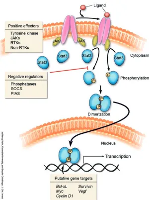
Stat’s (signal transducers and activa-tors of transcription) are a family of latent transcription factors that are acti-vated in response to many cytokines and growth factors. Stat activation is depen-dent upon tyrosine phosphorylation, which induces dimerization via reciprocal phosphotyrosine–src homology domain 2 (phosphotyrosine–SH2) interaction between two Stat molecules. Activated Stat’s translocate to the nucleus where they bind to consensus promoter sequences of target genes and activate their transcription (1) (Figure 1). Many tyrosine kinases, includ-ing JAKs (Janus kinases), RTKs (recep-tor tyrosine kinases), and non-RTKs can phosphorylate Stat proteins. In normal cells, Stat tyrosine phosphorylation is transient, lasting from 30 minutes to several hours.
However, in numerous cancer-derived cell lines or in primary tumors, Stat proteins (in particular Stat3) are persistently tyrosine phosphorylated either as a consequence of deregulated positive effectors of Stat acti-vation such as tyrosine kinases or negative regulators of Stat phosphorylation, e.g., phosphatases, suppressor of cytokine signal-ing, protein inhibitor of activated stats) (2). Inhibition of Stat3 activity in tumor-derived cell lines by the introduction of antisense, small interfering RNA, dominant-negative Stat3 constructs, and/or blockade of tyro-sine kinases has been associated with growth arrest and apoptosis (2). Furthermore, the introduction of a constitutively activated Stat3 molecule (Stat3C) into immortalized cell lines leads to transformation, indicating an oncogenic role for activated Stat3 (3, 4). A possible mechanism for transformation by activated Stat3 is the transcriptional upregulation of genes known to be involved in proliferation and apoptosis, including
Bcl-xL, c-Myc, cyclinD1, Vegf, and Survivin (3, 5–8). In addition to its role as a transcrip-tion factor, phosphorylated Stat3 has been described in a recent report as a component of focal adhesions (sites of cell contact with the extracellular matrix) that may contribute
to the invasiveness of ovarian cancer cells (9). The in vivo role of Stat3 in tumorigenesis has not been addressed until now. In this
issue of the JCI, Chan and colleagues
dem-onstrate in two different murine models of skin tumor development that Stat3 is required for de novo tumorigenesis (10).
One of the best-established model sys-tems for studying the mechanisms under-lying the process of malignant transforma-tion is the mouse skin model of multistage carcinogenesis (11, 12) (Figure 2). In this model, the process of skin tumor develop-ment can be subdivided into three differ-ent stages: initiation, promotion, and pro-gression. Initiation is typically induced by the topical application of the carcinogen 7,12-dimethylbenz[a]anthracene (DMBA). Interestingly, in DMBA-treated epidermal cells, one usually finds mutations within the Harvey rat sarcoma virus oncogene (Ha-ras) gene. These mutations, however, are not sufficient to induce de novo trans-formation. Promotion of tumorigenesis is generated by the topical application
of phorbol esters such as 12-O
-tetradec-anoylphorbol-13-acetate (TPA) to the skin, leading to epithelial cell proliferation with a concomitant increased expression of the ligand EGF as well as of cyclin D1, c-Jun, c-Fos, and c-Myc (13–16). TPA-treated mice form multiple benign papillomas within 10–20 weeks. Tumor progression is a spon-taneous process resulting in the formation of malignant squamous carcinomas.
Stat3 prevents apoptosis in the initiation phase of skin tumorigenesis
K5Cre.Stat3f l/f l transgenic mice, whose
However, they have defects in hair cycle processes as well as impaired wound heal-ing, and they develop spontaneous ulcers with age. In vitro, keratinocytes derived from these mice had no defects in pro-liferation, but growth factor–dependent migration was markedly impaired in con-trast to control keratinocytes (17). In the
study described in this issue of the JCI,
Chan et al. used K5Cre.Stat3fl/fl
transgen-ic mtransgen-ice to investigate the role of Stat3 in chemically induced carcinogenesis of the skin (10). The role of Stat3 in tumor ini-tiation, the first step of chemically induced carcinogenesis, was addressed both in vitro, in DMBA-treated primary keratinocytes, and in vivo, by topical treatment with this mutagen. Stat3 mutant keratinocytes underwent enhanced apoptosis follow-ing DMBA treatment, compared to
con-trol keratinocytes. Expression of Ha-ras
homolog (v-Ha-ras) into cultured primary
keratinocytes in vitro was used to gen-erate initiated keratinocytes. Upon the introduction of a Stat3 decoy molecule (a high-affinity double-stranded DNA bind-ing site for Stat3), the v-Ha-ras–initiated keratinocytes underwent apoptosis with a concomitant decrease in Bcl-xL levels. In general, inhibiting Stat3 function in can-cer-derived cell lines containing abundant phosphorylated Stat3 leads to apoptosis or growth arrest. In contrast, cell lines which contain low or no levels of detectable tyro-sine-phosphorylated Stat3 are relatively unaffected by Stat3 inhibitory therapies. It therefore remains unclear how Stat3 protects keratinocytes against DMBA-induced apoptosis, since DMBA does not induce tyrosine phosphorylation of Stat3 in primary keratinocytes, nor is it likely that v-Ha-ras–containing keratinocytes contain abundant levels of phosphorylated Stat3 (18, 19). Perhaps the low amounts
of phosphorylated Stat3 present in these cells are sufficient to drive transcription of
antiapoptotic genes such as Bcl-xL.
Alter-natively, nonphosphorylated Stat3 may be playing a role as a transcription factor as has been demonstrated for Stat1 (20). There are a few notable examples where relatively low levels of phosphorylated Stat3 are sufficient to mediate protection from growth arrest or apoptosis (21, 22). Thus, determination of the relative levels of phosphorylated Stat3 required to impart a phenotype is likely to be cell-type specific and remains an important objective. It has been shown that phosphorylated Stat1 levels are markedly enhanced in Stat3 null hepatocytes (23). Given that Stat1 activa-tion has been implicated in promoting growth arrest as well as apoptosis, it would be of interest to determine whether the enhanced apoptosis observed in the Stat3 null keratinocytes correlates with increased levels of activated Stat1.
It is hypothesized that keratinocyte stem cells, which are located mostly within the bulge region of the hair follicle, are the tar-get cells for two-stage carcinogenesis (24). Hair follicle stem cells have been identified within the label-retaining cells (LRCs), a population of cells that, following continu-ous administration of nucleotide analogs
such as BrdU or [3H]thymidine, retains the
[image:3.585.50.352.77.479.2]label for a sustained period of time, indicat-ing a very slow cyclindicat-ing frequency (25, 26).
Figure 1
Chan et al. (10) observed that the majority of the Stat3-deficient keratinocytes under-going apoptosis after exposure to DMBA were located primarily within the bulge region of the hair follicle in an area adja-cent to the LRC population. The authors suggest that the DMBA-sensitive cells may be keratinocyte stem cells, given their prox-imity to the LRCs. However, given the com-plete lack of overlap between the LRCs and the apoptotic cells, the cell type most sensi-tive to DMBA-induced apoptosis remains to be identified. The work of Chan et al. generates interesting questions regarding the mechanism(s) by which Stat3 affords protection against apoptosis and the deter-mination of which cell type(s) are most sen-sitive to the loss of Stat3.
Stat3 is required for keratinocyte proliferation during the promotion stage of chemically induced carcinogenesis
Tumor promotion, the second stage of chemically induced skin carcinogenesis, occurs following the topical application of TPA, leading to the proliferation of epi-dermal cells and the subsequent formation of benign papillomas in wild-type animals. Perhaps the most important observation made by Chan et al. (10) is that mice
defi-cient in Stat3 were completely resistant to skin tumor development. Given that DMBA treatment resulted in keratinocyte
apoptosis in the K5Cre.Stat3fl/fl animals,
there was undoubtedly a decreased num-ber of keratinocytes capable of proliferat-ing in response to TPA. Nevertheless, the role of TPA alone (in the absence of the initiation stage) was assessed in vivo by applying TPA topically. Marked epider-mal hyperproliferation and BrdU uptake was observed in wild-type animals. In con-trast, significantly less proliferation was
seen in the K5Cre.Stat3fl/fl animals. TPA
induced the tyrosine phosphorylation of Stat3, perhaps in part through increased production of EGF leading to activation of the EGFR (13). An increase in some of the known putative Stat3 targets, includ-ing cyclin D1 and c-Myc, was observed in the skin of wild-type animals while in
the K5Cre.Stat3fl/fl animals a delay in the
increased level of these proteins was seen. These data demonstrate that upregulation of these genes is partly dependent upon Stat3. The requirement of Stat3 in tumor promotion was also examined in trans-genic animals expressing v-Ha-ras targeted to keratinocytes. Topical TPA treatment of these animals led to benign papilloma formation, which was abrogated upon the
introduction of a Stat3 decoy molecule. The use of a Stat3 decoy was first described as an inhibitor of tubular morphogen-esis in cells grown in culture (27). It was subsequently shown to inhibit growth of squamous cell carcinoma–derived cell lines in vitro and more recently in vivo (28) (J. Grandis, personal communication).
The use of this molecule in vivo is indeed novel, as is the manner in which the Stat3 decoy was administered (10). The Stat3 decoy was applied topically to the mice on a weekly basis. It is remarkable that the DNA was absorbed in sufficient quantities to exert a profound inhibitory effect on papilloma formation. Intralesional injec-tion of the Stat3 decoy was also effective in decreasing the size of existing papillomas. It would be of interest to know whether an increase in apoptosis was observed in the Stat3 decoy–injected lesions, as has been described for preformed B16 melanoma tumors injected with a dominant negative Stat3 construct (29).
Decoy molecules against Stat3 may indeed be effective therapy for cutaneous or locally advanced cancers, including skin cancers, head and neck tumors, and meta-static breast cancers involving the skin. One potential caveat with the Stat3 decoy relates to the presumed concomitant
inhi-Figure 2
[image:4.585.53.529.79.281.2]bition of Stat1 activity. The decoy should bind equally well to the Stat3 and Stat1 dimers. Given the evidence that Stat1 may function as a tumor suppressor as well as a promoter of apoptosis, the sequestering of the Stat1 dimer may oppose the tumori-cidal effects of sequestering Stat3 (30–32). Nevertheless, this model system of skin tumorigenesis is an ideal one for develop-ing and testdevelop-ing anti-Stat3 drugs for the prevention and treatment of skin cancer.
Address correspondence to: Jacqueline Bromberg, Memorial Sloan Kettering Cancer Center, 1275 York Avenue, New York, New York 10021, USA. Phone: (212) 639-8191; Fax: (212) 422-2231; E-mail: [email protected].
1. Darnell, J.E., Jr. 1997. STATs and gene regulation.
Science.277:1630–1635.
2. Bromberg, J. 2002. Stat proteins and oncogen-esis. J. Clin. Invest.109:1139–1142. doi:10.1172/ JCI200215617.
3. Bromberg, J., et al. 1999. Stat3 as an oncogene. Cell.
98:295–303.
4. Dechow, T.N., et al. 2004. Requirement of matrix metalloproteinase-9 for the transformation of human mammary epithelial cells by Stat3-C. Proc. Natl. Acad. Sci. U. S. A.101:10602–10607. 5. Catlett-Falcone, R., et al. 1999. Constitutive
acti-vation of Stat3 signaling confers resistance to apoptosis in human U266 myeloma cells. Immu-nity.10:105–115.
6. Niu, G., et al. 2002. Constitutive Stat3 activity up-regulates VEGF expression and tumor angiogenesis.
Proc. Natl. Acad. Sci. U. S. A. 21:2000–2008. 7. Kanda, N., et al. 2004. STAT3 is constitutively
acti-vated and supports cell survival in association with
survivin expression in gastric cancer cells. Oncogene.
23:4921–4929.
8. Schlette, E.J., Medeiros, L.J., Goy, A., Lai, R., and Rassidakis, G.Z. 2004. Survivin expression predicts poorer prognosis in anaplastic large-cell lympho-ma. J. Clin. Oncol.22:1682–1688.
9. Silver, D.L., Naora, H., Liu, J., Cheng, W., and Mon-tell, D.J. 2004. Activated signal transducer and activator of transcription (STAT) 3: localization in focal adhesions and function in ovarian cancer cell motility. Cancer Res.64:3550–3558.
10. Chan, K.S., et al. 2004. Disruption of Stat3 reveals a critical role in both the initiation and the pro-motion stages of epithelial carcinogenesis. J. Clin. Invest.114:720–728. doi:10.1172/JCI200421032. 11. Angel, J.M., and DiGiovanni, J. 1999. Genetics
of skin tumor promotion. Prog. Exp. Tumor Res.
35:143–157.
12. Wu, X., and Pandolfi, P.P. 2001. Mouse models for multistep tumorigenesis. Trends Cell Biol.11:S2–S9. 13. Chan, K.S., et al. 2004. Epidermal growth factor
receptor-mediated activation of Stat3 during multi-stage skin carcinogenesis. Cancer Res.64:2382–2389. 14. Robles, A.I., and Conti, C.J. 1995. Early
overexpression of cyclin D1 protein in mouse skin carcinogenesis. Carcinogenesis.16:781–786. 15. Saez, E., et al. 1995. c-fos is required for malignant
progression of skin tumors. Cell.82:721–732. 16. Hsu, J.D., et al. 1999. Suppression of the
TPA-induced expression of nuclear-protooncogenes in mouse epidermis by crocetin via antioxidant activ-ity. Anticancer Res.19:4221–4227.
17. Sano, S., et al. 1999. Keratinocyte-specific abla-tion of stat3 exhibits impaired skin remodeling, but does not affect skin morphogenesis. EMBO J.
18:4657–4668.
18. Bromberg, J.F., Horvath, C.M., Besser, D., Lathem, W.W., and Darnell, J.E., Jr. 1998. Stat3 activation is required for cellular transformation by v-src. Mol. Cell. Biol.5:2553–2558.
19. Turkson, J., Bowman, T., Garcia, R., Caldenhoven, E., De Groot, R.P., and Jove, R. 1998. Stat3 activa-tion by Src induces specific gene regulaactiva-tion and is required for cell transformation. Mol. Cell. Biol.
18:2545–2552.
20. Chatterjee-Kishore, M., Wright, K.L., Ting, J.P., and Stark, G.R. 2000. How Stat1 mediates constitutive gene expression: a complex of unphosphorylated Stat1 and IRF1 supports transcription of the LMP2 gene. EMBO J.19:4111–4122.
21. Niu, G., et al. 2002. Roles of activated Src and Stat3 signaling in melanoma tumor cell growth. Onco-gene.21:7001–7010.
22. Li, L., and Shaw, P.E. 2002. Autocrine-mediated activation of STAT3 correlates with cell prolif-eration in breast carcinoma lines. J. Biol. Chem.
277:17397–17405.
23. Li, W., Liang, X., Kellendonk, C., Poli, V., and Taub, R. 2002. STAT3 contributes to the mitogenic response of hepatocytes during liver regeneration.
J. Biol. Chem.277:28411–28417.
24. Morris, R.J. 2000. Keratinocyte stem cells: targets for cutaneous carcinogens. J. Clin. Invest.106:3–8. 25. Morris, R.J., et al. 2004. Capturing and profiling adult
hair follicle stem cells. Nat. Biotechnol.22:411–417. 26. Tumbar, T., et al. 2004. Defining the epithelial stem
cell niche in skin. Science.303:359–363. 27. Boccaccio, C., et al. 1998. Induction of
epithe-lial tubules by growth factor HGF depends on the STAT pathway. Nature.391:285–288.
28. Leong, P.L., et al. 2003. Targeted inhibition of Stat3 with a decoy oligonucleotide abrogates head and neck cancer cell growth. Proc. Natl. Acad. Sci. U. S. A.
100:4138–4143.
29. Niu, G., et al. 1999. Gene therapy with domi-nant-negative Stat3 suppresses growth of the murine melanoma B16 tumor in vivo. Cancer Res.
59:5059–5063.
30. Bromberg, J.F., Horvath, C.M., Wen, Z., Schreiber, R.D., and Darnell, J.E., Jr. 1996. Transcription-ally active Stat1 is required for the antiproliferative effects of both IFN-a and IFN-g. Proc. Natl. Acad. Sci. U. S. A.93:7673–7678.
31. Kaplan, D.H., et al. 1998. Demonstration of an interferon gamma-dependent tumor surveillance system in immunocompetent mice. Proc. Natl. Acad. Sci. U. S. A.95:7556–7561.
32. Townsend, P.A., et al. 2004. STAT-1 interacts with p53 to enhance DNA damage-induced apoptosis.