Journal of Chemical and Pharmaceutical Research, 2014, 6(8):28-38
Research Article
CODEN(USA) : JCPRC5
ISSN : 0975-7384
Evaluation of antimicrobial activity of earthworm Eudrilus eugeniae
tissue extract
1*
Praval Singh Chauhan,
2Jyoti Tomar,
3G. B. K. S. Prasad and
1O. P. Agrawal
1
School of Studies in Zoology, Jiwaji University, Gwalior, MP, India
2
Department of Chemistry & Environmental Sciences, ITM University, Gwalior, India
3
School of Studies in Biochemistry, Jiwaji University, Gwalior, MP, India
_____________________________________________________________________________________________
ABSTRACT
As earlier reported that the defense system of earthworm is found in their coelomic fluid. Different proteins and peptides (lysozyme and fetidins) have been isolated in different species of earthworm, responsible for their antibacterial activity. Antibacterial & antifungal activities of the whole tissue extract of earthworm, Eudrilus eugeniae was tested using well diffusion method. Nine species were selected for antimicrobial screening and two for antifungal screening. The bacterial cultures were maintained on nutrient agar slant and the fungal strains were maintained on Sabouraud dextrose agar slant at 4°C as well as at -20°C by making glycerol stocks. Earthworm paste at a dose of 10mg/ml was able to inhibit the growth of bacteria. Among nine bacterial isolates only five species responded to earthworm extract. Maximum antibacterial activity was found with gram negative Pseudomonas aeruginosa (29 ± 1mm), which was almost similar to the standard antibiotic Streptomycin. (29.5±0.5 mm), Minimum antibacterial activity 21.5±1.5 mm was obtained with the gram positive Staphylococcus aureus. Earthworm extract didn’t not express any inhibition on Streptococcus pyrogenes, Enterococcus faecalis, Enterobacter aerogenes and Escherichia coli. No antifungal activity was observed against Candida albicans and Aspergillus niger. Total concentration of protein was found 70 mg/100 gm. Thus these results clearly indicates that the earthworm tissue extract have good antibacterial potential and the bioactive compounds to inhibit the growth of bacteria.
Key words: Eudrilus eugeniae, antimicrobial activity, fungal and bacteria strains.
_____________________________________________________________________________________________
INTRODUCTION
Earthworm increases the soil fertility and is often referred to as “farmer’s friend” or soil engineers. Earthworm has been recognized in oriental medicine as anti-inflammatory, analgesic and antipyretic agent [1-3]. In the 700 million years of existence, earthworms have evolved in the environment replete with microorganisms, some of which threatening their existence. To survive themselves in such an environment, earthworms possess humoral as well as cellular immune system against these microorganisms. Defense system of earthworm is found in their coelomic fluid [4]. Different proteins and peptides (lysozyme and fetidins) have been discovered in different species, are responsible for their antibacterial activity [5&6].
Earthworm Perionyx excavates contains amazing antimicrobial activity due to their high protein, nitrogen and fat content [7]. Cho et al. (1998) identified the first antimicrobial peptide (lumbricin I) from the earthworm Lumbricus
rubellus. Lumbricin I is considered as a proline-rich antimicrobial peptide containing 62 amino acids including
Pheretima tschiliensis and E. foetida respectively [10]. Balamurugan et al. (2008) found that earthworm coelomic
fluid contains biologically active molecules and leukocytes that participate in phagocytosis, encapsulation and killing of HeLa, HEp-2, PC-12 and PA317 cells in-vitro [2]. Presumably, earthworms synthesize and secrete several effective modulators of innate immune responses such as antibacterial molecules, cytotoxic proteins and cytokines. Increasing burden of antimicrobial resistance in clinically relevant bacteria is an emerging threat around the world. Discovery of new generation antibiotic is challenging task for the researchers as bacteria start resisting soon after introduction of the antibiotics. Furthermore newer drugs are less available and more expensive for resource poor communities. Antimicrobial potency of Eudrilus eugeniae extracts on certain plant pathogens were also studied [11]. Hence, in the present study the paste prepared from the earthworm Eudrilus eugeniae was tested for total protein content and antimicrobial & antifungal potential.
EXPERIMENTAL SECTION
2.1 Procurement of Materials:
All essential chemicals were procured from Hi-Media, SRL, Qualigens and Sigma Aldrich, U.S.A. and they were of high quality (AR grade). The glass wares used were of Corning, Borocil etc., Plastic wares of Tarsons and micro-pipettes of CE, Qualigens. Cattle dung required for vermicomposting was obtained from Buffalo Dairy Farms, Gwalior (M. P.) India.
2.2 Standardization of Techniques:
All the techniques used in the present study were first standardized in preliminary experiments in which, earthworm extract was found to be positive for different parameters of bioogical activities selected.
2.3 Procurement and Maintenance of Animals:
[image:2.595.138.480.444.699.2]Adult Wistar rats of requisite weight range used in different experiments were procured from Defence Research and Development Establishment (DRDE), Gwalior, India. They were maintained in animal house facility of School of Studies in Zoology of Jiwaji University, Gwalior under standard conditions (25±20 C, 55-60% RH and 12:12 hour light and dark cycling) and they were provided with standard laboratory rat feed pellets (Ashirvad Industries, Chandigarh, India) and water ad libitum. All experimental protocols and procedures were in accordance of institutional regulations and national criteria for animal experimentation and due approval was taken from the Institution Animal Ethics Committee.
Fig. 2: Large size vermin-tank used for mother culture of Eudrilus eugeniae at Vermicomposting Centre, Charak Udyan, Jiwaji University, Gwalior
2.4 Culture and Maintenance of Eudrilus eugeniae:
Adult earthworms, Eudrilus eugeniae (Annelida: Oligochaeta:Lumbricidae) were procured from the mother culture maintained in a large sized vermi-tank at Vermicomposting Center, located in Charak Udyan, Jiwaji University, Gwalior, (M. P.), India (Figs. 1& 2). The earthworm culture was maintained in a mixture of plant leaves and cattle dung (standard medium).
2.5 Preparation of Earthworm Extract:
Earthworm extract was prepared according to Hrzenjak et al. (1992) [12] with some modifications. Sexually mature clitellate worms (1.345 gm/worm approx.), collected from the stock culture, were thoroughly washed under running tap water to remove dung, debris etc. attached on their body surface. Then the worms were kept in 0.65% NaCl solution, at room temperature for two hours for removal of cast from the body. The solution of NaCl was changed after every hour. After two hours, NaCl solution was discarded and the animals were transferred in a glass tray. Tray was covered tightly by a cotton cloth through which air can penetrate. Earthworms were then left overnight for maximum release of cast from their body. Worms were then minced with a pair of scissors. Three grams of minced earthworm tissue was weighed and was mixed with 40 ml of chloroform-methanol (V/V) solution. The mixture was then homogenized and kept at 4oC overnight. The very next day, 16 ml of distilled water was added to the homogenate. It was mixed and centrifuged at 5000 RPM for 10 min. to obtain three clearly differentiated layers. The upper water - methanol layer was pipetted out and evaporated at 370 C in a water bath until brown colored paste was obtained. It was stored in Eppendorff (micro-centrifuge) tubes and kept at -20o C for further use.
2.6. Antimicrobial Activity of Earthworm Extract
Antimicrobial activity of earthworm extract was carried out by using well diffusion method of Shobha and Kale (2008) [13]. The test was conducted in duplicate for each isolate and the results were included in this study only for those which were showing same resistant pattern in both the plates. The following reagents are used 30% DMSO, Muller Hinton agar, Nutrient Agar, Nutrient Broth (Hi-Media), Earthworm Extract (10mg/ml), Barium Chloride (for McFarland standard), Sulfuric Acid (for McFarland standard), Antibiogram Scale (Hi-Media), Sabouraud Dextrose Agar (Hi-Media), Sabouraud Dextrose Broth (Hi-Media), Streptomycin (0.0125mg/ml) and Fluconazole (5mg/ml).
2.7 Maintenance of Bacterial Cultures:
Bacterial isolates related to skin and wound infections were obtained from Institute of Microbial Technology (IMTECH), Chandigarh, India. The reference strains of bacteria were cultured per week basis in routine basic media
slants at 4°C as well as at -20°C by making glycerol stocks. Different bacterial isolates, which were used for antibacterial activity, are as follows:
Gram positive Bacteria: Staphylococcus aureus (MTCC 87), Staphylococcus epidermidis (MTCC 435), Enterococcus faecalis (MTCC 439), Streptococcus pyogenes (MTCC 442).
Gram negative Bacteria: Escherichia coli (MTCC 118), Proteus mirabilis (MTCC 425), Proteus vulgaris (MTCC
426), Pseudomonas aeruginosa (MTCC 424), Enterobacter aerogenes (MTCC 2822).
(B) Preparation of Inoculum by Growth method:
According to this method few similar colonies were selected from an agar slant culture. Loop was touched on top of the selected colony and adhered bacteria were transferred into a tube containing 4 to 5 ml of nutrient broth medium. The culture was allowed to develop at 35°C until it achieved or exceeded the turbidity of the 0.5 McFarland standard (usually 2 to 6 hours). The turbidity of the broth culture was adjusted with sterile saline or broth, which was optically or photometrically comparable to the 0.5 McFarland standard. At this turbidity the culture concentration is nearly equal to 1x10⁸ CFU/ml.
(C) Methodology:
Muller Hinton agar (MHA) plates were used to evaluate antibacterial activity. After pouring media petri plates (90 mm) were kept at 370C for 24 hours for sterility checking. After assuring sterility, wells (6mm diameter X 7mm depth) were made through gel borer, which could bear 0.05 ml of sample. Then 0.02 ml suspension of respective bacterial culture was spread on the MHA surface evenly and left plate for drying. After drying, 0.05 ml of each sample viz. earthworm extract (10mg/ml), standard antibiotic streptomycin (positive control 0.0125mg/ml) and 30% DMSO (diluent of earthworm extract and as negative control) were poured in different wells on a petri plate and kept it at 370C for 16 to 18 hours for measuring antibacterial activity. After incubation, diameters of the zones of complete inhibition (as judged by the unaided eye) were measured including the diameter of the wells using Antibiogram scale. Clear inhibition zones around the wells indicated the presence of antibacterial activity.
2.7. Antifungal Activity:
For determination of antifungal activity of earthworm extract, Candida albicans and Aspergillus niger were used as test organisms.
(A) Maintenance of Fungal Culture and Preparation of Inoculum:
The desired species of fungus was cultured using similar procedure, as was used for the culture of bacteria. The only difference was use of sabouraud dextrose agar (SDA) in place of nutrient agar for slant preparation and sabouraud dextrose broth (SDB) was used for inoculum preparation. A satisfactory culture was obtained in 3 to 4 days after inoculation as indicated by turbidity of the culture.
(B) Methodology:
Same procedure, as for antibacterial activity, was employed for determination of antifungal activity but in this case standard antibiotic Fluconazole (5mg/ml) was used as positive control and fungal plates were incubated at 200C for 72 hours. Clear inhibition zones around the wells indicated the presence of antifungal activity
3.0 Total Protein Assay (Bradford, 1976) [14]:
Procedure: Total protein content of earthworm extract was determined with the help of spectrophotometer using
Bradford (1976) method. To 0.1 ml of the sample (1 mg/ml), 0.9 ml of PBS (0.1 molar, pH 7.5) was added to the test tube. Now, 3 ml of the dye (CBB G-250) reagent was mixed. For preparation of the stock reagent 100 mg of CBB G-250 was dissolved in a mixture of 50 ml of 95% ethanol and 100 ml of Ortho-phosphoric acid and then the volume was made to 200 ml by adding distilled water. Working reagent was prepared by adding 1 ml of stock dye with 4 ml of distilled water. The reaction mixtures were kept for 20 minutes at room temperature and then the optical density was measured at 595 nm in a spectrophotometer (SYSTRONICS, INDIA). The blank test tube included 1 ml PBS and 3 ml of Dye. In standard protein (known) test tube, 0.1-1ml of bovine serum albumin (BSA) solution (10%) was taken. The concentration of protein in unknown samples was calculated with the help of a standard graph of optical density versus concentrations, plotted using ten serial concentrations of BSA as samples. Sample (earthworm extract) was taken in triplicate.
4. Results of Antimicrobial Activity:
In this study nine isolates of both gram positive (Staphylococcus aureus, Staphylococcus epidermidis, Streptococcus
(Fig.3 –Fig. 10) and two species of pathogenic fungal strain (Candida albicans and Aspergillus niger) were used to evaluate antimicrobial activity of earthworm extract of Eudrilus eugeniae.
Antibacterial potency of earthworm extract was evaluated in duplicate by measurement of zone of inhibition on MHA plate after 16-18 hours incubation at 370C by well-diffusion method and photographed (Fig.3 -10). The results are tabulated in following manner:-
Table (1): Determination of Antibacterial activity by well-diffusion method
S.No. Bacterial Species MTCC Culture No.
Diameter of zone of inhibition (mm) EW Extract
(10mg/ml)
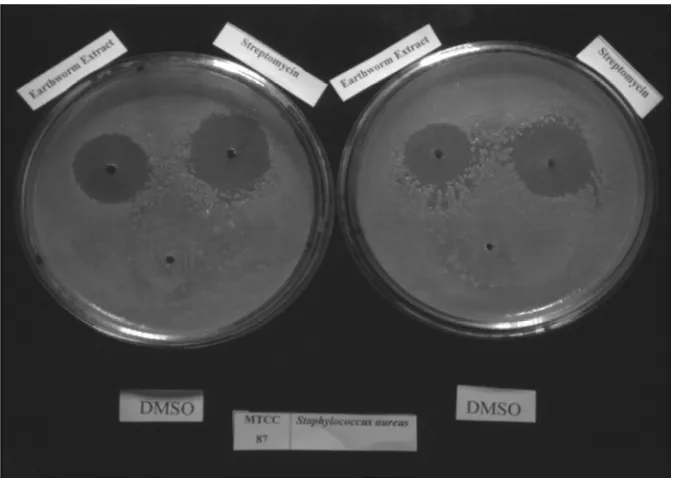
Streptomycin (0.0125mg/ml) 1 Staphylococcus aureus 87 21.5 ± 1.5 31 ± 1
2 Pseudomonas aeruginosa 424 29 ± 1 29.5 ± 0.5
3 Proteus mirabilis 425 23 ± 1 26.5 ± 1.5
4 Proteus vulgaris 426 24 ± 1 31 ± 1
5 Staphylococcus epidermidis 435 24 ± 3 26 ± 2
6 Streptococcus pyrogenes 442 - 23 ± 1
7 Enterococcus faecalis 439 - 35 ± 1
8 Enterobacter aerogenes 2822 - 21 ± 1
9 Escherichia coli 118 - 33.5 ± 0.5
Results are expressed as Mean ± SE of two observations of each isolate (n=2)
Among the nine bacterial isolates only five species responded to earthworm extract. Maximum antibacterial activity 29 ±1 mm was found with gram negative Pseudomonas aeruginosa, which was almost similar (29.5±0.5 mm) to the standard antibiotic Streptomycin(Fig .4). Minimum antibacterial activity 21.5±1.5 mm was obtained with the case of gram positive Staphylococcus aureus(Fig .4). Isolates of Proteus mirabilis showed zone of inhibition of 23±1 mm, while another species of this Proteus genus Proteus vulgaris were inhibited at 24±1 mm. Staphylococcus
epidermidis strain was also exhibited same inhibition value i.e. 24±3 mm as shown by Proteus vulgaris strain.
Earthworm extract didn’t not express any inhibition effect on remaining four strain of bacteria viz. Streptococcus
pyrogenes, Enterococcus faecalis, Enterobacter aerogenes and Escherichia coli.
Fig.(3): Antibacterial activity of Earthworm extract (10mg/ml) and antibiotic Streptomycin against Staphylococcus aureus. n=2 DMSO (Dimethyl Sulphoxide 30%): Negative control
[image:5.595.135.479.162.294.2] [image:5.595.126.463.407.646.2]Fig.(4): Antibacterial activity of Earthworm extract (10mg/ml) and antibiotic Streptomycin against Pseudomonas aeruginosa. n=2 DMSO (Dimethyl Sulphoxide 30%): Negative control
Results were expressed as Mean±SE
Fig.(5): Antibacterial activity of Earthworm extract (10mg/ml) and antibiotic Streptomycin against Enterobacter aerogenes. n=2 DMSO (Dimethyl Sulphoxide 30%): Negative control
Fig.(6): Antibacterial activity of Earthworm extract (10mg/ml) and antibiotic Streptomycin against Enterococcus faecalis. n=2 DMSO (Dimethyl Sulphoxide 30%): Negative control
Results were expressed as Mean±SE
Fig.(7): Antibacterial activity of Earthworm extract (10mg/ml) and antibiotic Streptomycin against Proteus mirabilis. n=2 DMSO (Dimethyl Sulphoxide 30%): Negative control
Fig.(8): Antibacterial activity of Earthworm extract (10mg/ml) and antibiotic Streptomycin against Escherichia coli. n=2 DMSO (Dimethyl Sulphoxide 30%): Negative control
Results were expressed as Mean±SE
Fig.(9): Antibacterial activity of Earthworm extract (10mg/ml) and antibiotic Streptomycin against Streptococcus pyrogenes. n=2 DMSO (Dimethyl Sulphoxide 30%): Negative control
Fig.(10): Antibacterial activity of Earthworm extract (10mg/ml) and antibiotic Streptomycin against Staphylococcus epidermidis. n=2 DMSO (Dimethyl Sulphoxide 30%): Negative control
Results were expressed as Mean±SE
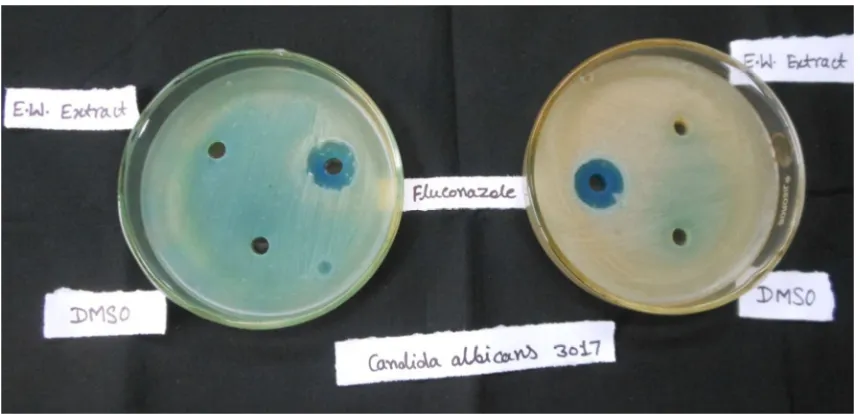
4.2 Results of Anti-fungal Activity:
As with antibacterial activity, anti-fungal activity was also evaluated in duplicate by measurement of zone of inhibition on SDA plate after 48 hours incubation at 250C by well-diffusion method and photographed (Fig.11 &12). The results are tabulated in following manner:-
Table 2): Determination of Antifungal activity by well-diffusion method
S. No. Fungal Species MTCC
Culture No.
Diameter of zone of inhibition (mm) EW Extract
(10mg/ml)
Fluconazole (5mg/ml) 1 Candida albicans 3017 - 17.0±0.35
2 Aspergillus niger 8189 - 17.5±0.50
Results are expressed as Mean ± SE of two observations of each organism (n=2)
Results of antifungal activity of earthworm extract showed that it was not able to inhibit fungal strains.
[image:9.595.146.469.68.312.2] [image:9.595.163.452.431.490.2] [image:9.595.93.523.522.729.2]Fig.(12): Antifungal activity of Earthworm extract (10mg/ml) and antibiotic Fluconazole (5mg/ml) against Aspergillus niger. n=2 DMSO (Dimethyl Sulphoxide 30%): Negative control
Results were expressed as Mean±SE
DISCUSSION
Earthworms kill microorganism by recognizing conserved molecular patterns (lipopolysaccharides (LPS) or peptidoglycans from bacterial cell walls, β-1, 3-glucan of fungal cell walls and double stranded RNA of viruses) on pathogen’s body surface. Recognition of molecules for foreign material has been named as pattern-recognition proteins (PRPs) [15] because the host’s primitive effecter cells would recognize molecular patterns rather than particular structures of the invading microorganisms. When bacteria invade into the coelomic cavity of earthworm, the coelomocytes starts to connect with each other by their adhesive structures around the bacteria and form “brown bodies” [3]. At the same time the coelomocytes intensively synthesize and secrete proteins that adhere to the bacteria, forming aggregations and may inhibit their further proliferation. These proteins attach to the lectin like monosaccharide of the cellular membrane of the bacteria. Different proteins and peptides of different species of earthworms have been extensively studied and different mechanisms of actions have been proposed [16]. Hence, it is very difficult to define which molecule and mechanism of coelomic fluid or extract of earthworms is responsible for its antibacterial activity.
In the present study, antimicrobial activity of earthworm extract was investigated using some species of fungi and bacteria. No antifungal activity could be demonstrated, but significant antibacterial activity to certain bacteria was noticed. While the other bacterial remained un-affected.
Out of eight bacterial isolates only four of them, gram positive Staphylococcus aureus and Staphylococcus
epidermidis, gram negative Proteus mirabilis and Pseudomonas aeruginosa were suppressed by earthworm extract.
No inhibitory effect was observed on remaining four strain of bacteria viz. Streptococcus pyrogenes, Enterococcus
faecalis, Enterobacter aerogenes and Escherichia coli. Here the explanation given by Valembois et al. (1982) may
be valid; earthworms have evolved specific antibacterial activity against the bacteria that are potential threat to them. In the present study earthworm Eudrilus eugeniae extract showed a strong significant antimicrobial activity. The results revealed that extract of Eudrilus eugeniae showed maximum antibacterial activity with gram negative
Pseudomonas aeruginosa. It showed 29±1 mm diameter of zone of inhibition, which was almost similar (29.5±0.5
mm) to the standard antibiotic streptomycin while minimum antibacterial activity of extract of E. eugeniae was obtained with gram positive, Staphylococcus aureus with the inhibiotion zone of 21.5±1.5 mm. Results of present study are in agreement of the findings of Prakash and Gunasekaran, (2011) [17]. They found that the earthworm powder of two species (Lampito maurtii and Perionyx excavatus) showed a strong antibacterial activity against the
S. aureus, P. mirabilis, and P. aeruginosa. Findings of this study are also evidenced by Hatti (2014), he has reported
that the coelomic fluid of earthworm, Polypheretima elongata exhibited highest antibacterial activity against
Staphylococcus aureus [18]. Khomyakov et al. (2007) [19] suggested that antimicrobial agents of earthworms
digestive fluid are formed in the earthworm body but not by the soil microorganisms entering their digestive tract.
supported by Wang et al., (2007). In the present study E. eugeniae extact didn’t show any antifungal activities [11]. Prakash (2013) [17] reported that earthworm powder was used against various ailments in indigenous system of medicine which was found to be fruitful against microorganisms.
According to the results, it can be concluded that extract of Eudrilus eugeniae can be used to formulate a new natural antimicrobial product for controlling infection of multidrug resistant bacteria where treatment is very difficult as the drug of choice for treating infection doesn’t work. The present study reported that earthworms can be used not only in environmental monitoring but also in the acquisition of novel molecules for human therapeutic purposes. This study may thus lead to formulation of new natural antimicrobial agent and thus may found beneficial in future prospects for mankind.
Acknowledgement
The authors are thankful to Defense Research Development Establishment (DRDE), Gwalior, M.P., India for financial support.
REFERENCES
[1]Yagnanarayan R, Sethi PP, Rajhan PA, Pulandiran, K and Ismail SA. Indian Journal of Pharmacology. 1987, 19:221-224.
[2]Balamurugan M, Parthasarathi K, Cooper E, Ranganathan LS. European Review for Medical and
Pharmacological Sciences. 2007; 11:77-90
[3]Valembios, P., Roch, P., Lassegues, M., Cassand, P. J. invertebr. Pathol. 1982; 40: 21-27.
[4]Engelmann, P., Kiss, J., Cooper, E.L., Nemeth, P., J. Biochem. Biophys. Methods 2004; 61:215–228
[5]Nagasawa H, Sawaki K, Fuji Y, Kobayashi M, Segawa T, Suzuki R, Inatomi H. Anticancer Res. 1991: 1061. [6]Wenli Li, Chong Wang and Zhenjun Sun. Pedobiologia, 2011; 54S: S49– S56.
[7]Prakash, M. and Gunasekaran, G. The Journal of Alternative and Complementary Medicine. 2011; 17(2): 167-170.
[8]Cho JH, Park CB, Yoon YG, Kim SC. Lumbricin I, Biochim. Biophys. Acta , 1998; 1408: 67-76.
[9]Shobha SV and Kale R. Antimicrobial potency of earthworm, Eudrilus eugeniae on certain plant pathogens. Administrator. 2007.
[10]Wang, F., Wang, C., Li, M., Gui, L., Zhang, J. and Chang, W. Biotechnology Letters. 2003; 25: 1105-1109. [11]Vasanthi, K., Chairman, K., Ranjit Singh, A. J. A. African Journal of Environmental Science and Technology.
2013; 7(8): 789-793.
[12]Hrzenjak, T. Hrzenjak, M. Kasuba, V. Efenberger-Marinculic, P. and Levanat, S. Comp. Biochem. Physiol. A.
1992; 102(3): 441–447.
[13]Shobha SV, Kale RD. In-vitro studies on control of soil-borne plant pathogens by earthworm Eudrilus eugeniae exudates. Green Pages, 2008. http://www.eco web.com/editorial/080106.html.
[14]Bradford, M. M. Analyt. Biochem. 1976; 72: 248
[15]Medzhitov R, Preston-Hurlburt P, Janeway CA Jr. Nature. 1997; 388 (6640):394-7.
[16]Marta J, Fiołka Mirosław P, Zagaja Tomasz D, Piersiak Marek Wróbel and Jarosław Pawelec. Journal of
Invertebrate Pathology, 2010, 105, 63-73. 254.
[17]Prakash, M. and Gunasekaran, G. The Journal of Alternative and Complementary Medicine.2011; 17 (2): 167-170.
[18]Hatti, S. S. Indian Journal of Applied Research.2014 ; 4(1): 541-544
[19]Khomyakov NV, Kharin SA, Nechitailo, TYu, Golyshin PN, Kurakov AV, Byzov BA, Zvyagintsev DG.
Microbiology, 2007, 76, 45-54.