77:17 (2015) 37–41 | www.jurnalteknologi.utm.my | eISSN 2180–3722 |
Jurnal
Teknologi
Full Paper
MINIATURE PARALLEL ECT: A PRELIMINARY
STUDY USING COMSOL
Wan Norhisyam Abd Rashid
a, Elmy Johana Mohmad
b*, Ruzairi
Abdul Rahim
c, Hanis Liyana Mahmad Ameran
b, Jaafar
Abdullah
d, Mimi Mohaffyza Mohamad
e, Hairulazwan Hashim
f,
Mohd Fadzli Shaib
b, Omar Mohd. Faizan Marwah
ga
Faculty of Engineering Technology, Universiti Teknikal Malaysia
Melaka, Malaysia
b
Faculty of Electrical and Electronics Engineering, Universiti Tun
Hussein Onn Malaysia, Batu Pahat, Malaysia
c
Process Tomography and Instrumentation Engineering Research
Group, Innovative Engineering Research Alliance, Faculty of
Electrical Engineering, Universiti Teknologi Malaysia, Johor,
Malaysia
d
Centre for Computed Tomography and Industrial Imaging
(CCTII), Malaysian Nuclear Agency, Bangi, Malaysia
e
Faculty of Technical Education and Vocational, Universiti Tun
Hussein Onn Malaysia, Batu Pahat, Malaysia
f
Faculty of Technology Engineering, Universiti Tun Hussein Onn
Malaysia, Batu Pahat, Malaysia
g
Faculty of Mechanical Manufacturing Engineering, Universiti Tun
Hussein Onn Malaysia, Batu Pahat, Malaysia
Article history Received
28 June 2015
Received in revised form
1 September 2015
Accepted
15 October 2015
*Corresponding author
[email protected]
Graphical abstract
Abstract
The usage of Electrical Capacitance Tomography (ECT) is not limited to the imaging of mixtures in process equipment, it also can be used to measure the concentration profiles of component, and in certain case ECT is being used to investigate the boundaries and phase sizes within vessels and pipelines. There are quite a number of type of ECT sensors such as circular structure, square structure, concentric -annulus and parallel structure which are used for particular applications. In this paper, a study was carried out in order to investigate the behavior of electric field on the miniature parallel ECT. Simulations using COMSOL Multiphysics are being done to study the effect of increasing the size of the test object and the effect of increasing the permittivity of the test object. The results show that (1) the electric field lines relatively follow the phantom shape corresponding to the increasing of the size of the phantom and (2) the electrical field lines seems to bend more around the when the permittivity increases. Presented simulations establish first step of investigation, however already proved its usefulness for ECT performance model validation.
Keywords: Miniature parallel ECT, COMSOL, parallel sensor
1.0 INTRODUCTION
Tomography is a process to obtain a cross sectional image of an enclosed object without ‘opening’ it. This technique is well known for its capability to help us investigate the internal structure or behavior of a subject [1]. In the beginning, tomography was well accepted in the medical field especially as one of the important tools to scan the body. Computed Tomography scanner is one of the examples of the kind which revolutionized the field of medical diagnostic imaging. It is best used in obtaining the good detail resolution of images and in the last two decades, has been used for non-medical application for example in investigating the purity of gold jewelries [2].
Nowadays, the science and engineering are moving towards small scale technology. The effort to develop such systems and technologies has started since the last two decades. With this new emerging technology, researchers are ready to control mater at the level of single atoms and small molecules and to describe the properties of materials and systems at that scale. Besides, they have made the astonishing discovery which conclude that the properties of small numbers of atoms or molecules – nanoscale clusters (such as strength, electrical resistivity and conductivity and optical absorption) are significantly different from the properties of the same matter at either the single-molecule scale or the bulk scale [3]. Presented analysis are the basic steps to find the best possible method to be used to investigate and measure the fineness of minted gold bar using miniature parallel ECT.
The capacitance between electrodes when an electric field is applied as well as the permittivity distribution between these two plates is acquired by applying the Linear Finite Element method (FEM) using COMSOL Multiphysics. COMSOL Multiphysics is very useful to perform finite element analysis for different kinds of physics and engineering applications. In this study, COMSOL Multiphysics is applied to obtain potential distribution calculation by means of FEM. Using the electrostatics module in COMSOL, the capacitance, C can be obtain from:
𝐶 =
𝑄
2
2𝑊
where W is the energy to charge a capacitor and Q is the electric charge.
There are a lot of studies which are focusing on the principal and the concept of ECT. Yang et al [4], [5] has clearly explained in details about the key issues and the concept to design the ECT sensors. S.M. Huang et al. [6] provide the detail information of designing the measuring circuit in order to process the signal obtained from the sensors. A few papers have been published on image reconstruction algorithms including Yang et al. [7], Soleimani et al. [8], Mou et al.
[9] and Weifu Fang et al.[10].
ECT is widely used in the medical and process industry. In medical field, a research has been done to observe the calcium of human bones through permittivity difference using ECT [11], [12]. ECT is also used in food manufacturing industry in order to monitor the quality of the products [13]. Besides, it is widely used in oil and gas industry especially for multiphase flow measurement [14], [15]. For the imaging of pneumatic conveyor and fluidization processes in fluidized bed, Jaworski and Dyakowski [16] , Yang and Liu [17], Wang
et al. [18] and Warsito and Fan [19], have explained
how ECT is successfully used to ease the monitoring and control process. In the building construction, study has been done to use the capacitance measurement techniques to monitor the structure of concrete beams [20]. Generally, ECT is best used for imaging materials such as oils, dry powders, plastics, bones and under favorable circumstances pure water, all which have low electrical conductivity.
The cross-sectional image of the material inside a closed enclosure is obtained by measuring the changes in the inter-electrode capacitance. These changes are caused by the change in the properties of dielectric materials inside a multi-electrode sensor [5]. Normally, density, permittivity and concentration of the material give the big influence to the measured signal [21]. Basically it visualizes the distribution or changes of dielectric properties of materials in the observed vessels or pipelines. It consists of 3 basic parts which are the sensors, the measuring circuits and the image reconstruction computer. The sensor should have the capability to measure multiple and localized measurement around the area of investigation. This is done by arranging the electrodes around the object properly. Normally, the number of electrodes in a set of sensors varies from 8, 12 and 16 electrodes based on the actual application. Using the concept of basic capacitor, one electrode will act as excitation electrode which is supplied with sinusoidal wave signal.
Besides, there are other types of sensor such as square sensor [22], conical sensor [23] and concentric-annulus sensors [24]. In this paper, a study was carried out in order to investigate the behavior of electric field on the miniature parallel ECT. There are three issues which will be discussed in this paper including the effect of increasing the size of the test object and the effect of increasing the permittivity of the test object.
2.0 METHODOLOGY
For this study, simulation is being done using COMSOL software. The numerical techniques involved in image reconstruction demand the use of a mathematical model of the sensor, even if some image-reconstruction techniques can be based purely on empirical data. It is difficult to calculate an inter-electrode capacitance which relate with the relative permittivity distribution and potential distribution, by using the Laplace equation is still too difficult to solve for the geometry and the boundary conditions, thus a finite element method (FEM) software simulation package is need to be used to find the electric field distributions [4].
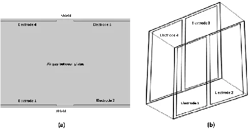
The real sensor will be simulated by the sensor model. This indicates that it can solve the problem of ECT forward which is to calculate the capacitances between all possible electrode pairs. The shape of the structure is drawn as the first step in developing the numerical modelling. The shape of the parallel structure is actually an illustration of 2 parallel plates with 4 electrodes on them. As for this case, the applied drawing geometry will have to follow the actual size and geometry of the hardware. Due to this, the building of the number of electrodes‟ alteration at the parallel structure can be done effortlessly.
The configurations of sensor are [25]: •Length of shielding layers and wall: 21mm •Electrode Length: 9mm
•Gap between adjacent electrodes: 1mm
•Material of shielding layers and electrode: copper •Material of wall: FR4 (ᶓr = 4.8)
•Number of electrodes: 4
The modelling approaches applied are listed as follows:
(i) The selection of the mode in the EM Module; (ii) The drawing process of the sensor geometries; (iii) The process of generating the mesh;
(iv)The setting of electrical properties in the domains; (v) The setting of the boundary conditions;
(vi) The solving and the finding of the field distribution;
(vii)The application of the post-processing capabilities in COMSOL to figure capacitance and voltage;
In the simulation, 2 studies have been done in order to investigate the effect of increasing the size of the test object and the effect of increasing the permittivity of the test object. Illustration in Figure 1 shows the parallel structure of ECT sensor.
Figure 1 (a) 2D and (b) 3D Geometry Shape of Miniature Parallel ECT
3.0 RESULTS AND DISCUSSION
3.1 Single Electrode Excitation
At the beginning, simulation is performed by setting each of electrode as exciting electrodes once at a time in order to observe the potential distribution and the electrical field lines between these 2 parallel structures. The concept which has been applied in ECT is by analysing the potential difference between the excited electrodes and the detecting electrode.
This can be done by setting electrode 1 as exciting electrode and automatically the rest of electrodes will become the detecting electrodes. Using this method, we can easily verify the potential difference between inter-electrode capacitance.
Figure 2 shows potential distribution between electrodes and electric field lines that present between any of the two electrodes which are curved and not straight.
Figure 1 Simulation result shows potential distribution and electric filed lines between electrodes
[image:3.612.313.558.75.202.2] [image:3.612.323.549.518.656.2]opposing electrode pair which resulting in the sensor to become more sensitive at the area of the wall rather than the central area. Therefore, this has become the reason of poor signal-to-noise ratio (SNR) in the central area. The mutual capacitances turn to be relatively small due to this effect which resulting to the possibility of very small electrode charges (and their change) and consequently, the measurements SNR inclines to be quite poor.
3.2 The Effect of Increasing the Size of Phantom
Next, simulation was being done in order to analyse the process of capacitance non-linear changes when the size of the higher dielectric material was increased. The capacitance is implemented between the model electrode-pairs. The effect of the permittivity size increase is analysed as due respect to a square. Figure 3 illustrates the results of the increasing of size of the higher dielectric material. The electric field lines as shown in the image relatively follow the square shape corresponding to the increasing of the dielectric size. The capacitance components reacting to the electrical field within the sensors will increase in proportion at all time to the material permittivity for the sensor. This is provided if the sensor is filled with uniformly higher permittivity material.
Figure 2 Image simulation when the size of object is increased
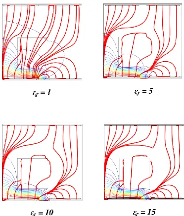
3.3 The Effect of Increasing the Permittivity of Phantom
[image:4.612.321.546.213.312.2]In this study, the permittivity distribution in the simulations was also verified. The ECT 2D which corresponds to the 3D geometries are illustrated in Figure 4. The capacitance is measured between the electric field distribution and the model electrode pairs for 2D and 3D simulations when electrode 1 was excited. A 5 mm cube was located at the centre of the parallel plate. The illustration shows that the electric field lines are deflected which relies on material distribution.
Figure 3 Results from (a) 2D and (b) 3D simulation respectively
Figures 5 (a)-(d) below reveals that the electrical field degree of penetration is subject to the material permittivity. The electrical fields seem to be reflected more if the permittivity is higher. This is corresponding to the permittivity of the test object where the reflected electrical field degree will seem to increase when the permittivity increases. Furthermore, when the phantom permittivity becomes higher due to the increasing of conductivity, the electrical field lines also seems to bend more around the test object.
[image:4.612.344.530.472.691.2]4.0 CONCLUSION
In this paper, the performance of miniature parallel ECT is being described based on the simulation using COMSOL software. The effect of increasing the size of the test object and the effect of increasing the permittivity of the test object have been investigated. As the size of the object increase, electric field lines relatively follow the square shape corresponding to the increasing of the dielectric size. Besides, the reflected electrical field degree will seem to increase when the permittivity increases. A lot of study need to be done in order to obtain more information about the potential of miniature parallel ECT. This kind of sensor would be used to sense any object with special geometry.
Acknowledgement
Authors are grateful to the financial support by Research University Grant of Universiti Tun Hussien Onn Malaysia (ERGS Grant: E041) and Universiti Teknikal Malaysia Melaka.
References
[1] Ruzairi Abdul Rahim. 2011. Electrical Capacitance Tomography Principal, Techniques and Applications. Johor Bahru: Penerbit UTM Press.
[2] J. Abdullah, A. Samsudin, N. Laili, and H. Abdul. 2015. Non Destructive Assaying Gold Jewellery Using Dual-Energy Micro-Computed Tomography, Jurnal Teknologi (Sciences & Engineering). 3: 25-28.
[3] S. I. Stupp, M. Bawendi, D. Beebe, R. Car, S. Chiang, D. Gray, M. Heller, K. Hess, G. Iafrate, L. Jelinski, T. S. Jenks, P. Kuekes, C. Murray, L. Sohn, T. Sudarshan, and T. N. Theis. 2002. Small Wonders, Endless Frontiers: A Review of the National Nanotechnology Initiative. United States of America: National Academy Press.
[4] W. Q. Yang. April 2010. Design of Electrical Capacitance Tomography Sensors. Measurement Science Technology. 21(4): 1-13.
[5] W. Q. Yang. 2006. Key issues in Designing ECT. IEEE Sensors. 497-505.
[6] S. M. Huang, C. G. Xie, M. S. Beck, R. Thorn, D. Snowden, M. S. Beck, R. Thorn, and D. Snowden. 1992. Design of Sensor Electronics for Electrical Capacitance Tomography. IEE Proceeding Circuits, Devices Systems. 139(1): 83.
[7] W. Q. Yang, D. M. Spink, T. A. York, and H. McCann. 1999. An Image-Reconstruction Algorithm Based On Landweber’s Iteration Method For Electrical-Capacitance Tomography. Measurement Science and Technology.10(11): 1065-1069. [8] M. Soleimani and W. R. B. Lionheart. 2005. Nonlinear Image
Reconstruction For Electrical Capacitance Tomography
Using Experimental Data. Measurement Science
Technology. 16(10): 1987-1996.
[9] C. Mou, L. Peng, D. Yao, and D. Xiao. 2005. Image Reconstruction Using a Genetic Algorithm for Electrical
Capacitance Tomography. Tsinghua Science Technology. 10: 587-592.
[10] W. Fang. 2004. A Nonlinear Image Reconstruction Algorithm For Electrical Capacitance Tomography. Measurement Science and Technology. 15: 2124-2132.
[11] B. B. Abraham and G. Anitha. 2012. Designing of Lab View Based Electrical Capacitance Tomography System for the Imaging. Bonfring International Journal Power System Integrated Circuits. 2(2): 1-6.
[12] Y. C. Liang, D. Tien, S. Tang, Y. C. Liang, D. Tien, and C.-H. Wang. 2011. Development of a Portable Electrical Capacitance Tomography System. IECON 2011-37th Annual Conference IEEE Industrial Electronics Society. 2634-2638. [13] G. Bolton and U. Sharif. 2001. Process Tomography for
Contamination Detection in Liquid Foods : A Feasibility Study. In 2nd World Congress on Industrial Process Tomography. August: 719-725.
[14] I. Ismail, A. Shafquet, and M. N. Karsiti. April 2011. Application of Electrical Capacitance Tomography And Differential Pressure Measurement In An Air-Water Bubble Column For Online Analysis Of Void Fraction. Fourth International Conference Modeling Simulation Application Optimization. 1: 1-6.
[15] I. Ismail, J. C. Gamio, S. F. a. Bukhari, and W. Q. Yang. April 2005. Tomography for Multi-Phase Flow Measurement In The Oil Industry. Flow Measurement Instrumentation. 16(2-3): 145-155.
[16] A. J. Jaworski and T. Dyakowski, April 2005. Measurements of Oil–Water Separation Dynamics In Primary Separation Systems Using Distributed Capacitance Sensors. Flow Measurement Instrumentation. 16(2-3): 113-127.
[17] W. Q. Yang and S. Liu. September 2000. Role of
Tomography In Gas/Solids Flow Measurement. Flow
Measurement Instrumentation. 11(3): 237-244.
[18] S. Xin and H. Wang. 2011. Extensible Electrical Capacitance Tomography System For Gas Liquid Two-Phase Flow. Image Processing IET. 5: 500-507.
[19] W. Warsito and L.-S. Fan. February 2003. ECT Imaging Of Three-Phase Fluidized Bed Based On Three-Phase Capacitance Model. Chemical Engineering Science. vol. 58(3-6): 823-832.
[20] M. Niedostatkiewicz, K. Grudzie, Z. Chaniecki, and A. Romanowski. 2005. Application of the Capacitance And X-Ray Measurement Techniques For Monitoring The Structure Of Concrete Beams. 6th World Congress on Industrial Process Tomography 2: 1353-1367.
[21] A. Azmi, R. A. Rahim, P. S. Chee, S. M. Din, N. Muzakkir, N. Ayob, and P. L. Leow. 2014. Miniaturized Planar Sensor Development. Jurnal Teknologi (Sciences Engineering). 8: 101-105.
[22] W. Q. Yang and S. Liu. 1999. Electrical Capacitance Tomography With Square Sensor. Electronics Letter. 35(4): 295.
[23] S. Liu, Q. Chen, H. G. Wang, F. Jiang, I. Ismail, and W. Q. Yang. April 2005. Electrical Capacitance Tomography For Gas–Solids Flow Measurement For Circulating Fluidized Beds. Flow Measurement Instrumentation. 16(2-3): 135-144. [24] J. Ye, Y. Li, H. Wang, R. Ge, and W. Yang. September 2013.
Concentric-annulus Electrical Capacitance Tomography Sensors. Measurement Science Technology. 24(9):095403. [25] Z. Ren and W. Yang. 2014. A Simulation Study Of A Miniature