Journal of Chemical and Pharmaceutical Research, 2016, 8(7):499-508
Research Article
CODEN(USA) : JCPRC5
ISSN : 0975-7384
499
Purification and characterization of
α
-Galactosidase produced
from mutant bacterial strain
El-Sayed O. H.*, El-Shibawi K. H.*, El-Kady E. M.*, Afify A. E.** and Mohamed S. S.*
*Microbial Biotechnology Department, National Research Centre, Cairo, Egypt **Biochemistry Department, Faculty of Agriculture, Cairo University, Giza, Egypt
ABSTRACT
An organism producing extracellular α-Galactosidase was isolated from soil and identified as Bacillus circulans. An extracellular α-Galactosidase from Bacillus circulans has been purified to homogeneity and purity by chromatographic steps, using Sephadex G-100, DEAE-cellulos, and Sephadex G-200. The specific activity of the enzyme was increased approximately 9.5 fold. The molecular weight of the purified enzyme was determined by SDS-PAGE was 81 kDa. The optimum pH and temperature for the purified enzyme were 4.5 and 40ºC, respectively. α -Galactosidase is stable over abroad pH rang (3.0-10.0). Km values for p-nitrophenyl-α-D-galactopyranoside,
melibiose, and raffinose were determined 2.00, 50.0, and 97.9 mM, respectively. α-Galactosidase activity was strongly inhibited by Ag+ and Hg2+ metal ions. The enzyme hydrolyzes flatulence causing galactooligosaccharides, viz. raffinose and stachyose present in the soybean
Key words: α-Galactosidase -production- Bacillus circulans mutant
INTRODUCTION
α-Galactosidase (EC 3.2.1.22, α-D-galactoside galactohydrolase) is an exo-type glycoside hydrolase that catalyzes
the hydrolysis of α-1,6-galactosidic linkages in galactose-containing oligosaccharides such as melibiose , raffinose ,
and stachyose and in galactomannan, which is commonly found in legumes and seeds. Many sources including
plants and microorganisms can produce α-Galactosidase by de nova synthesis depending on environmental
conditions and its requirements. Hence bacteria, actinomycetes, yeasts, and fungi were used for enzyme production (Singh and Kayastha, 2013). However, purification to homogeneity and characterization of this enzyme have only been done for a few organisms (Liu et al., 2014).
On the other hand, α-Galactosidase that catalyzes the hydrolysis of simple and complex oligo- and poly-saccharides
containing terminal α-D-galactosyl groups and can be found in microorganisms (Ulezlo and Zaprometova,1982) and
plants (Chinen et al., 1981). Several industrial applications of α-D-Galactosidases are known, mainly in food
industry. α-D-Galactosidase s are used for hydrolysis of raffinose and stachyose present in leguminous food.
(Ohtakara and Mitsutomi, 1987) it can be also used for hydrolysis of raffinose, stachyose, and leguminous
polysaccharides present in soybean milk (Kotwal et al., 1998). In sugar industey, α-Galactosidases improve
crystallization of sucrose by hydrolysis of raffinose content in molasses during the manufacture process (Linden, 1982). Moreover, they can enhance the bleaching effect in pulp and paper industry (Ratto et al., 1993). The underlying pathology of many human genetic diseases involves molecular changes related to carbohydrate metabolism, largely the complex sugars. Fabry's disease (abnormal sphingolipid metabolism) is due to a deficiency
in thermolabile lyososomal α-Galactosidase . Recently, α-D-Galactosidase s are used for medical purposes, e.g. for
treatment of Fabry's disease by enzyme replacement therapy (Fuller et al., 2004) or blood type conversion (Olsson
et al., 2004).
500
EXPERIMENTAL SECTION
Microorganism sources
The mutant Bacillus circulans was isolated and identificated from Egyptain soil, the strain was mutant by exposed to gamma irradiation (3.0 kGy).
Production of α-Galactosidase enzyme
The basal growth medium for enzyme production had the following composition: 1g K2HPO4, 3g KH2PO4, 3g yeast
extract, 20g raffinose. The ingredient were dissolved in one liter distilled water. The pH was adjusted to 7.0, for preparation of the inoculum, 1.0 ml of suspension was transferred to 50 ml of the basal medium in a 250 ml Erlenmeyer flask and incubated in a rotary shaker (150 rpm) at 37°C for 24 h. Cultivation was also done in 250 ml Erlenmeyer flask, each containing 50 ml of sterile medium. One milliliter of the inoculum was transferred to the growth medium, then incubated at 37°C in the rotary shaker at 150 rpm., the culture was centrifuged at 5000 rpm for 10 min. and the clear culture filtrate was taken for enzyme assay.
Enzyme assay
α-Galactosidase activity was assayed by measuring the release of p-nitrophenol from chromogenic substrate
((p-nitrophenyl-α-D-galactopyranoside) in citrate phosphate buffer. One unit of α-Galactosidase activity was
expressed as the amount of enzyme that liberates 1 µmol of product (p-nitrophenol) per minute under assay conditions.
Fractionation by salting-out with ammonium sulfate
The method described by Dixon (1953) was used for fractionation of α-Galactosidase by salting-out with
ammonium sulfate. In this method, the culture medium was centrifuged at 5000 rpm for 10 minutes in a refrigerated centrifuge to separate the cells. The whole enzyme solution kept in an ice bath then followed by adding solid ammonium sulfate slowly until the required saturation of ammonium sulfate was reached (20, 40, 60, and 80%; w/v). The solution was left overnight at 4ºC then centrifuged at 5000 rpm for 25 minutes in a refrigerated centrifuge, each precipitate was dissolved in 10 ml distilled water. The precipitate was dialyzed against distilled water in a cellophane bag (at 4ºC) until the water outside the bag gave no precipitation with 1% barium chloride solution. This indicated that the enzyme solution inside the bag became free from excess sulfate. The water outside the bag should
be changed several times. For each concentration of ammonium sulfate, the protein content and α-Galactosidase
activity were determined in each dialysate.
Gel filtration chromatography
The method described by Wong et al. (1986) was used for gel filtration chromatography. In this method, the dialysate of ammonium sulfate fraction was applied to a column (2.6 × 75 cm) of Sephadex G-100, equilibrated with
0.2 M Na2HPO4-0.1 M citric acid buffer solution (pH 5.0), and eluted with the same buffer at flow rate 60 ml/h. The
active fractions were pooled and dialyzed against distilled water.
Ion exchange chromatography (DEAE)
The fractions were concentrated and dialyzed against 0.2 M Na2HPO4-0.1 M citric acid buffer solution (pH 5.0) then
adsorbed on a diethylaminoethyl-cellulose (DEAE-cellulose) column (1.5 × 40 cm) equilibrated with the same buffer solution. Non-absorbed proteins were washed with the same buffer solution and the absorbed proteins were eluted with stepwise of NaCl solution (0.1-0.5 M). The column was eluted at flow rate 60 ml/h. The active fractions were pooled and dialyzed against distilled water (Whistler,1965).
Fractionation by Sephadex G-200 gel filtration
The most active fractions from DEAE-cellulose column were pooled, dialysed as described in previous step and loaded on a (2.5 × 75 cm) column of Sephadex G-200 equilibrated and eluted with the same buffer solution as described previously. The active fractions were pooled and dialyzed against distilled water.
Electrophoretic techniques
Native-PAGE (without SDS-PAGE and reducing agent) and SDS-PAGE were performed in Bio Rad Mini-Protein II Dual-Slab apparatus according to the method described by Laemmli (1970) .
Properties of purified α-Galactosidase
Effect of temperature and pH
The effect of temperature on the activity of the purified α-Galactosidase was determined by standard assay at 30, 35,
40,45,50,55 and 60ºC. The α-Galactosidase stability was assessed by incubation of the enzyme for 30 min at the
501
method described by (Ohtakara and Mitsutomi, 1984). The effect of pH on enzyme activity was determined using casein as the substrate, which was dissolved in different buffers of pH 3-9. The enzyme stability at various pH values was determined by pre-incubating the enzyme with an equal volume of each buffer for 30 min at 30ºC .The
residual α-Galactosidase activity was assayed under standard conditions at an optimized temperature of 30ºC
(Ohtakara and Mitsutomi, 1984)
Effect of metal ions
The purified enzyme was diluted with an equal volume of metal salts (Ag, NO3, MnCl2, MgSO4, HgCl2 and CaCl2
solution) with concentrations ranging from 0.1-1mM and incubated for 30min at 30ºC. The residual α-Galactosidase
activity was assayed under standard conditions.
RESULTS AND DISCUSSION
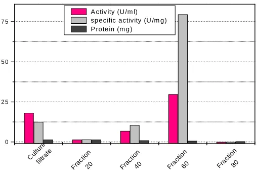
Four fractions of crude α-Galactosidase produced by mutant B. circulans were obtained by four different
concentrations of ammonium sulfate ( 20, 40, 60, and 80 %, w/v). Results obtained was illustrated in Fig.(1). The
fraction obtained by 60% (w/v) of ammonium sulfate showed low protein content, high α-Galactosidase activity
and specific activity comparing with the crude of α-Galactosidase and other fractions. Therefore, the fraction
obtained by 60% (w/v) ammonium sulfate was used for further purification studies.
0 2 5 5 0 7 5
Fra ctio
n
60 Fra
ctio n
80 Frac
tion 40 Fra
ctio n
20 Cul
ture
filtra te
[image:3.595.178.432.307.484.2]A c tiv ity (U /m l) sp e c ific a ctiv ity (U /m g ) P ro te in (m g )
Fig. 1. Protein content, Enzyme activity and specific activity of different fractions of mutant B. circulans α-Galactosidase obtained by
ammonium sulfate fractionation
In respect of fractionation of α-Galactosidase by salting-out with ammonium sulfate, Wong et al. (1986)
precipitated α-Galactosidase from Monscus pilosus by 50% (w/v) saturation of ammonium sulfate. Moreover, α
-Galactosidase produced by Bacillus stearothermophilus was precipitated by 50% (w/v) saturation of ammonium
sulfate. Rezessy-Szabo et al. (2006); Katrolia et al., (2012) isolated thermostable α-Galactosidase from the
thermophilic Thermomyces lanuginosus CBS 395.62/b. The enzyme was precipitated at 70% concentrated
ammonium sulphate, α-Galactosidase produced by mutant B. circulans after partial purification with 60% (w/v)
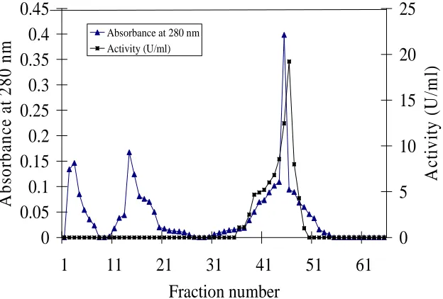
ammonium sulfate was applied to a Sephadex G-100. The elution diagram of the enzyme was illustrated graphically
in Fig.(2). α-Galactosidase active fractions (from fraction No. 17 to 22) were pooled and concentrated then used for
further purification studies. The pooled fractions (from fraction No. 17 to 22) obtained from the previous step (Sephadex G-100) were adsorbed on a DEAE-cellulose. The elution diagram of enzyme purification was illustrated graphically in Fig (3), it was observed that the enzyme was eluted effectively in seven fractions namely from fraction No. 41 to 47. The fractions were pooled, concentrated then used for further purification study. The concentrated enzyme solution obtained from the previous step (DEAE-cellulose) was subjected to the final purification on Sephadex G-200. The elution diagram of the enzyme was illustrated graphically in Fig.(4), i000t was
noticed that the enzyme was eluted effectively in seven fractions namely from fraction No. 9 to 15. The α
-Galactosidase active fractions were pooled and dialyzed against distilled water then used for electrophoresis
502
Fig. 2. Elution diagram for purification of mutant B. circulans α-Galactosidase obtained by ammonium sulfate precipitation (60%
(NH4)2SO4 fraction) on Sephadex G-100. The column (2.6 × 75 cm) was equilibrated with 0.2 M Na2HPO4-0.1 M citric acid buffer
[image:4.595.151.483.108.286.2]solution (pH 5.0). Elution was carried out with 500 ml of the same buffer. The flow rate was 1 ml/min and fraction volume 5 ml
Fig. 3. Elution diagram for purification of mutant B. circulans α-Galactosidase on DEAE-cellulose. The pooled fractions (from 17 to 22)
obtained from Sephadex G-100 were purified. The column (40 × 1.5 cm) was equilibrated with 0.2 M Na2HPO4-0.1 M citric acid buffer
solution (pH 5.0). Elution was carried out with 500 ml of a linear stepwise of NaCl (0.0-0.5 M). The flow rate was 1 ml/min and fraction volume 5 ml
0
0.05
0.1
0.15
0.2
0.25
0.3
0.35
0.4
0.45
1
11
21
31
41
51
61
Fraction number
A
b
so
rb
an
ce
a
t
2
8
0
n
m
0
5
10
15
20
25
A
ct
iv
it
y
(
U
/m
l)
[image:4.595.157.469.345.556.2]503
Fig. 4. Elution diagram for purification of mutant B. circulans α-Galactosidase on Sephadex G-200. The pooled fractions (from 41 to 47)
obtained from DEAE-cellulose were purified. The column (2.5 × 75 cm) was equilibrated with 0.2 M Na2HPO4-0.1 M citric acid buffer
solution (pH 5.0). Elution was carried out with 500 ml of the same buffer. The flow rate was 1 ml/min and fraction volume 5 ml
Total enzyme activity, total protein content, and specific enzyme activity of the proteins at each step of fractionation
and purification are summarized in Table (1). The purified α-Galactosidase had a specific activity of 118.27 U/mg
protein with 9.5-fold purification. In the present study, the value of specific activity of purified α-Galactosidase
(118.27 U/mg protein) was differed from the obtained by Durance and Skura (1985) for Clostridium perfringens
(1.61 U/mg protein), Talbot and Sygusch (1990) for Bacillus stearothermophilus (160 U/mg protein), and Gote et al. (2006) for Bacillus stearothermophilus (NCIM-5146) (400 U/mg protein).
Table 1. Summary of steps of fractionation and purification of α- Galactosidase from mutant B. circulans
Step Volume
(ml) Total activity (U) Total protein (mg) Specific activity (U/mg protein) Culture filtrate 10000 18190.0 1446 12.500 60% ammonium sulfate fraction 10.00 296.750 3.730 79.650 Sephadex G-100 25.00 265.00 2.500 106.00 DEAE-cellulose 35.00 142.86 1.250 114.29 Sephadex G-200 35.00 92.250 0.780 118.27
5. Electrophoresis a. Native-PAGE.
The efficiency of the purification process of each step separately (60% ammonium sulfate fraction, Sephadex G-100, DEAE-cellulose, and Sephadex G-200) was evaluated through the Native-PAGE. Native-PAGE was performed
under non-denaturing conditions, i.e. in the absence of β-mercaptoethanol and SDS without heating. Gel was
conducted using a 12% (w/v) polyacrylamide gel. Fig.(5) shows the resolution patterns of each fraction, obtained
data clearly showed that 60% ammonium sulfate fraction (lane 1) consisted of eight bands with Rf value of 0.125,
0.187, 0.387, 0.45, 0.582, 0.75, 0.812, and 0.85. Partially purified protein obtained by Sephadex G-100 (lane 2)
consisted of three bands with Rf value of 0.125, 0.387, and 0.812. Semi-final purified protein obtained by
DEAE-cellulose (lane 3) consisted of two bands with Rf value of 0.125 and 0.387. Final purified protein obtained by
Sephadex G-200 (lane 4) consisted of one band with Rf value of 0.387.
0
0.2
0.4
0.6
0.8
1
1.2
1
11
21
31
41
51
61
Fraction number
A
b
so
rb
an
ce
a
t
2
8
0
n
m
0
1
2
3
4
5
6
7
A
ct
iv
it
y
(
U
/m
l)
[image:5.595.144.467.428.498.2]504
Fig. 5. Native of α-Galactosidase enzyme produced by mutant B. circulans at various stages of purification. Analysis performed on a
polyacrylamide and stained with Comassie Brilliant Blue R-250. From left to right: Lane 1, 60% ammonium sulfate fraction; Lane 2,
partially purified protein obtained by Sephadex G-100; Lane 3, semi-final purified protein obtained by DEAE-cellulose and Lane 4, final purified protein obtained by Sephadex G-200
b. SDS-PAGE
Final purified protein obtained by Sephadex G-200 was subjected to SDS-PAGE. Gel was conducted using a 12%
(w/v) polyacrylamide gel gave a single band MW approximately 81 kDa by SDS-PAGE Fig.(6). This single subunit
indicating the purity and homogeneity of α-Galactosidase . This molecular weight is slightly differed to those of
other reported by Talbot and Sygusch (1990) for Bacillus stearothermophilus (73 kDa) and Gote et al. (2006) for
Bacillus stearothermophilus (NCIM-5146) (79.9 kDa). However, the subunit molecular weight of α-Galactosidase from present strain and from other reported strains of the same species (Bacillus sp.) ranges between 80 and 84 kDa, suggesting that the gene size could be similar in all these species (Granter et al.,1988; Talbot and Sygusch,1990; Fridjonsson et al.,1999; and Gote et al.,2006)
Fig. 6. SDS-PAGE of α-Galactosidase enzyme produced by mutant B. circulans. Analysis performed on a polyacrylamide and stained
with silver staining technique. From left to right: Lane 1, standard molecular weight markers (β-Galactosidase , 116; phosphorylase b,
97; bovine serum albumin, 66; ova albumin, 45; soybean inhibitors, 20; and α-lactalbumin, 14 kDa) and Lane 2, final purified protein
obtained by Sephadex G-200
Properties of purified α-Galactosidase
a. Effect of pH
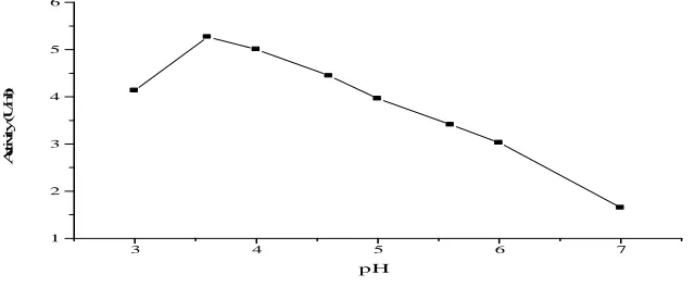
The aim of the present experiment was to find out the pH value at which α-Galactosidase exhibits its optimum
activity. In this respect, different buffers with various pH values, namely citrate buffer (pH 3.0-6.0) and citrate-phosphate buffer (pH 4.0-7.0) were used. In all reaction mixtures prepared, the same amounts of substrate and buffers were added, but each was adjusted at a specific pH value. The reaction mixtures were incubated at 40ºC for 30 min. Thereafter, the reaction was stopped and the released glucose was measured. Results obtained are presented as enzyme activity (U/ml) in Fig.(7). The present results indicated that the enzyme activity in citrate-phosphate
[image:6.595.197.433.415.595.2]505
were recorded with this buffer followed by citrate-buffer (5.8). From these results, pH 4.5 seems to be the most
suitable pH for the enzyme activity. The same findings were observed by Wong et al. (1986) who showed that α
-Galactosidase activity of Monococus pilosus assayed at 40ºC in citrate-phosphate and acetate buffers showed an
optimum pH at 4.5-5.0. On the other hand, the optimum pH of enzyme activity from Aspergillus saitoi was found at
a wide range 4.0 to 8.0 (Sugimoto, and Van-Buren, 1970). Purified α-Galactosidase from Bacillus
stearothermophilus (NCIM-5146) exhibited maximum activity (more than 70%) in the pH range 5.5-8.0 with
[image:7.595.160.448.181.374.2]optimum at 6.5-7.0
Fig. 7. Effect of pH on the activity of purified mutant B. circulans α-Galactosidase
b. Effect of temperature.
The enzyme activity was assayed under the standard conditions, purified enzyme incubated 30 min at pH 4.6 at various temperatures (30, 40, 50, 60, 70, and 80ºC) Fig.(8). The enzyme showed maximum activity at 40°C, after
this degree a decrease in activity was observed. In general, the optimum temperature for most α-Galactosidase is in
the range of 37-40°C (Ulezlo, and Zaprometova, 1982).
Fig. 8. Effect of temperature on the activity of purified mutant B. circulans α-Galactosidase
c. Effect of time.
The effect of incubation time at 40°C on α-Galactosidase activity was examined to establish the assay procedure.
The enzyme assay mixture was incubated for 10, 20, 30, 40, 50, and 60 minutes. The results obtained were collected and illustrated in Fig.(9). The results showed that the enzyme activity increased linearly with the time increase and
3.0 3.5 4.0 4.5 5.0 5.5 6.0
0 1 2 3 4 5 6 7
A
ct
iv
ity
(
U
/m
l)
pH
Citrate-buffer
Citrate phosphate-buffer
30 40 50 60 70 80
0 1 2 3 4 5 6
A
ct
iv
ity
(
U
/m
l)
[image:7.595.169.433.495.681.2]506
[image:8.595.163.430.137.313.2]reached the maximum after 30 min at 40°C. The results also showed that the hydrolysis of melibiose reached its maximum after 30 min of incubation. Incubation of enzyme mixture for more than 30 min did not influence the enzyme activity.
Fig. 9. Effect of time on the activity of purified mutant B. circulans α-Galactosidase at 40°C d. pH stability
In this experiment, identical purified enzyme in 0.2 M citrate-phosphate buffer at pH values ranging from 3.0 - 7.0 were incubated at 40°C for 24 h. Thereafter, the pH value of the purified enzyme was directly readjusted to pH 4.5 and added to the mixture containing the substrate then the enzyme activity was determined. Results obtained were presented in Fig.(10). The present results indicated that the enzyme was stable around pH 3.5 at 40°C for 24 h. The
same findings were observed by Wong et al. (1986) who found that the α-Galactosidase of Monascus pilosus was
stable between pH range 3.0-8.0. On the other hand, Rezessy-Szabo et al. (2006) reported that the Thermomyces
lanuginosus CBS 395.62/b α-Galactosidase is stable for at least 24 h at 55 ºC in the pH range 6.4-8.0.
Fig. 10. Effect of pH on the stability of purified B. circulans α-Galactosidase incubated at 40ºC for 24 h
e. Thermal stability
This experiment was carried out to investigate the heat stability of the purified enzyme (Liu, 2014). Purified enzyme was heated in water bath with different temperatures (30-60°C) for different periods of time up to one hour.
Thereafter, the tubes were then rapidly cooled and assayed for α-Galactosidase activity using standard procedures.
α-Galactosidase was stable at 30 and 40°C at different time limits as shown in Fig.(11). The residual activity was
decreased to 10.22% when the enzyme was stored at 60°C for 30 minutes and completely distorted at 60°C for 40 minutes.
0 10 20 30 40 50 60
0 1 2 3 4 5 6
A
ct
iv
ity
(
U
/m
l)
Time (min)
3 4 5 6 7
1 2 3 4 5 6
A
ct
iv
it
y
(
U
/m
l)
[image:8.595.140.460.453.585.2]507
Fig. 11. Effect of temperature on the stability (thermal stability) of the purified mutant B. circulans α-Galactosidase
Metal ions and EDTA
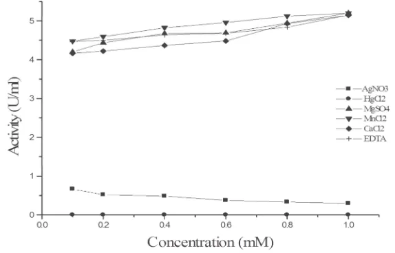
The effect of different concentrations of metal ions (AgNO3, MnCl2, MgSO4, HgCl2, and CaCl2) and EDTA on the
α-Galactosidase activity is shown in Fig.(12), α-Galactosidase was strongly inhibited by Ag+ and Hg2+ metal salts,
while EDTA did not affect the α-Galactosidase activity. These findings were similar to that obtained by Gote et al.
(2006) and Wong et al. (1986) for Monscus pilosus and Bacillus stearothermophilus (NCIM-5146), respectively.
The inhibition by silver ions may be attributed to their reaction with the carboxyl group or histidine residues, and that by mercury ions may be attributed to their binding of thiol group of the enzyme.
Fig. 12. Effect of different concentrations of metal ions and EDTA on the activity purified α-Galactosidase
CONCLUSION
After studying the properties of α-Galactosidase enzyme was fixed at a temperature of 40°C and also had the
highest activity at pH 4.5. Then when estimated Km and Vmax found that the enzyme was active in the presence of
Ag+2 and Hg+2
Acknowledgements
Thanks are due to the National Research Centre, Cairo, Egypt for the facilities that enabled the authors to accomplish this work.
REFERENCES
[1] I Chinen; T Nakamura; and N Fukuda. J. Biochem. , 1981, 90, 1453-1461. [2] M Dixon, J. Biochem., 1953, 54, 457-9.
[3] T Durance and B Skura, J. Food Sci., 1985, 50(2), 518-22.
10 20 30 40 50 60
0 1 2 3 4 5
A
ct
iv
ity
(
U
/m
l)
Time (min)
[image:9.595.160.447.384.568.2]508
[4] C Ganter; A Bock;P Buckel and R Mattes, J. Biotechnol., 1988, 8: 301-10.
[5] O Fridjonsson; H Watzlawick; A Gehweiler; T Rohrhirsch; R Mattes, Appl. Environ. Microbiol., 1999, 65:
3955-63.
[6] M Fuller; M Lovejoy; DA Brooks; ML Harkin; JJ Hopwood, Clin. Chem., 2004, 50, 1979-85. [7] MM Gote; ML Khan; DV Gokhale; KB Bastawde; JM Khire, Process Biochem., 2006, 41, 1311-17 . [8] P Katrolia; HY Jia; QJ Yan; S Song; ZQ Jiang; HB Xu, Bioresour. Technol., 2012,110, 578–586. [9] SM Kotwal; MM Gote; SR Sainkar; MI Khan; JM Khire, Process Biochem., 1998, 33, 337-343. [10] UK Laemmli, Nature, 1970, 227, 680–5.
[11] JC Linden, Enzyme Microb. Technol., 1982, 4: 130-136.
[12] X Liu; PC Claude; BH Lee; Joyce . Boye; Michel Casgrain, Biotechnoi.Resh.International 2014,2014,1-7. [13] A Ohtakara and M Mitsutomi, Agric. Biol. Chem.,1984, 48(5), 1319-27.
[14] A Ohtakara and M Mitsutomi, J. Ferment. Technol., 1987 65(4): 493-98.
[15] ML Olsson; CHA Hill; H Vega; QP Liu; MR Stroud, J Valdinocci; S Moon; H Clausen; MS Kruskall, Clinique
et Biologique., 2004, 11, 33-39.
[16] M Ratto; M Siika-aho; J Buchert; A Valkeajarvi; L Viikari, Appl. Microbiol. Biotechnology, 1993, 40: 449-454. [17] JM Rezessy-Szabo; QD Nguyen; A Hoschke; C Braet; G Hajos; M Claeyssens, Biochem. Biophys. Acta, 2006, 1-8.
[18] N Singh; AM Kayastha, J. Plant Biochem. Biotechnol. , 2013,22, 353–356. [19] H Sugimoto and JP Van-Buren, J. Food Sci., 1970 35, 655-60.
[20] G Talbot and J Sygusch, Appl. Environ. Microbiol., 1990,56: 3505-10. [21] IV Ulezlo and OM Zaprometova, Appl. Biochem. Microbiol., 1982 , 18:, 1-12. [22] LR Whistler, Academic press. Inc., New York and London, 1965 p. 14-17.