Simultaneous Identification of
Staphylococcus aureus
and
Coagulase-Negative Staphylococci Directly from Blood Culture Bottles in Less
than 30 Minutes
Melissa K. Deck,aErica S. Anderson,aRebecca J. Buckner,bGeorgia Colasante,cJames M. Coull,aBenjamin Crystal,a
Phyllis Della Latta,dMartin Fuchs,aDeanna Fuller,bWill Harris,aKevin Hazen,eLisa L. Klimas,aDaniel Lindao,cMichelle C. Meltzer,a Margie Morgan,fJaneen Shepard,aSharon Stevens,aFann Wu,dand Mark J. Fiandacaa
AdvanDx Inc., Woburn, Massachusetts, USAa
; Indiana University School of Medicine, Indianapolis, Indiana, USAb
; Health Network Laboratories, Allentown, Pennsylvania, USAc
; Columbia University Medical Center, NewYork-Presbyterian Medical Center, New York, New York, USAd
; University of Virginia, Charlottesville, Virginia, USAe ; and Cedars-Sinai Medical Center, Los Angeles, California, USAf
A novel rapid peptide nucleic acid fluorescencein situhybridization (FISH) method,Staphylococcus QuickFISH, for the direct detection ofStaphylococcusspecies from positive blood culture bottles was evaluated in a multicenter clinical study. The method utilizes a microscope slide with predeposited positive- and negative-control organisms and a self-reporting 15-min hybridiza-tion step, which eliminates the need for a wash step. Five clinical laboratories tested 722 positive blood culture bottles containing Gram-positive cocci in clusters. The sensitivities for detection ofStaphylococcus aureusand coagulase-negative staphylococci (CoNS) were 99.5% (217/218) and 98.8% (487/493), respectively, and the combined specificity of the assay was 89.5% (17/19). The combined positive and negative predictive values of the assay were 99.7% (696/698) and 70.8% (17/24), respectively. Studies were also performed on spiked cultures to establish the specificity and performance sensitivity of the method.Staphylococcus QuickFISH has a turnaround time (TAT) of<30 min and a hands-on time (HOT) of<5 min. The ease and speed of the method have the potential to improve the accuracy of therapeutic intervention by providingS. aureus/CoNS identification simultane-ously with Gram stain results.
S
taphylococci are the organisms most frequently isolated fromblood cultures, withStaphylococcus aureusbeing the most
fre-quently isolated pathogen in true bacteremia (20,22). Each year in
the United States, more than 300,000 patients contractS. aureus
infections, leading to more than 12,000 deaths, 2.7 million excess days of hospitalization, and close to $9.5 billion in excess hospital
charges (8,11). Coagulase-negative staphylococci (CoNS) are the
predominant organisms isolated from positive blood cultures (7),
60 to 80% of which are the result of contamination of the sample
during the blood draw due to inadequate antiseptic techniques (2,
17,21). Discrimination betweenS. aureusand CoNS has
signifi-cant clinical value, as it allows clinicians to differentiate between likely contamination and true bacteremia, thereby enabling more
informed and appropriate patient management decisions (1,2).
The proven value of this information creates a need for a rapid, accurate, and simple method to identify and distinguish between
S. aureus, CoNS, and other Gram-positive cocci in clusters (GPCC) in the clinical microbiology lab.
There are a number of methods currently used for identifica-tion of staphylococci in clinical samples, and they vary greatly in accuracy, speed, and cost. The tube coagulase test detects the pres-ence of free coagulase directly from a blood culture and has a high
specificity (⬎99%), but sensitivities range from 62 to 100%,
de-pending on the duration and dilution method (9,19). The
prin-cipal disadvantage of latex agglutination coagulase tests, tradi-tional growth-dependent culture methods, or automated broth microdilution identification systems is the prolonged turnaround time (TAT) for results. In contrast, rapid molecular techniques,
such as peptide nucleic acid (PNA) fluorescencein situ
hybridiza-tion (FISH), provide results in⬍2 h. Assays that generate
identi-fications directly from positive blood cultures in real time are likely to have a significant clinical impact and become mainstream diagnostic tools of clinical microbiology laboratories.
The PNA FISH method has been used in clinical microbiology labs for over 10 years for identification of a variety of organisms
(12,13, 18). In clinical evaluations, the PNA FISH method has
demonstrated sensitivities and specificities ranging from 96 to
100% (6,15,16). Other studies have measured the value of PNA
FISH in preventing overuse of antibiotics and unnecessary
hospi-talization (5,10). Unfortunately, clinical laboratories have
incen-tives to perform PNA FISH testing in batches to minimize the total number of control samples and maximize labor efficiency, which creates delays in reporting results and prevents the full realization
of the value of the method for the hospital and for the patient (3,
4). In response to this need, a second-generation assay,
Quick-FISH, was developed that implements several innovations de-signed to simplify the method and decrease TAT.
Like PNA FISH,QuickFISH takes advantage of the highly
spe-cific and rapid binding properties of PNA probes to detect species-specific rRNA sequences. Where PNA FISH incorporates a
30-Received25 January 2012 Returned for modification21 February 2012
Accepted29 March 2012
Published ahead of print4 April 2012
Address correspondence to Mark J. Fiandaca, [email protected].
Supplemental material for this article may be found athttp://jcm.asm.org/.
Copyright © 2012, American Society for Microbiology. All Rights Reserved.
doi:10.1128/JCM.00225-12
on May 16, 2020 by guest
http://jcm.asm.org/
min hybridization step and a 30-min wash step to remove unbound
fluorescent probe, QuickFISH introduces a novel self-reporting
probe design which enables a 15-min hybridization without the need
for a wash step. Elimination of the wash inQuickFISH is achieved by
quenching excess PNA fluorescence through formation of stable hy-brids with complementary quencher-labeled sequences. Another
QuickFISH innovation is an improved slide design that incorporates built-in positive and negative controls on the same slide as the sam-ple. Fixed organisms are predeposited into control wells and serve as controls for the hybridization step of the assay. The presence of con-trols on each sample slide reduces the number of slides a technologist needs to handle and track to process a sample and eliminates the need for the laboratory to grow and maintain control organisms.
TheStaphylococcus QuickFISH method is designed to rapidly
differentiateS. aureusfrom otherStaphylococcusspecies directly
from positive blood culture bottles (the method does not
differ-entiate methicillin-resistantS. aureus[MRSA] from
methicillin-sensitive S. aureus [MSSA]). The purpose of this study was to
determine whether theStaphylococcus QuickFISH method is
ro-bust and specific for identification ofS. aureusand CoNS in the
clinical microbiology laboratory setting.
MATERIALS AND METHODS
Clinical study.Samples were tested and results recorded in five hospital microbiology laboratories located in New York, Pennsylvania, Virginia, California, and Indiana. Each site received the same materials and train-ing. All sites worked under a protocol that included blinding and discrep-ant resolution methods that were approved by their institutional review boards. Study samples were classified as leftover clinical material.
Clinical specimens.A total of 722 GPCC-positive blood culture bot-tles from five clinical laboratories were included in the study. Two sites used the BacT/Alert system (bioMérieux, Durham, NC) blood culture bottles (n⫽236), and the other three sites used Bactec systems (Becton Dickinson, Sparks, MD) (n⫽486). Samples from all available BacT/Alert and Bactec blood culture bottle types were included in the study, except for BacT/Alert PF, FA, and FN FAN types (prior to initiation of the stud-ies, BacT/Alert bottles containing charcoal were found to interfere with theQuickFISH method). Bactec samples included 203 Lytic Anaerobic, 230 Plus Aerobic, 34 Peds Plus, and 19 Plus Anaerobic bottles; BacT/Alert samples included 101 SN (anaerobic) and 135 SA (aerobic) bottles. Sam-ples in bottles that signaled positive by the automated blood culture mon-itoring instrument and showed GPCC upon Gram staining were included in the study.
Routine identification of bacteria.Routine identification methods varied across the participating clinical laboratories. Of the three sites using Bactec blood culture systems, one site used theS. aureus/CoNS PNA FISH system (AdvanDx) combined with conventional subculture and the BD Phoenix system (BD Diagnostic Systems, Sparks, MD) for speciation of the CoNS isolates; the second site used Xpert MRSA/SA (Cepheid) with subculture followed by Staphaurex latex agglutination (Remel) if the Xpert MRSA/SA was negative; the third site also used Xpert MRSA/SA and conventional subculture as well as MicroScan (Siemens Healthcare). The BacT/Alert sites employedS. aureus/CoNS PNA FISH, conventional sub-culture, and Vitek or Vitek 2 (bioMérieux, Durham, NC) as routine lab-oratory identification methods.
Non-StaphylococcusGPCC-spiked blood cultures.There is a low prevalence of GPCC other thanS. aureusand CoNS species in blood culture samples (22). In order to broaden the number of organisms with a GPCC morphotype, additional testing was performed on spiked clinical isolates, including Aerococcus, Gemella, Kocuria, Micrococcus, Rothia, Granulicatella, andLactococcusspecies. All species tested were preserved strains isolated at the institution that performed the testing, with the ex-ception ofMicrococcus luteusATCC 4698, which was purchased from
ATCC (Manassas, VA). Isolates were inoculated into aerobic and anaer-obic BacT/Alert and Bactec blood culture bottles that had failed to signal growth after incubation for 7 days. Approximately 0.5 ml of bacteria sus-pended in 0.9% saline, initially equivalent to a 0.5 McFarland standard, then diluted 1:10, was inoculated into a blood culture bottle. The blood culture bottle was returned to the automated blood culture machine and incubated until the instrument reported a positive signal. One smear was prepared from each blood culture bottle. Cultures were assumed to con-tain the spiked bacteria and were not reidentified.
Reference strain-spiked blood cultures.A wide variety of microor-ganisms, including non-GPCC ormicroor-ganisms, were screened to examine the specificity and performance sensitivity of the method. The rationale for expansion of screening beyond the GPCC morphotype was 2-fold. In the clinical microbiology lab, there is an expectation of a low rate of Gram stain error (14), which requires the specificity of the method to be inde-pendent of the Gram stain result. Additionally, there is also a low preva-lence of polymicrobial blood cultures (22) with a likelihood of one strain being the GPCC morphotype; therefore, there is the potential for non-GPCC organisms to be found in a sample that will be tested by the Staph-ylococcus QuickFISH method.
Simulated blood culture samples were prepared at AdvanDx for 116 reference strains and 26 preserved clinical isolates (142 samples in total), including 40S. aureusstrains and 34 strains of otherStaphylococcus spe-cies, as well as other Gram-positive and Gram-negative bacteria and yeasts. Thirty-two of the 44 recognized species of staphylococci were rep-resented in the study. Samples were cultured and Gram stained, with the operator blinded to species identifications. BacT/Alert SA bottles were inoculated with 8 ml of sterile human donor blood; 3-ml aliquots were transferred into sterile test tubes. Each tube was inoculated with one to two colonies selected from agar plates for each strain and incubated ap-propriately for growth. Gram stains were performed on all samples to verify adequate growth.
Staphylococcus QuickFISH method.Fixation solutions (QuickFix-1 andQuickFix-2), hybridization solutions (StaphylococcusPNA Blue and StaphylococcusPNA Yellow), andQuickFISH slides and coverslips were provided by AdvanDx. All reagents were provided in dropper bottles. Instrumentation included a digitally controlled heat block and a fluores-cence microscope equipped with a custom dual-band filter and a 60⫻or 100⫻oil immersion objective. AdvanDx filter vials were provided to test-ing sites ustest-ing Bactec Plus blood culture bottles and were used to eliminate medium resin from the sample. Processed slides were examined by fluo-rescence microscopy.
Preparation of smears.QuickFISH slides were placed on a heat block at 55⫾1°C. A small volume (⬍0.5 ml) of each blood culture sample was transferred to a secondary vessel (e.g., microcentrifuge tube) via an aspi-rating needle. Ten microliters of sample was transferred from the second-ary vessel to the center of theQuickFISH slide sample area. One drop of QuickFix-1 was immediately added on top of the 10-l sample, and the mixture was spread evenly throughout the sample area with an inoculat-ing needle. The slide remained on the slide warmer until the smear was visibly dry (1 to 3 min). Two drops ofQuickFix-2 were added to the center of the sample area and allowed to dry (⬍1 min).
Hybridization and scoring.The hybridization reagent is a two-part mixture that is separately prepared for each slide prior to use. For each fixed sample, one drop ofStaphylococcusPNA Blue was applied to the center of a 25- by 50-mm coverslip, followed by one drop of Staphylococ-cusPNA Yellow. The blue and yellow hybridization reagents were thor-oughly mixed with an inoculating needle until a uniform green color was observed (approximately 5 to 10 s/coverslip). Coverslips were inverted and applied to theQuickFISH slides to simultaneously cover the control wells and the fixed samples with mixed hybridization reagents. Hybrid-ization was performed for 15 min on a heat block at 55⫾1°C. Slides were transferred immediately to a fluorescence microscope for scoring. The presence of multiple bright green fluorescent cocci in multiple fields of view indicated anS. aureus-positive sample, red fluorescent cocci
on May 16, 2020 by guest
http://jcm.asm.org/
cated a CoNS-positive sample, and no fluorescence indicated a negative sample.
RESULTS
Clinical study.Sensitivities of 99.5% (217/218) forS. aureusand 98.8% (487/493) for CoNS were obtained; the combined specific-ity for the assay was 89.5% (17/19). The combined positive and negative predictive values of the assay were 99.7% (696/698) and 70.8% (17/24), respectively. In total, 722 clinical samples were tested; 8 specimens were positive for both green and red fluores-cent cocci. The results of the clinical evaluation are displayed in
Table 1. The amount of time that passed between the bottle
sig-naling positive and the initiation of testing by theStaphylococcus
QuickFISH method was recorded for 537 samples. Thirteen per-cent of samples (73/537) were tested within 2 h, 47% (254/537) within 24 h, and 97% (522/537) within 48 h.
A total of 211 samples were identified asS. aureus, of which 41
were further described as methicillin resistant or methicillin sus-ceptible (18 MRSA and 23 MSSA). A total of 485 samples were
identified as non-S. aureusstaphylococci, of which 185 were
iden-tified to the species level, whereas 302 were simply ideniden-tified as
CoNS. Sixteen differentStaphylococcusspecies were identified in
total. Only 18 samples (2.5%) included in the clinical study con-tained nonstaphylococcal species, and of these, only 12 samples
(1.7%) contained GPCC phenotypes (MicrococcusandKocuria).
Non-StaphylococcusGPCC-spiked blood cultures. Twenty-nine seeded blood cultures representing 21 separate GPCC-posi-tive, nonstaphylococcal strains were included in a supplementary study performed at two of the clinical sites; the data are displayed inTable 2. All samples in the seeded culture experiment tested
negative, including 11 strains ofMicrococcus.
Reference strain-spiked blood cultures.Blinded screening of simulated blood culture samples inoculated with reference strains
was performed. AllS. aureussamples produced a green positive
result (15 reference strains and 25 clinical isolates), and all CoNS samples (34 strains, representing 31 species) produced a red
pos-itive result, except forS. simulansandS. felis. A variety of other
organisms were screened, and all had negative test results. Samples
included less-common GPCC genera (12), species identified as
Gram-positive cocci in pairs in chains (11), Gram-positive bacilli
(5), yeast (7), Gram-negative cocci (1), Gram-negative bacilli
(23), Gram-variable bacilli (6), Gram-negative coccobacilli (2),
and Gram-variable coccobacilli (1). Full screening data are
pro-vided in Table S3 in the supplemental material.
DISCUSSION
Our study presents the first description of a next-generation PNA
FISH technology, the Staphylococcus QuickFISH method. The
method provides a presumptive identification ofS. aureusand
classifies most non-S. aureusstaphylococci as CoNS. Clinical trial
results demonstrated high sensitivity and specificity for detection of staphylococci in blood cultures and rapid and easy
discrimina-tion ofS. aureusand CoNS.
The study’s discrepant resolution protocol required reevalua-tion of routine identificareevalua-tions and retesting of discrepant samples. Eleven discrepant results were reported, including two misidenti-fications (upon reexamination of the routine identimisidenti-fications, the
Staphylococcus QuickFISH results were determined to be correct). Two false-positive and seven false-negative scores were recorded.
One sample was scored as a dual positive (green/red) by
Staphy-lococcus QuickFISH but identified only as S. aureusby routine methods; this sample was not available to retest and was scored as a CoNS false positive. Another discrepant sample was determined
to be aMicrococcusspecies by conventional methods but produced
a green positive result byStaphylococcus QuickFISH. This result
was not repeated upon retesting and was recorded as anS. aureus
[image:3.585.41.284.90.341.2]false-positive result. A sample identified asS. aureusby routine
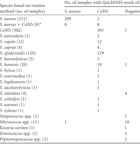
TABLE 1Performance of theStaphylococcus QuickFISH method with 722 blood cultures positive for GPCC tested at five hospital laboratories
Species based on routine method (no. of samples)
No. of samples withQuickFISH result of:
S. aureus CoNS Negative
S. aureus(211) 209 2
S. aureus⫹CoNS (8)a 8 8
CoNS (302) 301 1
S. auricularis(2) 2
S. capitis(12) 12
S. caprae(4) 4
S. epidermidis(129) 129
S. haemolyticus(5) 5
S. hominis(20) 19 1
S. hyicus(1) 1
S. intermedius(1) 1
S. lugdunensis(1) 1
S. saccharolyticus(1) 1
S. simulans(4) 4
S. schleiferi(1) 1
S. warneri(1) 1
S. xylosus(1) 1
Streptococcusspp. (2) 2
Micrococcusspp. (11) 1 10
Kocuria varians(1) 1
Enterococcusspp. (2) 2
Peptostreptococcusspp. (2) 2
aIncludes one sample identified asS. aureusandS. epidermidis, one asS. aureusandS.
[image:3.585.299.545.98.299.2]saprophyticus, and one as MSSA andS. epidermidis.
TABLE 2Performance of theStaphylococcus QuickFISH method with 29 blood cultures inoculated with nonstaphylococcal GPCC tested at two hospital laboratories
Species based on routine method (no. of strains, no. of samples)
Blood culture system
Bottle type(s)a
No. of samples with QuickFISH result of:
S. aureus CoNS Negative
Aerococcus urinae(1, 2) BacT/Alert B 2 Aerococcus viridans(1, 2) BacT/Alert B 2
Gemellaspp. (1, 2) Bactec B 2
Granulicatella adiacens (1, 2)
BacT/Alert B 2
Kocuriaspp. (2, 2) BacT/Alert A 2
Lactococcus garvieae (1, 2)
BacT/Alert B 2
M. luteusATCC 4698 (1, 1)
BacT/Alert A 1
Micrococcusspp. (4, 4) BacT/Alert A 4 Micrococcusspp. (2, 4) BacT/Alert B 4
Micrococcusspp. (4, 4) Bactec A 4
Rothia mucilaginosa (1, 2)
Bactec A 2
Rothiaspp. (2, 2) BacT/Alert B 2
a
A, aerobic bottle only; B, both aerobic and anaerobic bottles.
on May 16, 2020 by guest
http://jcm.asm.org/
methods was scored as a red positive by initial Staphylococcus QuickFISH testing, and the sample was recorded as false negative forS. aureus; however, the expected greenQuickFISH result was confirmed upon retest. Six CoNS false-negative results were re-corded: two samples produced the correct red result upon
retest-ing, and the other four samples were identified asS. simulans. All
discrepant results were reported from sites using BacT/Alert sys-tems only. Among the discrepant results, no patterns emerged to suggest a consistent assay failure mode. The rare number and var-ied types of discrepant results and reconciliation of discrepancies upon retesting support the conclusion that these results were due to human error and not related to the assay itself.
S. simulansandS. feliswere the only CoNS species tested that produced a negative result in the reference strain screening exper-iment. This limitation was predicted by sequence alignment of the probes used in the test. Four negative results were recorded at
clinical sites for samples identified asS. simulans by Vitek or
Vitek-2. No clinical samples were identified asS. felis. The fourS.
simulansfalse-negative results represented 0.81% (4/493) of all non-S. aureusstaphylococci. These fourS. simulans-positive bot-tles occurred on separate days and appeared to be independent,
with no clear clinical relevance. SinceS. simulansproduced an
expected negative result, it could be interpreted as a true negative instead of a false negative. Recalculation of the CoNS sensitivity,
excluding S. simulans, increased the sensitivity to 99.6% (487/
489).
Reference strain screening data of spiked simulated blood cul-tures expanded the breadth of organisms evaluated. Forty strains ofS. aureus(15 strains and 25 clinical isolates) and 34 non-S. aureusstaphylococci, representing 31 different species, were in-cluded in the study. Eleven strains of Gram-positive cocci in pairs and chains, 5 strains of Gram-positive bacilli, and 12 nonstaphy-lococci GPCC strains all produced the expected negative result. The results indicate high specificity and analytic sensitivity of the method performed on true samples containing phylogenetically diverse organisms.
Although this study included a large number of clinical sam-ples, a limitation of the study was the small number of true-nega-tive samples encountered during the trial. Despite GPCC being
the most common morphotype identified in blood cultures (8),
the vast majority of study isolates were staphylococci, and only 18 were nonstaphylococcal GPCC. Nonstaphylococci isolates
deter-mined in the study as GPCC included 11Micrococcusspp., 1
Ko-curia varians, 2Enterococcusspp., 2Peptostreptococcusspp., 1
al-pha-hemolytic Streptococcus, and 1Streptococcus parasanguinis.
Spiked negative blood cultures of 29 organisms were tested at two clinical labs to augment the variety of GPCC genera in the study. All clinical spiked isolates of nonstaphylococci GPCC tested
neg-ative byStaphylococcus QuickFISH. These data demonstrated the
broad specificity of theStaphylococcus QuickFISH assay. The
ad-dition of results from the seeded samples increased the specificity of the assay from 89.5% to 95.8% (46/48), demonstrating a
per-formance similar to PNA FISH (96 to 100% specificity [3,6,12,15,
16]).
The results of this study, although not a direct comparison to PNA FISH, demonstrate sensitivity and specificity values
compa-rable to previous reports on PNA FISH assays (15,16). The
Staph-ylococcus QuickFISH method eliminates the wash step, integrates controls, and shortens the fixation and hybridization steps, thus reducing hands-on time and TAT compared to PNA FISH. The
integration of theStaphylococcus QuickFISH method into the lab
workflow will hasten availability of species identification, provid-ing a greater potential clinical impact than previously obtained by the PNA FISH method.
As GPCC is the most commonly encountered bacterial mor-photype in blood cultures, laboratory personnel and clinicians are often challenged with determining whether the organism is a true
pathogen or a culture contaminant.Staphylococcus QuickFISH
will enable a clinical microbiology laboratory to accurately iden-tifyS. aureusfrom CoNS in blood cultures in⬍30 min. The rapid
presumptive identification of CoNS with theQuickFISH method
provides valuable information regarding possible contamination.
On the other hand, anS. aureus-positive result confirms to the
clinician that the organism represents a true infection that re-quires further testing to determine the strain’s susceptibility pro-file (MRSA or MSSA). The method has the potential to revolu-tionize standard testing and reporting protocols for blood cultures, as it will provide laboratories with the ability to report Gram stain and species identification results for Gram-positive
cocci at the same time.Staphylococcus QuickFISH has the potential
to significantly impact individual patient outcomes and overall bloodstream infection treatment costs by providing physicians with information to allow earlier implementation of targeted an-tibiotic therapy.
ACKNOWLEDGMENTS
We thank Jehan Farah, Tiffany S. Kidd, Sarah Regi, and Morgan Santham-oorthy for their excellent work.
None of the authors from the clinical laboratories involved in this study hold any financial interest in AdvanDx, Inc.
Kits and disposable materials for this work were provided by Ad-vanDx, Inc. All AdvanDx authors made critical contributions to the de-sign and development of theStaphylococcus QuickFISH kit; all are Ad-vanDx employees.
REFERENCES
1.Bates D, Goldman L, Goldman T.1991. Contaminant blood cultures and resource utilization. JAMA265:365–369.
2.Beekmann SE, Diekema DJ, Doern GV.2005. Determining the clinical significance of coagulase-negative staphylococci isolated from blood cul-tures. Infect. Control Hosp. Epidemiol.26:559 –566.
3.Forrest GN, et al.2006. Impact of rapid in situ hybridization testing on coagulase-negative staphylococci positive blood cultures. J. Antimicrob. Chemother.58:154 –158.
4.Forrest GN, et al.2006. Peptide nucleic acid fluorescence in situ hybrid-ization-based identification of Candida albicans and its impact on mor-tality and antifungal therapy costs. J. Clin. Microbiol.44:3381–3383. 5.Forrest GN, et al.2008. Peptide nucleic acid fluorescent in situ
hybrid-ization for hospital-acquired enterococcal bacteremia: delivering earlier effective antimicrobial therapy. Antimicrob. Agents Chemother. 52: 3558 –3563.
6.Hensley DM, Tapia R, Encina Y.2009. An evaluation of the AdvanDx Staphylococcus aureus/CNS PNA FISH assay. Clin. Lab. Sci.22:30 –33. 7.Karlowsky JA, et al.2004. Prevalence and antimicrobial susceptibilities of
bacteria isolated from blood cultures of hospitalized patients in the United States in 2002. Ann. Clin. Microbiol. Antimicrob.3:7.
8.Klein E, Smith DL, Laxminarayan R.2007. Hospitalizations and deaths caused by methicillin-resistant Staphylococcus aureus, United States, 1999 –2005. Emerg. Infect. Dis.12:1840 –1846.
9.Lagace-Weins PRS, Alfa MJ, Manickam K, Karlowsky JA.2007. Ther-mostable DNase is superior to tube coagulase for direct detection of Staphylococcus aureus in positive blood cultures J. Clin. Microbiol.45: 3478 –3479.
10. Ly T, Gulia J, Pyrgos V, Waga M, Shoham S.2008. Impact upon clinical outcomes of translation of PNA FISH-generated laboratory data from the
on May 16, 2020 by guest
http://jcm.asm.org/
clinical microbiology bench to bedside in real time. Ther. Clin. Risk Man-age.4:637– 640.
11. Noskin G, et al.2005. The burden ofStaphylococcus aureusinfections on hospitals in the United States: an analysis of the 2000 and 2001 Nationwide Inpatient Sample Database. Arch. Intern. Med.165:1756 –1761. 12. Oliveira K, Procop GW, Wilson D, Coull J, Stender H.2002. Rapid
identification ofStaphylococcus aureus directly from blood cultures by fluorescence in situ hybridization with peptide nucleic acid probes. J. Clin. Microbiol.40:247–251.
13. Perry-O’Keefe H, et al.2001. Identification of indicator microorganisms using a standardized PNA FISH method. J. Microbiol. Methods47:281– 292.
14. Rand KH, Tillan M.2006. Errors in interpretation of Gram stains from positive blood cultures. Am. J. Clin. Pathol.126:686 – 690.
15. Shepard JR, et al.2008. Multicenter evaluation of the Candida albicans/ Candida glabrata peptide nucleic acid fluorescent in situ hybridization method for simultaneous dual-color identification of C. albicans and C. glabrata directly from blood culture bottles. J. Clin. Microbiol.46:50 –55. 16. Søgaard M, Hansen DS, Fiandaca MJ, Stender H, Schønheyder HC.
2007. Peptide nucleic acid fluorescence in situ hybridization for rapid
detection of Klebsiella pneumoniae from positive blood cultures. J. Med. Microbiol.56:914 –917.
17. Souvenir D, et al.1998. Blood cultures positive for coagulase-negative staphylococci: antisepsis, pseudobacteremia, and therapy of patients. J. Clin. Microbiol.36:1923–1926.
18. Stender H.2003. PNA FISH: an intelligent stain for rapid diagnosis of infectious diseases. Expert Rev. Mol. Diagn.3:649 – 655.
19. Sturm PDJ, Kwa D, Vos FJ, Bartels CJM, Schulin T.2008. Performance of two tube coagulase methods for rapid identification ofStaphylococcus aureusfrom blood cultures and their impact on antimicrobial manage-ment. Clin. Microbiol. Infect.14:510 –513.
20. Styers D, Sheehan DJ, Hogan P, Sahm DF. 2006. Laboratory-based surveillance of current antimicrobial resistance patterns and trends among Staphylococcus aureus: 2005 status in the United States. Ann. Clin. Microbiol. Antimicrob.5:2. doi:10.1186/1476-0711-5-2.
21. Uslan D, et al. 2007. Age- and sex-associated trends in bloodstream infection. Arch. Intern. Med.167:834 – 839.
22. Weinstein MP, et al. 1997. The clinical signicance of positive blood cultures in the 1990s: a prospective comprehensive evalution of the mi-crobiology, epidemiology, and outcome of bacteremia and fungemia in adults. Clin. Infect. Dis.24:584 – 602.