Differences in Community Structure between Fungi and Bacteria
Reveal Predominance of Transient Fungal Elements
Rolf Kramer,aAnnette Sauer-Heilborn,bTobias Welte,b,cCarlos A. Guzman,aWolf-Rainer Abraham,aManfred G. Höflea Department of Vaccinology and Applied Microbiology, Helmholtz Centre for Infection Research, Braunschweig, Germanya
; Department of Pneumology, Hanover Medical School, Hannover, Germanyb
; German Centre for Infection Research and German Centre for Lung Research, Hannover, Germanyc
The respiratory mycobiome is an important but understudied component of the human microbiota. Like bacteria, fungi can cause severe lung diseases, but their infection rates are much lower. This study compared the bacterial and fungal communities of sputum samples from a large cohort of 56 adult patients with cystic fibrosis (CF) during nonexacerbation periods and under continuous antibiotic treatment. Molecular fingerprinting based on single-strand conformation polymorphism (SSCP) analysis revealed fundamental differences between bacterial and fungal communities. Both groups of microorganisms were taxonomi-cally classified by identification of gene sequences (16S rRNA and internal transcript spacer), and prevalences of single taxa were determined for the entire cohort. Major bacterial pathogens were frequently observed, whereas fungi of known pathogenicity in CF were detected only in low numbers. Fungal species richness increased without reaching a constant level (saturation), whereas bacterial richness showed saturation after 50 patients were analyzed. In contrast to bacteria, a large number of fungal species were observed together with high fluctuations over time and among patients. These findings demonstrated that the mycobiome was dominated by transient species, which strongly suggested that the main driving force was their presence in inhaled air rather than colonization. Considering the high exposure of human airways to fungal spores, we concluded that fungi have low coloni-zation abilities in CF, and colonicoloni-zation by pathogenic fungal species may be considered a rare event. A comprehensive under-standing of the conditions promoting fungal colonization may offer the opportunity to prevent colonization and substantially reduce or even eliminate fungus-related disease progression in CF.
H
umans are constantly exposed to environmental fungi, and many body surfaces, e.g., the human upper respiratory tract, are colonized by fungal commensals (1). Already in the 1970s, culturable fungi were observed in⬎80% of sputum and lung sam-ples (2). A healthy oral mycobiome was recently identified, and the first comparisons with lung transplant recipients were made (3,4). Apparently, the mycobiome has a significant impact on human health, but our understanding of the underlying mecha-nisms ruling the mycobiome structure in different body sites and under different pathological conditions lags behind understand-ing of the bacterial microbiome (5). The same is true for the over-all role played by fungal species as part of the human airway mi-crobiome in health and disease (6).Lung diseases due to respiratory infections are the major cause of death in patients with cystic fibrosis (CF). CF is a systemic inherited metabolic disorder that predisposes the human body to microbial colonization of the airways. This in turn results in tissue damage and then lethal respiratory failure (7). Bacteria, such as
Pseudomonas aeruginosaandStaphylococcus aureus, are
consid-ered to be responsible for the majority of respiratory tract infec-tions in adult CF patients (8,9). In addition, fungi exhibit the potential to cause lung diseases, but the mycobiome was only re-cently addressed in CF microbiome studies (5,10). Fungi consid-ered pathogens in CF are mainly filamentous species from the classesSordariomycetesandEurotiomycetes, namely,Aspergillus
fu-migatus,Scedosporium apiospermum (S. apiospermum complex,
including Pseudallescheria boydii), Rasamsonia argillacea, and
Exophiala dermatitidis(8,11,12). However, despite high
preva-lence rates, the numbers of reported fungal lung infections are remarkably low. Therefore, the impact of fungal pathogens and
the cause of fungus-related lung disease progression are still mat-ters of debate (13). Fungi are part of the biosphere, and being orders of magnitude smaller than the bacterial microbiome in terms of total cell numbers of the human microbiome, the struc-ture and diversity of the mycobiome is poorly understood (5). Comparative analyses of the community structures between fungi and bacteria in human airways are rare, and comprehensive data sets from large cohort studies are missing. It is our understanding that culture-independent molecular fingerprinting targeting only abundant bacteria and fungi in the individual respiratory speci-men will reveal the nature and structure of both communities in a comparable manner. In single-strand conformation polymor-phism (SSCP) electrophoresis, identification and detection accu-racy of sequence variation consists of two elements: (i) measurable
Received23 April 2015Returned for modification7 June 2015 Accepted22 June 2015
Accepted manuscript posted online1 July 2015
CitationKramer R, Sauer-Heilborn A, Welte T, Guzman CA, Abraham W-R, Höfle MG. 2015. Cohort study of airway mycobiome in adult cystic fibrosis patients: differences in community structure between fungi and bacteria reveal predominance of transient fungal elements. J Clin Microbiol 53:2900 –2907.
doi:10.1128/JCM.01094-15. Editor:D. J. Diekema
Address correspondence to Rolf Kramer, [email protected].
Supplemental material for this article may be found athttp://dx.doi.org/10.1128 /JCM.01094-15.
Copyright © 2015, American Society for Microbiology. All Rights Reserved.
doi:10.1128/JCM.01094-15
on May 16, 2020 by guest
http://jcm.asm.org/
differences in mobility through the gel and (ii) sequence analyses of the individual bands.
Characterization of fungal and bacterial communities in spu-tum from CF patients has been minimal (10,14). This study pro-vides the first taxonomic survey of the structure and composition of bacterial and fungal communities in a large cohort of adult CF patients. Prevalence of the most abundant species and dynamics of communities are presented, thereby offering the opportunity to directly compare both parts of the airway microbiome. Conclu-sions on colonization and infection abilities of fungi in lower re-spiratory tracts were drawn based on correlations with fungal prevalences in ambient air.
MATERIALS AND METHODS
Patient cohort and sample collection.Ethical clearance and sputum sam-ple collection from CF patients enrolled in a longitudinal study of micro-bial diagnostics were described in detail in previous publications (15,16). In brief, 72 sputum samples were collected in sterile containers from 56 CF patients recruited in the CF ambulatory center of the Hannover Med-ical School (MHH; Hannover, Germany). This includes a subcohort of 13 patients who provided sputum two (n⫽10) or three (n⫽3) times within the 2-year sampling period. Patients were between 18 and 51 years old, and a median age of 31 years was calculated for the cohort. The genders were equally represented (48.2% female, 51.8% male). No pulmonary exacerbation was observed by the time of sputum collection. Sputum samples were collected in duplicate and stored at⫺20°C. All patients were in clinically stable condition during the time of our study. Administration of antibiotics to the CF patients followed the recommendations of the International Standard of Care for adult CF patients.
Sample preparation and DNA extraction.Microbial DNA from spu-tum was extracted according to the protocol described by Kramer et al. (16). Briefly, sputum sample preparation consisted of cysteine buffer (2% NaOH, 1.45% sodium citrate, and 0.5%N-acetylcysteine) and enzymatic lysis, including lyticase (Sigma-Aldrich, Germany). DNA extractions were performed with the GeneMatrix tissue and bacteria DNA purification kit (EurX Roboklon; Berlin, Germany).
PCR amplification and preparation of single-strand DNA (ssDNA).
For bacteria, the primers 27F (5=-AGAGTTTGATCMTGGCTCAG-3=) and 521R (5=-ACCGTGGCTGCTGGCAC-3=) were used to amplify parts of the 16S rRNA gene (17), including the variable regions V1 to V3 of the small subunit of the ribosome (SSU), with an amplicon size between 459 bp forNocardiasp. and 505 bp forVeillonellasp. PCR was carried out using 50 ng DNA of the sputum extractions in a final volume of 50l, starting with an initial denaturation for 15 min at 95°C. A total of 30 cycles (1 min at 95°C, 40 s at 56°C, and 1 min at 72°C) was followed by a final elongation for 10 min at 72°C. For fungi, a seminested PCR strategy was chosen, using the forward primer internal transcript spacer 1 (ITS1) (5= -TCCGTAGGTGAACCTTGCGG-3=) and the reverse primers ITS4 (5=-T CCTCCGCTTATTGATATGC-3=) and ITS2 (5=-GCTGCGTTCTTCATC GATGC-3=) (18). Length heterogeneity for the ITS region is known, and amplicon sizes in the current study varied from 134 bp forYarrowia lipo-lyticato 475 bp forCandida glabrata(19). The first PCR was carried out using 50 ng DNA from sputum extract in a final volume of 50l. By adding 3l of MgCl2(25 mM), PCR efficiency was substantially
im-proved. Starting with an initial denaturation for 15 min at 95°C, 20 cycles (1 min at 95°C, 1 min at 58°C, and 1 min at 72°C) were followed by a final elongation for 10 min at 72°C. Amplification of the final target region, the ITS between the 18S and 5.8S rRNA genes, was achieved by using 1l of the previous PCR in a final volume of 50l. Initial denaturation for 15 min at 95°C was followed by a total of 30 cycles (30 s at 95°C, 20 s at 62°C, and 30 s at 72°C) and a final elongation for 10 min at 72°C. A total of 1.5 U HotStarTaq DNA polymerase was used for all amplifications (Qiagen Hilden, Germany). For single-strand DNA (ssDNA) preparation, the re-verse primers 521R and ITS2 were 5=-biotin labeled, and magnetic
streptavidin-coated beads (Promega, Madison, WI) were applied to ob-tain ssDNA from the PCR amplicons according to Eichler et al. (20).
SSCP fingerprints and sequencing of individual ssDNA bands.
Dried pellets of ssDNA were resuspended in 7l of gel loading buffer (95% formamide, 10 mM NaOH, 0.25% bromophenol blue, 0.25% xy-lene cyanol). After incubation for 10 min at 95°C, the ssDNA samples were stored on ice and loaded onto a nondenaturing polyacrylamide-like gel (0.6⫻MDE gel solution; Cambrex BioScience, Rockland, ME) for SSCP electrophoresis. SSCP fingerprints of bacterial and fungal DNA were ob-tained at 20°C and 400 V for 21 h on 20-cm glass plates and at 20°C and 700 V for 20 h DNA on 55-cm glass plates, respectively. The gel was silver stained according to the method described by Bassam et al. (21). All bands with an estimated intensity ofⱖ0.5% of the total lane were considered for further analysis and excised from the SSCP gel, boiled in Tris buffer (10 mM Tris-HCl, 5 mM MgCl2, 5 mM KCl, 0.1% Triton X-100, pH 9.0), and
reamplified according to the conditions described above. Data of only the most abundant species of the bacterial and fungal microbiome (ⱖ0.5% of at least one sample) were considered in this comparative study. Purified amplicons were sequenced by cycle sequencing (ABI Prism BigDye Ter-minator cycle sequencing ready reaction kit; Applied Biosystems, Foster City, CA). Products were purified again with the BigDye Terminator pu-rification kit (Qiagen Hilden, Germany) and analyzed using capillary elec-trophoresis with fluorescence detection (ABI Prism 3100 genetic ana-lyzer). More details on the SSCP fingerprints and gene sequencing were published by Eichler et al. (20).
Data processing and phylogenetic analysis.Phylogenetic analysis of sequences was performed with the NCBI tool BLAST/blastn and the Ri-bosomal Database Project (RDP) Seqmatch tool (only for bacteria) (22). The closest taxonomic groups were determined by sequence similarity and the height of ssDNA bands in the molecular fingerprints. Operational taxonomic units (OTUs) with ambiguous BLAST results were defined according to the closest shared taxonomic level. Taxonomic classification of fungi followed the outline of fungal phylogeny published by Hibbett et al. (23). For identification at the species level, sequence similarity was defined as a minimum of 97%. Additionally, ssDNA bands with equal running distances were affiliated to the same species. Phylogenetic dis-tances of sequences were calculated with the Jukes-Cantor algorithm and displayed in neighbor-joining trees using MEGA version 5 (24). Species accumulation curves were calculated using AccuCurve 1.0 (25). Inci-dence-based data sets were applied and permutated 250 times, and the resultant curves were averaged.
Accession numbers.The complete ITS sequence data set for the iden-tification of fungal OTUs is available in the European Nucleotide Archive (ENA) under accession numbers LN835855to LN836015. Bacterial 16S rRNA sequence data are available in the supplemental material of Kramer et al. (16).
RESULTS
Comparison of fungal and bacterial species richness in the CF co-hort.The prevalence of bacterial and fungal species was assessed in 72 individual sputum samples collected over a period of 2 years from 56 CF patients (sputum from 13 patients was collected repeatedly, at different time points). Molecular fingerprints of the bacterial and fungal communities were generated using SSCP gel electrophoresis, followed by sequencing of all major bands (see Fig. S1 in the supple-mental material). In total, 60 fungi and 38 bacteria were considered OTUs, and species affiliation was identified by comparative sequence analysis (see Tables S1 and S2 in the supplemental material).Figure 1 illustrates the individual species counts per sample, showing a median of 4 bacterial and 2 fungal OTUs per sputum. In most of the 72 sputum samples, 3 to 6 bacterial species and 1 to 3 fungal species were observed. For bacteria and fungi, a maximum number of 11 species in one sample was detected. At least 1 bacterial OTU was detected in
on May 16, 2020 by guest
http://jcm.asm.org/
each sample, whereas 3 sputum samples did not contain any fungal species.
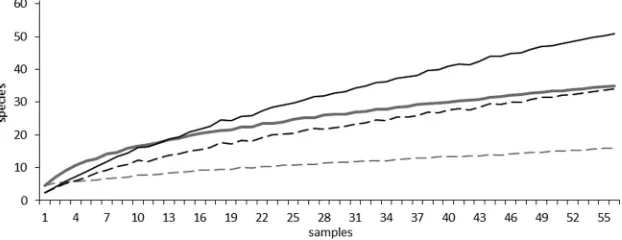
Species accumulation curves were calculated using observed spe-cies counts (richness) and counts for spespe-cies only occurring in one sample (unique species) to determine the total number of bacterial and fungal species in the cohort (Fig. 2). Overall, opposing trends were observed for fungi and bacteria. For bacteria, the curve strongly rose at the beginning, indicating a high number of bacterial species in a relatively small number of samples. The curve flattened with in-creasing number of samples analyzed, indicating, in the end, only a small incremental increase in species number when more samples were analyzed. For fungi, no flattening of the curve was observed. A small number of samples showed a low incidence of species, but a continuous rise of the curve indicated a steady increase in fungal OTUs. Considering that the number of unique species should be as low as possible in a comprehensive sampling, the trends for the curves of unique species are more informative for the estimation of species richness. For bacteria, a flat curve similarly shows only a small in-crease in unique species when taking more samples into account, indicating, in the end, no substantial increase of unique species with further sampling. In 56 sputum samples (only the first collected sam-ple from each patient), 16 bacterial species were occurring only in one
sample (unique species). Again, a rather different picture was ob-served for fungi: the curve for unique species showed a continuous rise with increasing sample numbers. After analyzing 56 sputum sam-ples, we found that 34 out of 51 fungal OTUs occurred only once. Overall, the number of bacterial species counts seemed to stabilize beyond 56 samples, whereas new fungal species had to be continu-ously considered with increasing sample numbers. Species accumu-lation curves for all collected sputum samples (n⫽72) were likewise calculated and confirmed these results (see Fig. S2 in the supplemen-tal material). In conclusion, a greater richness in fungal species in contrast to a relatively smaller number of bacteria was determined in this CF cohort study.
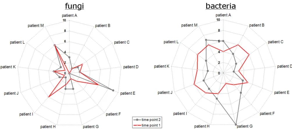
Comparing individual species richness at two time points. From a subcohort of 13 CF patients, sputum samples were repeat-edly collected for 2 years. Comparing the species richness in the individual samples of this subgroup, again a different picture emerged for fungi and bacteria, as illustrated inFig. 3. Differences were observed among the individual patients and between differ-ent time points for the same patidiffer-ents. For fungi, the starlike ap-pearance of the curves indicated major differences in the number of observed OTUs. Individual numbers were rather diverse among the 13 patients and between the different time points of the same patient. In contrast, the circlelike appearance of the radar plots for bacteria indicated a relatively stable number of OTUs among the patients and between the different time points of the same patient. One exception was patient G, in whom samples showed only 2 bacterial OTUs at the first collection, whereas 10 OTUs were detected in the second sputum sample. Neither fur-ther correlations between the individual numbers of fungal and bacterial OTUs nor correlations between specific fungal species and bacterial pathogens were observed.
Special emphasis was given to the dynamics of known fungal pathogens. The CF pathogenExophiala dermatitidiswas found in two samples collected within 3 months from patient C. With two and three fungal OTU counts in total, a relatively constant number of species was observed for this patient.Scedosporium apiospermum
was detected in two samples collected within 3 months from patient E, in whom ABPA (allergic bronchopulmonary aspergillosis) was di-agnosed. Six fungal species were observed in the first sputum sample and nine in the second sputum sample (Fig. 3). In the latter sample, several filamentous fungi were detected, such asPenicilliumsp. (
Eu-rotiomycetes), Fusarium oxysporum (Sordariomycetes), and two
CladosporiumOTUs, whereasAspergillus fumigatus(
Eurotiomyce-FIG 1Boxplots of species counts per sample for bacteria and fungi. Boxes are defined by upper and lower quartiles, whereas bars within boxes indicate the median number of OTUs. The ends of the whiskers represent the minimum and maximum species counts found in individual samples.
FIG 2Species accumulation curves are shown for bacterial and fungal OTUs detected for 56 CF patients. Only the first sample of each patient was considered. Averaged curves show the number of detected species versus the number of samples. Gray curves, bacteria; black curves, fungi; solid lines, observed species counts (richness); dashed lines, unique species counts.
on May 16, 2020 by guest
http://jcm.asm.org/
[image:3.585.42.287.63.239.2] [image:3.585.138.449.573.693.2]tes), another major CF pathogen, was only detected in the first sam-ple. With the exception of this case of ABPA, no relevant correlation between clinical data or other host factors and the presence of single fungal species was observed for the patients in our cohort. In this regard, an overview of host factors, clinical data, and results from microbial diagnoses is given in Table S3 in the supplemental material for all patients of the subcohort.
Prevalent bacteria in the cohort’s microbiota.Bacterial spe-cies from nine major taxonomic groups at the class level were observed in the sputum samples of the CF cohort. An overview is given in Table S4 in the supplemental material.Bacilliwith 14 OTUs were the most prevalent class, including alpha-hemolytic
Streptococci, such asS. salivariusandS. parasanguinis, which were
both detected in 57% of the samples. ThreeStaphylococcaceae spe-cies were observed, includingS. aureus, which, with 25% positive samples, was the most prevalent non-Streptococcusorganism from the classBacilli. Regarding the individual prevalence of all bacte-rial species, all other taxonomic classes, with the exception of
Bac-teroidia, were dominated by a single species.
Gammaproteobacte-ria were observed in 71% of the samples, P. aeruginosa was detected in 69.4%, and Stenotrophomonas maltophiliain 4.1%. Three species ofBetaproteobacteriawere detected.Achromobacter
xylosoxidanswas the most prevalent, with 8.3% positive samples,
followed byBurkholderia cepaciaandBordetella petrii.
Actinobac-teriawith 6 detected species were dominated byRothia
mucilagi-nosa, which was found in 43% of the samples. Four taxonomic classes with a single OTU each were observed.Fusobacterium
nu-cleatum(Fusobacteria) was detected in 6.9% of the samples and
Veillonellasp. (Negativicutes) in 5.5%. Unique classes found in
just one sample were represented byCapnocytophaga gingivalis
(Flavobacteria) and an oral TM7 candidate taxon (see Fig. S3 in the
supplemental material). The most diverse genera were repre-sented by 9 OTUs each in the genusPrevotellaand the genus
Strep-tococcus. More details on the bacterial community composition
and its dynamics were published previously (16).
Prevalent fungi in the cohort’s microbiota.Ten classes from the two fungal phyla,AscomycotaandBasidiomycota, were identi-fied (Table 1). One OTU could not be taxonomically assigned, but partial 18S and 5.8S sequences of the amplicon exhibited typical fungal structures and were therefore considered. Additionally, one OTU in each division did not exhibit sufficient similarity to be further assigned to a specific class. The prevalence of each fungal class in the cohort is shown in Fig. S4in the supplemental material, including the number of OTUs per class.Saccharomyceteswere the most frequently found fungi, and this class comprised the five most prevalent species. Overall, 89% of samples were positive for 19 OTUs. The polymorphic yeastsCandida albicansandC.
dub-liniensiswere the dominant fungal species in the CF cohort, with
44.4% and 23.6% positive samples, respectively, followed by
Sac-charomyces cerevisiae, with 19.4%, andC. parapsilosis, with 13.8%
positive samples (Table 1). Several filamentous mold species from the classes Dothiomycetes and Eurotiomycetes, including four
Aspergillus-like species (A. fumigatus,A. conicus,A. versicolor, and
Neosartorya pseudofischeri), were observed in the CF cohort. From
Dothiomycetes, 10 species in 25% of the samples were observed.
Two OTUs were dominant in this class: theCladosporium
cla-dosporioidescomplex, with 11.1%, and theCladosporium
herba-rumcomplex, with 9.7%. The basidiomycete classPucciniomycetes
showed only two species: the yeastsSporobolomyces roseusand
Sporobolomyces ruberrimus, which were detected in 11.1% and
5.5% of sputum samples, respectively (for details on phylogeny, see Fig. S5 and S6 in the supplemental material). These two species were repeatedly detected together in the same samples (n⫽3). Within the other taxonomic classes, individual prevalence of the associated OTUs was more equally distributed.
Exposure of human airways to fungal spores.Overall, a high species diversity of fungi was observed in sputum samples from the CF cohort. Both major fungal divisions were represented by several classes and species. Yeastlike fungi showed the highest diversity. Polymorphic and dimorphic fungal species were
FIG 3Differences in individual bacterial (right) and fungal (left) OTU counts for a subcohort of 13 CF patients with repeated sputum collection. For each patient, the first two samples were considered. Radar plots show patients A to M on angular axes. Numbers of detected species are given in radial axes. Red and gray symbols indicate values for first and second sputum samples, respectively. Symbols are accordingly combined with red and gray lines to locate the correct values for each patient.
on May 16, 2020 by guest
http://jcm.asm.org/
[image:4.585.40.542.63.286.2]found, as well as species limited to a single type of growth. Further comparisons with fungal diversity from urban and ru-ral outdoor air from a comparable region in Germany revealed close similarity in terms of taxonomy in both types of samples (data for outdoor air was retrieved from Fröhlich-Nowoisky et al. [26]) (Fig. 4). Although individual numbers were different, fungi with a considerable proportion in outdoor air were also present in the sputum samples of the cohort. While yeasts such
asSaccharomycetesor thePucciniomycetesspecies were dominant
in the sputum samples,Agaricomyceteswere dominant in mixed rural and urban air. However, the same fungus classes were found in sputum and in outdoor air.
DISCUSSION
In this study, we did an extensive comparative analysis of the bac-terial and fungal communities of the airway microbiome in a co-hort of 56 adult CF patients. In addition to the assessment of structural differences, we presented a taxonomic overview of the observed mycobiomes and correlated our results with fungi pres-ent in the air. Thus, this study was a comprehensive effort to eval-uate the airway mycobiome in CF and may serve as a basis for more detailed studies on the infection ecology of fungi in the lower respiratory tract. Our findings suggest that colonization and in-fection of CF airways by pathogenic (filamentous) fungi are ex-ceptional events that are triggered by unknown factors. Collec-tively, a more comprehensive understanding of the ecological processes during fungal infection may offer new opportunities in CF health care to prevent fungi-related diseases progression.
Our findings demonstrated that there are major differences between the structures of the fungal and bacterial communities. A considerably smaller number of bacterial species was observed in relation to the broad range of fungi. The individual bacterial mi-crobiome is dominated by only a few species with high relative abundances (5), whereas the rare biosphere, including fungi, is known to be more diverse (27). In a simplistic model, Fuhrman
(28) reasoned that abundant microorganisms are more likely to be detected, whereas rare microorganisms in one place are more likely below the detection limit in other locations. Consequently, individual communities might partly be diverse, but the more frequently samples in a cohort are analyzed, the more often pre-viously observed species will be detected. Assuming that fungi are generally rare members of the human microbiome, the probabil-ity of detection is much lower, resulting in a higher number of observed species, each represented by only a few cells in the indi-vidual communities. This is further supported by our observation that, on average, twice as many bacterial species than fungal spe-cies were detected per sample, thereby indicating more complex bacterial microbiota in the lungs of individual CF patients. Fur-thermore, in the current study, bacteria were detected in every sample, whereas this was not always the case for fungi. Collec-tively, these findings suggest that the fungal communities are dominated by transient members and not by persistent colonizers. Another important observation for the two communities was that the temporal dynamics of the bacteria and fungi present in CF spu-tum samples gave very different pictures. Relatively constant num-bers of bacterial species were detected, indicating stable communities with permanent colonizers. In contrast, high fluctuations were ob-served for fungi among the different time points and patients, sug-gesting dominance of transient community members instead of sta-ble fungal communities. Drawing attention to the dynamics of fungal pathogens, such asA. fumigatus,S. apiospermum, orE. dermatitidis, we observed a persistence of fungal species, which in turn con-firmed the accuracy of our detection method.A. fumigatuswas observed in the only patient of the cohort with diagnosed ABPA. In the same patient,S. apiospermumwas repeatedly detected. This is in agreement with the previously reported association betweenS.
apio-spermumandA. fumigatus(13). The pathogenE. dermatitidiswas
[image:5.585.40.549.76.284.2]also repeatedly found in sputum from the same patient within 3 months, suggesting permanent colonization by this fungus.
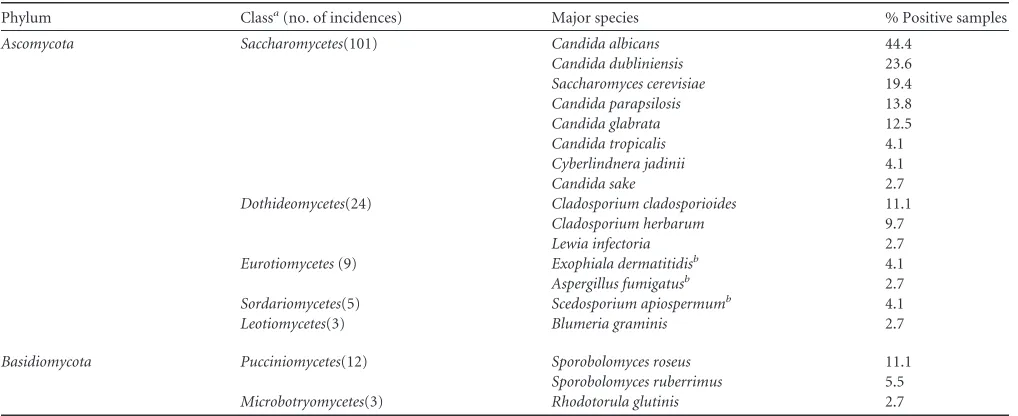
TABLE 1Prevalence of major fungal OTUs identified by their ITS sequences in 72 CF sputum samples from 56 patients
Phylum Classa(no. of incidences) Major species % Positive samples
Ascomycota Saccharomycetes(101) Candida albicans 44.4
Candida dubliniensis 23.6
Saccharomyces cerevisiae 19.4
Candida parapsilosis 13.8
Candida glabrata 12.5
Candida tropicalis 4.1
Cyberlindnera jadinii 4.1
Candida sake 2.7
Dothideomycetes(24) Cladosporium cladosporioides 11.1
Cladosporium herbarum 9.7
Lewia infectoria 2.7
Eurotiomycetes(9) Exophiala dermatitidisb 4.1
Aspergillus fumigatusb 2.7
Sordariomycetes(5) Scedosporium apiospermumb 4.1
Leotiomycetes(3) Blumeria graminis 2.7
Basidiomycota Pucciniomycetes(12) Sporobolomyces roseus 11.1
Sporobolomyces ruberrimus 5.5
Microbotryomycetes(3) Rhodotorula glutinis 2.7
a
Table includes only fungal OTUs detected more than once. Fungal OTUs are grouped according to taxonomic classification and ranked according to number of positive samples. The absolute number of detected incidences for each class is given in parentheses (considering all detected OTUs, including unique species). The following classes ofBasidiomycetes
were represented by unique OTUs only and are therefore not considered in this table:Agaricomycetes,Tremellomycetes,Exobasidiomycetes.
bEmerging or acknowledged pathogenic species for CF patients.
on May 16, 2020 by guest
http://jcm.asm.org/
A major strength of this study is the comprehensive taxonomic overview of the mycobiome by a molecular approach that detects all major fungal taxa. By analyzing the phylogeny, we attempted to gain insights into the origin of the microbial community mem-bers. For bacteria, oral cavities potentially act as a reservoir and “stepping stone” for subsequent migration to the CF lung (29). The healthy bacterial microflora of the oral cavity recently
pre-sented by the Human Microbiome Project revealed the genera
Streptococcus,Haemophilus,Actinomyces, andPrevotellato be the
most abundant taxa andS. parasanguinisandS. salivariusto be the most dominant species (30). Likewise, the generaPrevotellaand
Streptococcuswere observed to be the most diverse in our study,
with high prevalences ofS. parasanguinisandS. salivarius. These similarities suggest that the upper and lower respiratory tract
mi-FIG 4Comparison of relative proportion of fungi in outdoor air and in sputum of CF patients. Pie charts for outdoor air are based on data from Fröhlich-Nowoisky et al. (26), and these data were compared with the mycobiome present in 72 samples of the cohort. Each color represents one taxonomic unit further subdivided in the figure. (A) Different phyla. (B) Different classes ofAscomycota. (C) Different classes ofBasidiomycota.
on May 16, 2020 by guest
http://jcm.asm.org/
[image:6.585.74.508.63.586.2]croflora are closely related. For CF patients, a notably high species diversity for the familiesPrevotellaceaeandStreptococcaceaewas recently described, which matches the outcome of our study (31). CF end-stage lungs have been shown to harbor relatively low numbers of species, and 16S rRNA gene analysis of explanted lung tissue samples revealed the same four phyla of the five bacterial classes with more than one OTU found in the current study:
Pro-teobacteria,Bacteroidia,Firmicutes, andActinobacteria(32). Great
similarity was observed between the dominant OTUs in the cur-rent and other studies; therefore, the communities in CF airways are presumably greatly influenced by these species, e.g.,P.
aerugi-nosa,S. aureus,Rothia mucilaginosa, andStreptococcusspp. (33–
35). Occurrence frequencies of other bacterial pathogens, such as
A. xylosoxidans,S. maltophilia, andB. cepacia, were in the ranges
reported in the literature (36).
Aerial transport is considered a major mechanism of dispersal for many fungal species. Consequently, we compared our results with those of a previous study of airborne fungal presence in a comparable geographic area (26). All fungal classes identified in the current study were also observed in outdoor air and some, such asDothideomycetes andEurotiomycetes, even with similar prevalence (26). Therefore, air can be considered a major source for the diversity of fungal species. Despite the high exposure, the number of reported fungal lung infections is remarkably low (13). However, the real prevalence and role of fungal pathogens in CF patients remain to be elucidated. Apparently, a few species find conditions to colonize the respiratory tracts under certain circum-stances. In a first study analyzing the fungal respiratory micro-biome, the bronchoalveolar lavage (BAL fluid) of healthy individ-uals exhibited only low fungal presence, whereas high levels were observed in the BAL fluid of lung transplant recipients, with
Can-didaandAspergillusbeing the dominant genera (4). These
find-ings support the hypothesis that fungal colonization of healthy lungs is inhibited by a host defense, including bacterial micro-biome-mediated resistance (5). We propose that fungal coloniza-tion of the CF lung by environmental fungi can be considered a rare event triggered by exceptional conditions that promote infec-tion and remain to be elucidated.
Although there is no reliable estimate of the fungal load of the human body, nobody is fungus free (5). Particularly, the oral cavity is known to harbor a mycobiome, and the genusCandidarepresents the most prevalent species of this microbiome (3). In comparing the proportion of fungi in outdoor air and in the sputum of CF patients, yeastlike species were found to make the largest differ-ence. We hypothesize that if the oral cavities potentially act as a reservoir and stepping stone for bacteria (29), similar mechanisms might be involved for migration of yeasts into the airways.
Saccha-romycetesand particularlyCandidawere the most frequently
de-tected fungi in our study and are presumably (temporary) colo-nizers of the CF airways. Likewise, C. albicans was previously shown to be the most isolated fungus from CF patients (37–39), followed byC. dubliniensisin the current study.C. dubliniensisis rarely found in non-CF individuals, which may be due to its ability to display cell surface hydrophobicity-associated adherence at 37°C, which enables the fungus to take advantage of the dehy-drated respiratory secretion of these patients (40,41). The other importantCandidaspecies, namely,C. parapsilosis,C. glabrata,
andC. tropicalis, were observed with lower occurrence rates, but
different CF studies showed a ranking of occurrence surprisingly similar to that found in our study (11,42). This conformity in
observations supports the hypothesis of similar mechanisms for the transport ofCandidafrom oral cavities to the lower respira-tory tract, although to a minor extent compared with bacteria. BesidesCandida, several others of the observed classes were yeast-like species, including the most prevalentBasidiomycetes, repre-sented by twoSporobolomycesspecies. Whether the same trans-port mechanism can likewise be assumed for these and other yeasts is unclear, since nothing is known about their prevalence in the oral microbiome.Sporobolomyces roseus together with
Rho-dotorulaandCryptococcusspecies (the latter belonging to the class
Tremellomycetesrepresented by unique OTUs in our study) are
considered the most important viable yeasts in outdoor air, al-though generally only⬃2% of all viable spore counts are yeast species (43). However, the pathogenicity ofCandidaand other yeastlike species in CF airways remains to be elucidated.
A limitation of our study might be the low number of patients colonized by known fungal pathogens. Future studies focusing on a cohort of patients in the exacerbation phase with reported fungal infections will be necessary to analyze the dynamics and to char-acterize the ecological changes before and during colonization by pathogenic (filamentous) fungi. This will bring insights into the unknown triggers and factors that promote fungal persistence. Additionally, BAL fluid analyses, the gold standard for respiratory specimens, may bring further insights into the infection ecology of fungi. Despite these limitations, we were able to clearly demon-strate the different community structures of bacteria and fungi in the CF airways. In summary, our study indicates a strong predom-inance of transient elements in the mycobiome, including patho-gens, despite a high exposure of human airways to fungal contam-inants. Only yeast species, particularly Candida species, were detected frequently. Collectively, circumstances favoring fungal colonization seem to be rare events, and fungal contaminants are presumably immediately cleared out of the lung. The knowledge derived from this study is crucial, since a deeper understanding of the airway microbiome and its infection ecology is critical to sub-stantially improve CF health care.
ACKNOWLEDGMENTS
We thank the volunteers from the CF outpatient clinic of the Medical School Hannover for participating in the study.
We also thank Josefin Koch and Verena Maiberg for excellent assis-tance with the SSCP fingerprinting.
REFERENCES
1.Iliev ID, Underhill DM.2013. Striking a balance: fungal commensalism versus pathogenesis. Curr Opin Microbiol16:366 –373.http://dx.doi.org /10.1016/j.mib.2013.05.004.
2.Mullins J, Seaton A.1978. Fungal spores in lung and sputum. Clin Allergy
8:525–533.http://dx.doi.org/10.1111/j.1365-2222.1978.tb01506.x. 3.Ghannoum MA, Jurevic RJ, Mukherjee PK, Cui F, Sikaroodi M, Naqvi
A, Gillevet PM.2010. Characterization of the oral fungal microbiome (mycobiome) in healthy individuals. PLoS Pathog6:e1000713.http://dx .doi.org/10.1371/journal.ppat.1000713.
4.Charlson ES, Diamond JM, Bittinger K, Fitzgerald AS, Yadav A, Haas AR, Bushman FD, Collman RG.2012. Lung-enriched organisms and aberrant bacterial and fungal respiratory microbiota after lung transplant. Am J Respir Crit Care Med186:536 –545.http://dx.doi.org/10.1164/rccm .201204-0693OC.
5.Huffnagle GB, Noverr MC. 2013. The emerging world of the fungal microbiome. Trends Microbiol21:334 –341.http://dx.doi.org/10.1016/j .tim.2013.04.002.
6.Huang YJ, Charlson ES, Collman RG, Colombini-Hatch S, Martinez FD, Senior RM.2013. The role of the lung microbiome in health and disease: a National Heart, Lung, and Blood Institute workshop report. Am
on May 16, 2020 by guest
http://jcm.asm.org/
J Respir Crit Care Med187:1382–1387.http://dx.doi.org/10.1164/rccm .201303-0488WS.
7.Davies JC, Alton EWFW, Bush A.2007. Cystic fibrosis. BMJ335:1255– 1259.http://dx.doi.org/10.1136/bmj.39391.713229.AD.
8.Lipuma JJ.2010. The changing microbial epidemiology in cystic fibrosis. Clin Microbiol Rev23:299 –323.http://dx.doi.org/10.1128/CMR.00068-09. 9.Hauser AR, Jain M, Bar-Meir M, McColley SA.2011. Clinical
signifi-cance of microbial infection and adaptation in cystic fibrosis. Clin Micro-biol Rev24:29 –70.http://dx.doi.org/10.1128/CMR.00036-10.
10. Delhaes L, Monchy S, Fréalle E, Hubans C, Salleron J, Leroy S, Prevotat A, Wallet F, Wallaert B, Dei-Cas E, Sime-Ngando T, Chabé M, Vis-cogliosi E.2012. The airway microbiota in cystic fibrosis: a complex fungal and bacterial community–implications for therapeutic management. PLoS One7:e36313.http://dx.doi.org/10.1371/journal.pone.0036313. 11. Müller F-MC, Seidler M.2010. Characteristics of pathogenic fungi and
antifungal therapy in cystic fibrosis. Expert Rev Anti Infect Ther8:957– 964.http://dx.doi.org/10.1586/eri.10.72.
12. Giraud S, Favennec L, Bougnoux M-E, Bouchara J-P.2013.Rasamsonia argillaceaspecies complex: taxonomy, pathogenesis and clinical relevance. Future Microbiol8:967–978.http://dx.doi.org/10.2217/fmb.13.63. 13. Pihet M, Carrere J, Cimon B, Chabasse D, Delhaes L, Symoens F,
Bouchara J-P.2009. Occurrence and relevance of filamentous fungi in respiratory secretions of patients with cystic fibrosis–a review. Med Mycol
47:387–397.http://dx.doi.org/10.1080/13693780802609604.
14. Willger SD, Grim SL, Dolben EL, Shipunova A, Hampton TH, Morri-son HG, Filkins LM, O’Toole GA, Moulton LA, Ashare A, Sogin ML, Hogan DA.2014. Characterization and quantification of the fungal mi-crobiome in serial samples from individuals with cystic fibrosis. Micro-biome2:40.http://dx.doi.org/10.1186/2049-2618-2-40.
15. Kramer R, Sauer-Heilborn A, Welte T, Guzman CA, Höfle MG, Abra-ham WR.2015. A rapid method for breath analysis in cystic fibrosis patients. Eur J Clin Microbiol Infect Dis34:745–751.http://dx.doi.org/10 .1007/s10096-014-2286-5.
16. Kramer R, Sauer-Heilborn A, Welte T, Jauregui R, Brettar I, Guzman CA, Höfle MG.2015. High individuality of respiratory bacterial commu-nities in a large cohort of adult cystic fibrosis patients under continuous antibiotic treatment. PLoS One10:e0117436.http://dx.doi.org/10.1371 /journal.pone.0117436.
17. Wos-Oxley ML, Plumeier I, von Eiff C, Taudien S, Platzer M, Vilchez-Vargas R, Becker K, Pieper DH.2010. A poke into the diversity and associations within human anterior nare microbial communities. ISME J
4:839 – 851.http://dx.doi.org/10.1038/ismej.2010.15.
18. Schoch CL, Seifert KA, Huhndorf S, Robert V, Spouge JL, Levesque CA, Chen W, Fungal Barcoding Consortium.2012. Nuclear ribosomal inter-nal transcribed spacer (ITS) region as a universal DNA barcode marker for fungi. Proc Natl Acad Sci U S A109:6241– 6246.http://dx.doi.org/10.1073 /pnas.1117018109.
19. Chen YC, Eisner JD, Kattar MM, Rassoulian-Barrett SL, Lafe K, Bui U, Limaye AP, Cookson BT.2001. Polymorphic internal transcribed spacer region 1 DNA sequences identify medically important yeasts. J Clin Microbiol
39:4042– 4051.http://dx.doi.org/10.1128/JCM.39.11.4042-4051.2001. 20. Eichler S, Christen R, Höltje C, Westphal P, Bötel J, Brettar I, Mehling
A, Höfle MG.2006. Composition and dynamics of bacterial communities of a drinking water supply system as assessed by RNA- and DNA-based 16S rRNA gene fingerprinting. Appl Environ Microbiol72:1858 –1872. http://dx.doi.org/10.1128/AEM.72.3.1858-1872.2006.
21. Bassam BJ, Caetano-Anollés G, Gresshoff PM.1991. Fast and sensitive silver staining of DNA in polyacrylamide gels. Anal Biochem196:80 – 83. http://dx.doi.org/10.1016/0003-2697(91)90120-I.
22. Cole JR, Wang Q, Cardenas E, Fish J, Chai B, Farris RJ, Kulam-Syed-Mohideen AS, McGarrell DM, Marsh T, Garrity GM, Tiedje JM.2009. The Ribosomal Database Project: improved alignments and new tools for rRNA analysis. Nucleic Acids Res37:D141–D145.http://dx.doi.org/10 .1093/nar/gkn879.
23. Hibbett DS, Binder M, Bischoff JF, Blackwell M, Cannon PF, Eriksson OE, Huhndorf S, James T, Kirk PM, Lücking R, Thorsten Lumbsch H, Lutzoni F, Matheny PB, McLaughlin DJ, Powell MJ, Redhead S, Schoch CL, Spatafora JW, Stalpers JA, Vilgalys R, Aime MC, Aptroot A, Bauer R, Begerow D, Benny GL, Castlebury LA, Crous PW, Dai Y-C, Gams W, Geiser DM, Griffith GW, Gueidan C, Hawksworth DL, Hestmark G, Hosaka K, Humber RA, Hyde KD, Ironside JE, Kõljalg U, Kurtzman CP, Larsson K-H, Lichtwardt R, Longcore J, Miadlikowska J, Miller A, Moncalvo J-M, Mozley-Standridge S, Oberwinkler F, Parmasto E, Reeb
V, Rogers JD, Roux C, Ryvarden L, Sampaio JP, Schüssler A, Sugiyama J, Thorn RG, Tibell L, Untereiner WA, Walker C, Wang Z, Weir A, Weiss M, White MM, Winka K, Yao YJ, Zhang N.2007. A higher-level phylogenetic classification of the fungi. Mycol Res111:509 –547.http://dx .doi.org/10.1016/j.mycres.2007.03.004.
24. Tamura K, Peterson D, Peterson N, Stecher G, Nei M, Kumar S.2011. MEGA5: molecular evolutionary genetics analysis using maximum likeli-hood, evolutionary distance, and maximum parsimony methods. Mol Biol Evol28:2731–2739.http://dx.doi.org/10.1093/molbev/msr121. 25. Drozd P, Novotny V.2010. AccuCurve. Version 1.http://prf.osu.cz/kbe
/dokumenty/sw/AccuCurve/AccuCurve.xls.
26. Fröhlich-Nowoisky J, Pickersgill DA, Després VR, Pöschl U.2009. High diversity of fungi in air particulate matter. Proc Natl Acad Sci U S A
106:12814 –12819.http://dx.doi.org/10.1073/pnas.0811003106. 27. Pedrós-Alió C.2012. The rare bacterial biosphere. Annu Rev Mar Sci
4:449 – 466.http://dx.doi.org/10.1146/annurev-marine-120710-100948. 28. Fuhrman JA.2009. Microbial community structure and its functional
impli-cations. Nature459:193–199.http://dx.doi.org/10.1038/nature08058. 29. Rogers GB, Carroll MP, Serisier DJ, Hockey PM, Jones G, Kehagia V,
Connett GJ, Bruce KD.2006. Use of 16S rRNA gene profiling by terminal restriction fragment length polymorphism analysis to compare bacterial communities in sputum and mouthwash samples from patients with cys-tic fibrosis. J Clin Microbiol 44:2601–2604.http://dx.doi.org/10.1128 /JCM.02282-05.
30. The Human Microbiome Project Consortium.2012. Structure, function and diversity of the healthy human microbiome. Nature486:207–214. http://dx.doi.org/10.1038/nature11234.
31. Sibley CD, Grinwis ME, Field TR, Eshaghurshan CS, Faria MM, Dowd SE, Parkins MD, Rabin HR, Surette MG.2011. Culture enriched mo-lecular profiling of the cystic fibrosis airway microbiome. PLoS One
6:e22702.http://dx.doi.org/10.1371/journal.pone.0022702.
32. Rudkjøbing VB, Thomsen TR, Alhede M, Kragh KN, Nielsen PH, Johan-sen UR, Givskov M, Høiby N, Bjarnsholt T.2011. True microbiota involved in chronic lung infection of cystic fibrosis patients found by culturing and 16S rRNA gene analysis. J Clin Microbiol49:4352– 4355.http://dx.doi.org/10 .1128/JCM.06092-11.
33. Guss AM, Roeselers G, Newton ILG, Young CR, Klepac-Ceraj V, Lory S, Cavanaugh CM.2011. Phylogenetic and metabolic diversity of bacteria associated with cystic fibrosis. ISME J5:20 –29.http://dx.doi.org/10.1038 /ismej.2010.88.
34. van der Gast CJ, Walker AW, Stressmann FA, Rogers GB, Scott P, Daniels TW, Carroll MP, Parkhill J, Bruce KD.2011. Partitioning core and satellite taxa from within cystic fibrosis lung bacterial communities. ISME J5:780 –791.http://dx.doi.org/10.1038/ismej.2010.175.
35. Zhao J, Schloss PD, Kalikin LM, Carmody LA, Foster BK, Petrosino JF, Cavalcoli JD, VanDevanter DR, Murray S, Li JZ, Young VB, LiPuma JJ.
2012. Decade-long bacterial community dynamics in cystic fibrosis air-ways. Proc Natl Acad Sci U S A109:5809 –5814.http://dx.doi.org/10.1073 /pnas.1120577109.
36. Cystic Fibrosis Foundation.2012. Patient registry 2011 annual data re-port. Cystic Fibrosis Foundation, Bethesda, MD.
37. Bakare N, Rickerts V, Bargon J, Just-Nübling G.2003. Prevalence of Aspergillus fumigatusand other fungal species in the sputum of adult pa-tients with cystic fibrosis. Mycoses46:19 –23.http://dx.doi.org/10.1046/j .1439-0507.2003.00830.x.
38. Valenza G, Tappe D, Turnwald D, Frosch M, König C, Hebestreit H, Abele-Horn M.2008. Prevalence and antimicrobial susceptibility of mi-croorganisms isolated from sputa of patients with cystic fibrosis. J Cyst Fibros7:123–127.http://dx.doi.org/10.1016/j.jcf.2007.06.006.
39. Muthig M, Hebestreit A, Ziegler U, Seidler M, Müller F-MC.2010. Persis-tence ofCandidaspecies in the respiratory tract of cystic fibrosis patients. Med Mycol48:56 – 63.http://dx.doi.org/10.3109/13693780802716532. 40. Hazen KC, Wu JG, Masuoka J.2001. Comparison of the hydrophobic
properties ofCandida albicansandCandida dubliniensis. Infect Immun
69:779 –786.http://dx.doi.org/10.1128/IAI.69.2.779-786.2001.
41. Peltroche-Llacsahuanga H, Döhmen H, Haase G. 2002. Recovery of Candida dubliniensisfrom sputum of cystic fibrosis patients. Mycoses45:
15–18.http://dx.doi.org/10.1046/j.0933-7407.2001.00719.x.
42. Doern GV, Brogden-Torres B.1992. Optimum use of selective plated media in primary processing of respiratory tract specimens from patients with cystic fibrosis. J Clin Microbiol30:2740 –2742.
43. Rantio-Lehtimäki A.1985. Mould spores and yeasts in outdoor air. Al-lergy40(Suppl 3):S17–S20.