0095-1137/08/$08.00⫹0 doi:10.1128/JCM.01611-07
Copyright © 2008, American Society for Microbiology. All Rights Reserved.
Evaluation of Eight Different Bioinformatics Tools To Predict Viral
Tropism in Different Human Immunodeficiency Virus
Type 1 Subtypes
䌤
Carolina Garrido,
1Vanessa Roulet,
2,3Natalia Chueca,
4Eva Poveda,
1Antonio Aguilera,
5Katharina Skrabal,
2,3Natalia Zahonero,
1Silvia Carlos,
4Federico Garcı´a,
4Jean Louis Faudon,
2Vincent Soriano,
1and Carmen de Mendoza
1*
Hospital Carlos III, Madrid, Spain1; Eurofins Viralliance Inc., Kalamazoo, Michigan2; Bioalliance Pharma, Paris, France3;
Hospital Clı´nico Universitario San Cecilio, Granada, Spain4; and Hospital de Conxo—CHUS,
Santiago de Compostela, Spain5
Received 13 August 2007/Returned for modification 30 September 2007/Accepted 19 December 2007
Human immunodeficiency virus type 1 (HIV-1) tropism can be assessed using phenotypic assays, but this is quite laborious, expensive, and time-consuming and can be made only in sophisticated laboratories. More accessible albeit reliable tools for testing of HIV-1 tropism are needed in view of the prompt introduction of CCR5 antagonists in clinical practice. Bioinformatics tools based on V3 sequences might help to predict HIV-1 tropism; however, most of these methods have been designed by taking only genetic information derived from HIV-1 subtype B into consideration. The aim of this study was to evaluate the performances of several genotypic tools to predict HIV-1 tropism in non-B subtypes, as data on this issue are scarce. Plasma samples were tested using a new phenotypic tropism assay (Phenoscript-tropism; Eurofins), and results were compared with estimates of coreceptor usage using eight different genotypic predictor softwares (Support Vector Machine [SVM], C4.5, C4.5 with positions 8 to 12 only, PART, Charge Rule, geno2pheno coreceptor, Position-Specific
Scoring Matrix X4R5 [PSSMX4R5], and PSSMsinsi). A total of 150 samples were tested, with 115 belonging
to patients infected with non-B subtypes and 35 drawn from subtype B-infected patients, which were taken as controls. When non-B subtypes were tested, the concordances between the results obtained using the phenotypic assay and distinct genotypic tools were as follows: 78.8% for SVM, 77.5% for C4.5, 82.5% for
C4.5 with positions 8 to 12 only, 82.5% for PART, 82.5% for Charge Rule, 82.5% for PSSMX4R5, 83.8% for
PSSMsinsi, and 71.3% for geno2pheno. When clade B viruses were tested, the best concordances were seen
for PSSMX4R5 (91.4%), PSSMsinsi (88.6%), and geno2pheno (88.6%). The sensitivity for detecting X4
variants was lower for non-B than for B viruses, especially in the case of PSSMsinsi(38.4% versus 100%,
respectively), SVMwetcat(46% versus 100%, respectively), and PART (30% versus 90%, respectively). In
summary, while inferences of HIV-1 coreceptor usage using genotypic tools seem to be reliable for clade B viruses, their performances are poor for non-B subtypes, in which they particularly fail to detect X4 variants.
To enter human cells, the human immunodeficiency virus (HIV) type 1 (HIV-1) gp120 envelope glycoprotein interacts with the cellular CD4 receptor and two main coreceptors, CCR5 and/or CXCR4. Based on the ability to use CCR5 or CXCR4, HIV-1 variants are classified as CCR5 tropic (R5), CXCR4 tropic (X4), or dual-mixed tropic (X4R5) (2). R5 strains are generally responsible for the establishment of early infection, while the use of the CXCR4 coreceptor is generally seen in more advanced disease stages. The emergence of X4 variants has generally been associated with a faster CD4 de-cline and progression to AIDS (15, 28). In addition to the relationship between coreceptor use and HIV pathogenesis, the interest in HIV tropism has recently been raised because of the introduction of CCR5 antagonists in the HIV armamen-tarium. Two agents, maraviroc and vicriviroc, are the agents
within this family in the latest steps of clinical development (25, 36). Since these drugs show activity against R5 viruses only, viral tropism testing should be made before their prescription and eventually during treatment in order to exclude the pres-ence of X4 viruses.
Although there are several methods to determine HIV-1 tropism, it is unclear at the moment which is the most appro-priate for routine clinical use (26). Phenotypic assays based on recombinant viruses seem to be the most accurate methods (36); however, these methods are quite laborious and expen-sive and can be used only in sophisticated facilities. Therefore, more rapid and easier tools are desirable. The HIV-1 envelope V3 region has been shown to be the major determinant of viral tropism (5, 30), and accordingly, prediction of coreceptor us-age based on the interpretation of V3 sequences using bioin-formatics tools could be a good alternative to infer tropism in the clinical routine. The first genotypic algorithm designed to predict HIV-1 tropism took into account only the charge of amino acids at two key residues located within the V3 loop (amino acids 11 and 25). Estimates using the “11/25 charge rule” are relatively satisfactory (3), but other positions within
* Corresponding author. Mailing address: Department of Infectious Diseases, Hospital Carlos III, Sinesio Delgado 10, 28029 Madrid, Spain. Phone: 34 91 4532500. Fax: 34 91 7336614. E-mail: cmendoza @teleline.es.
䌤Published ahead of print on 16 January 2008.
887
on May 16, 2020 by guest
http://jcm.asm.org/
the V3 region are currently known to influence viral tropism as well, causing occasional disagreements (9, 11, 26). For this reason, most current genotypic bioinformatics tools consider the entire V3 sequence (27). Moreover, positions outside the V3 loop may also influence viral tropism (21). More impor-tantly, most genotypic predictors have been designed based on the genetic characteristics of HIV-1 clade B (25, 33). Since non-B subtypes show a wide genetic variability in the V3 region and since X4 viruses might be more prevalent in some clades than others (1, 17, 22, 34, 35), there is an urgent need to know the reliability of genotypic tools for inferring HIV-1 tropism in non-B subtypes, especially in regions where these HIV-1 vari-ants are quite prevalent and may soon have access to CCR5 antagonists.
The aim of this study was to evaluate the concordance be-tween eight distinct bioinformatics tools to estimate HIV-1 coreceptor usage by taking the results of a phenotypic tropism assay in a relatively large population of patients infected with non-B subtypes as a reference.
MATERIALS AND METHODS
Study population. Stored plasma specimens from HIV-1-positive patients known to be infected with non-B subtypes based on reverse transcriptase (RT)
and protease (PR) sequences along thepolgene were identified at three HIV
clinics located in Spain: Hospital Carlos III in Madrid, Hospital Clinico San Cecilio in Granada, and Hospital de Conxo—CHUS in Santiago de Compostela. As controls, stored plasma samples matching the same years of collection but derived from patients infected with clade B viruses were selected in these clinics. Demographic information as well as viral load, CD4 counts, and antiretroviral treatment exposure were recorded at the time of analysis. HIV-1 subtyping had been determined in all samples by phylogenetic analyses of the RT/PR genes using the PHYLIP software package (version 3.5c; J. Felsenstein, University of Washington, Seattle, WA). Evolutionary distances were estimated with Dnadist (Kimura two-parameter method), and phylogenetic relationships were deter-mined by Neighbor (neighbor-joining method). The branch reproducibility of trees was evaluated using Seqbot (1,000 replicates) and Consense.
HIV-1 coreceptor usage.Phenotypic coreceptor usage was determined using the Phenoscript-tropism assay (Eurofins, Kalamazoo, MI). Briefly, amplification of HIV-1 envelope sequences derived from plasma was initially carried out,
followed by homologous recombination betweenenvamplicons and a deleted
NL43 plasmid of the corresponding region during cotransfection of producer cells. Recombinant viruses were used to infect indicator cells expressing CCR5 or
CXCR4, besides CD4, and carrying thelacZgene under control of the viral long
terminal repeat. The specificity of the infection was assessed in each assay by incubation of indicator cells in the presence and absence of coreceptor inhibitors and five controls. The sensitivity of detection of minor quasispecies and the specificity of the Phenoscript-tropism assay were previously reported for clinical specimens with different viral loads and different HIV-1 non-B variants (29, 33). Inference of viral tropism using genotypic tools was carried out in parallel in the same plasma specimens. V3 sequences were amplified by nested PCR using E80 and E105 as outer primers and ES7 and E125 as inner primers; primers and PCR conditions were previously described (7, 26). Sequences were edited with
SeqScape v2.5 (Applied Biosystems). Nucleotide mixtures were considered if the
second highest peak in the electropherogram wasⱖ25%. Codons containing
mixtures were translated into every possible amino acid, and for each sample, all possible combinations of V3 amino acid sequences were considered for further analyses. The V3 sequences generated were then interpreted according to eight different genotypic predictors available at three Web pages: C4.5, C4.5 with positions 8 to 12 only, PART, SVM, and Charge Rule (http://genomiac2.ucsd
.edu:8080/wetcat/v3.html) (23); PSSMX4R5and PSSMsinsi(http://ubik.microbiol
.washington.edu/computing/pssm/) (12, 13); and geno2pheno coreceptor (http: //coreceptor.bioinf.mpi-sb.mpg.de/cgi-bin/coreceptor.pl) (32). webPSSM may produce predictions based on subtype B or C (13). In our study, we used predictions based on subtype B only. For the geno2pheno website, the maximum sensitivity value for recognizing X4 variants was chosen for predictions. In all cases, HIV-1 variants were classified as being R5 or X4 tropic, with the latter including both pure X4 and dual-mixed X4R5 variants.
Statistical analysis.Results were given as percentages or median values with their interquartile ranges (IQR). Comparisons were performed using nonpara-metric or chi-square tests. Logistic regression models were used for multivariate
analyses. All reportedP values were two sided and were considered to be
significant only whenPvalues were below 0.05. All data were analyzed using
SPSS 13.0 statistical software (SPSS Inc., Chicago, IL).
RESULTS
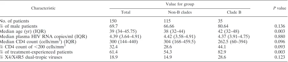
A total of 150 plasma specimens collected from HIV-1-infected individuals with enough volume available were iden-tified at three sites in Spain. Of them, 115 belonged to subjects infected with non-B subtypes and 35 belonged to patients in-fected with clade B viruses. The main baseline characteristics of the study population are summarized in Table 1. Overall, 69.7% were male and 61.4% were antiretrovirally experienced patients. The median viral load value was 4.4 log HIV RNA copies/ml, and the median CD4 count was 300 cells/mm3. The
proportion of treatment-experienced patients was significantly higher in the group infected with clade B than in the group infected with non-B viruses (82.9% versus 54.3%, respectively;
P ⫽ 0.003). Moreover, treatment-experienced patients had significantly lower CD4 counts (271 versus 364 cells/mm3,
[image:2.585.45.542.81.189.2]re-spectively;P⫽0.012) and lower plasma HIV RNA levels (4.33 versus 4.54 log copies/ml, respectively; P ⫽ 0.033) than did drug-naı¨ve subjects. HIV-1 clade distribution was as follows: 13 A (8.7%), 35 B (23.3%), 13 C (8.7%), 7 D (4.7%), 4 F (2.7%), 24 G (16%), 2 H (1.3%), 2 J (1.3%), 7 CRF01_AE (4.7%), 24 CRF02_AG (16%), 3 CRF11 (2%), 4 CRF12_BF (2.7%), 3 CRF14_BG (2%), 8 URF (5.5%), and 1 undetermined (0.7%). The phenotypic assay could be successfully performed for 122 (81.3%) specimens, while results using the genotypic tools could be obtained from 130 (86.7%) specimens. Paired phe-notypic and gephe-notypic results were available for 115 samples, 80 belonging to non-B subtypes (69.5%) and all 35 clade B
TABLE 1. Baseline characteristics of the study population
Characteristic
Value for group
Pvalue
Total Non-B clades Clade B
No. of patients 150 115 35
% of male patients 69.7 66.66 80.64 0.136
Median age (yr) (IQR) 39 (34–45.75) 38 (32–44) 42 (32–48) 0.003
Median plasma HIV RNA copies/ml (IQR) 4.39 (3.64–4.91) 4.42 (3.58–4.91) 4.37 (3.91–4.75) 0.880 Median CD4 count (cells/mm3) (IQR) 300 (144–440) 304 (168–459.5) 262.5 (60–394) 0.096
% CD4 count of⬍200 cells/mm3 32.4 28.6 44.1 0.093
% of treatment-experienced patients 61.4 54.3 82.9 0.003
% X4/X4R5 dual-tropic viruses 18.9 14.9 28.6 0.123
on May 16, 2020 by guest
http://jcm.asm.org/
specimens. Failure of amplification was the main reason for the lack of results for non-B subtypes, most likely due to the high genetic variability within the envelope region in these variants.
Some differences in coreceptor usage based on the pheno-typic information, considering baseline characteristics of the study population, were found. Overall, 18.9% of samples had X4 or X4R5 dual-tropic viruses. There were no significant differences in coreceptor usage when non-B and B samples were compared (14.9% versus 28.6%, respectively;P⫽0.123). However, a higher prevalence of X4 variants was observed in subjects with CD4 counts below 200 cells/mm3 than in those
with higher CD4 counts (32.4% versus 14.7%, respectively;
P⫽0.045). X4 variants also tended to be more prevalent in treatment-experienced than in drug-naı¨ve patients (26.5% ver-sus 10.6%, respectively;P⫽0.056). In the multivariate analy-sis, a 3.6-fold-higher probability of X4 viruses was recognized for patients with CD4 counts below 200 cells/mm3regardless of
treatment exposure and HIV-1 clade (P⫽0.012) [odds ratio⫽ 3.6; 95% confidence interval, 1.33 to 9.82]. Consideration of each of the different HIV-1 subtypes separately did not provide any remarkable finding, although numbers were too low for most non-B subtypes tested.
Reliability of genotypic tools to predict coreceptor usage.
The overall concordance between the results obtained using the distinct genotypic tools taking the phenotypic results as a reference was always over 76%. Overall, genotypic tools performed slightly better in testing clade B than non-B viruses (mean concordance of 85.7% versus 80.2%,
respec-tively). For testing of subtype B samples, the most accurate bioinformatics tools were PSSMX4R5 (91.4%), PSSMsinsi (88.6%), and geno2pheno (88.6%), while the best concor-dance for testing non-B variants was seen with PSSMsinsi (83.8%), which was closely followed by Charge Rule, PART, C4.5 with positions 8 to 12 only, and PSSMsinsi, with a concordance of 82.5% for each one. Overall, no significant differences in concordance were found by comparing B and non-B groups, although the most remarkable difference was found using geno2pheno (88.6% versus 71.3%, respectively;
P⫽0.056) (Table 2).
SVM, PSSMR5X4, PSSMsinsi, and geno2pheno discordances generally resulted in an overestimation of X4 viruses, while other bioinformatics tools tended to underestimate X4 viruses more frequently. Geno2pheno was the genotypic predictor more prone to overestimation of X4 tropism (up to 22.5% in non-B samples), and C4.5 with positions 8 to 12 only was the genotypic tool that more frequently underestimated X4 (14.8% of all samples). Of note, a careful examination of subjects with discordances did not reveal that disagreements between phe-notypic and gephe-notypic tools could be attributed to differences in patient characteristics such as viral load, CD4 counts, or antiretroviral treatment exposure.
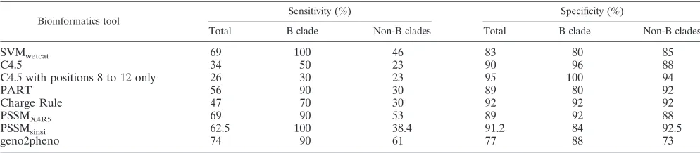
[image:3.585.44.542.80.214.2]The sensitivities and specificities for detecting X4 variants using distinct genotypic tools are summarized in Table 3. All genotypic tools showed a higher sensitivity for testing of clade B than non-B viruses, while the specificity differed according to the method used. Of note, while geno2pheno showed a good sensitivity for inferring X4 tropism for clade B viruses, its
TABLE 2. Concordance between results of HIV-1 tropism obtained using genotypic tools and a reference phenotypic assaya
Genotypic tools
Total Clade B Non-B clades
Concordance (%)
X4/R5 discordance
(%)
R5/X4 discordance
(%)
Concordance (%)
X4/R5 discordance
(%)
R5/X4 discordance
(%)
Concordance (%)
X4/R5 discordance
(%)
R5/X4 discordance
(%)
SVMwetcat 80.9 6.1 13 85.7 0 14.3 78.8 8.8 12.5
C4.5 79.1 13 7.8 82.9 14.3 2.9 77.5 12.5 10
C4.5 with positions 8 to 12 only
81.7 14.8 3.5 80 20 0 82.5 12.5 5
PART 82.6 8.7 8.7 82.9 2.9 14.3 82.5 11.3 6.3
Charge Rule 83.5 10.4 6.1 85.7 8.6 5.7 82.5 11.3 6.3
PSSMX4R5 85.2 6.1 8.7 91.4 2.9 5.7 82.5 7.5 10
PSSMsinsi 85.2 7 7.8 88.6 0 11.4 83.2 10 6.3
geno2pheno 76.5 5.2 18.3 88.6 2.9 8.6 71.3 6.3 22.5
aFor X4/R5 discordance, tropism determines X4 dual tropism, while the bioinformatic tool predicts R5 tropism. For R5/X4 discordance,
Phenoscript-tropism determines R5 Phenoscript-tropism, while the bioinformatic tool predicts X4 dual Phenoscript-tropism.
TABLE 3. Sensitivities and specificities of distinct bioinformatics tools to predict X4 tropism using the results obtained using a phenotypic assay as a reference
Bioinformatics tool
Sensitivity (%) Specificity (%)
Total B clade Non-B clades Total B clade Non-B clades
SVMwetcat 69 100 46 83 80 85
C4.5 34 50 23 90 96 88
C4.5 with positions 8 to 12 only 26 30 23 95 100 94
PART 56 90 30 89 80 92
Charge Rule 47 70 30 92 92 92
PSSMX4R5 69 90 53 89 92 88
PSSMsinsi 62.5 100 38.4 91.2 84 92.5
geno2pheno 74 90 61 77 88 73
on May 16, 2020 by guest
http://jcm.asm.org/
[image:3.585.43.540.616.725.2]specificity was the worst for non-B subtypes. An overestimation of X4 viruses, mainly for non-B subtypes, was the main reason for this behavior.
DISCUSSION
The imminent approval of CCR5 antagonists for the treat-ment of HIV-1 infection has significantly raised interest in viral tropism. In general, coreceptor usage must be known before these drugs are prescribed. While phenotypic tests are consid-ered to be the “gold standard,” they are laborious and costly and require sophisticated facilities and experienced personnel. Genotypic predictors of HIV-1 tropism have been developed to circumvent these disadvantages; however, the high genetic variability of HIV-1 may represent a serious obstacle to ensure their reliability. In addition to these diagnostic obstacles, it is important to better understand HIV pathogenesis and explain why some HIV-1 clades seem to be more frequently X4 tropic than others, as recently claimed for subtypes A, D, and CRF14_BG (1, 10, 14, 17, 22), since patients infected with these variants might progress faster to AIDS and will benefit less from CCR5 antagonists.
In this study, we evaluated the prevalence of X4 or X4R5 dual-tropic viruses in a relatively large population of non-B HIV-1 clinical specimens. Up to 19% of these individuals car-ried X4/X4R5 dual-tropic viruses, with no significant differ-ences with a control group of patients infected with clade B viruses. In other studies that included a larger number of samples from clades C and D, it was noted that clade C might less frequently harbor X4 viruses, even in late disease stages (8, 16, 20, 24, 31), while conversely, clade D could be X4 tropic more frequently (10, 14). The small number of samples with these variants in our study precluded the examination of this aspect more appropriately.
A significantly higher prevalence of X4 and X4/R5 dual-tropic viruses was found in patients with lower CD4 counts, which is in agreement with previously reported observations and with the notion that these HIV-1 variants tend to be selected as immunodeficiency progresses (3, 14, 15, 31). A higher prevalence of X4/X4R5 viruses also tended to be asso-ciated with prior exposure to antiretroviral therapy, which is also in agreement with previously reported observational data from cross-sectional surveys (11, 19).
The accuracy of genotypic tools to assess viral tropism based on V3 sequences was previously examined mostly in samples collected from patients infected with clade B viruses, using phenotypic tests as a comparison (4, 18, 26). To our knowl-edge, this is the first study to assess the performances of var-ious genotypic bioinformatics tools to predict HIV-1 tropism in non-B subtypes using the results obtained with a phenotypic test as a reference. In the present study, we have sequenced the V3 region for the bulk viral population. Moreover, we also considered minority species by assessing nucleotide mixtures in the electropherogram. Molecular cloning of each sample seems to be the best method to detect mixed populations, but it is much more complicated, and it seems to produce very similar results (6).
By comparing the results obtained using genotypic tools and the phenotypic assay in the whole population, there was a relatively good concordance (mean of 81.3%, ranging from
76% using geno2pheno to 85.5% using the charge rule). Most genotypic algorithms, however, performed better for testing clade B than non-B viruses. The most pronounced differences were found using PSSM and geno2pheno, most likely because they are designed exclusively based on clade B strains, while the wetcat system has been built with both B and non-B se-quences.
Considering that the main application for the V3 genotypic algorithms in clinical settings will be for excluding the presence of X4 or X4R5 dual-tropic variants in patients in whom CCR5 antagonists might be considered as potential therapy, it is man-datory to know the sensitivity and specificity of each of these genotypic tools precisely. Our results showed that the specific-ity was above 80% when either B or non-B variants were tested by all the different genotypic tools except geno2pheno (77%). Moreover, geno2pheno showed the highest differences when B and non-B samples were compared, obtaining a specificity of 73% for non-B and 88% for B specimens. However, the sen-sitivity for the detection of X4 variants differed substantially when distinct genotypic tools were used according to HIV-1 subtype. The sensitivities of SVM and PSSMsinsiwere 100% for clade B viruses, while they were only 46% and 38.4%, respec-tively, for non-B clades. The highest sensitivity for recognizing X4 viruses when testing non-B subtypes was seen for geno2pheno, although it was only 61%, which may be consid-ered to be unacceptable for clinical use to replace phenotypic tests.
This study has several limitations. First, although the entire set of non-B subtypes examined included a relatively large number of clinical samples, some HIV-1 clades were not or only scarcely represented. Second, only one phenotypic assay was used as a reference, and disagreement between distinct phenotypic tests for determining HIV-1 coreceptor usage in pairwise plasma samples has recently been highlighted, being of up to 15% in one recent study (33). Third, phenotypic and genotypic results could not be obtained from 14 to 19% of specimens belonging to patients infected with non-B subtypes, and therefore, the concordance between phenotypic and ge-notypic tropism results could have been slightly overestimated. In conclusion, estimations of HIV-1 tropism using bioinfor-matics tools based on V3 sequences are better for testing clade B than non-B viruses. Moreover, the sensitivity for detecting X4 variants differed substantially according to HIV-1 subtype. In light of their low sensitivity, it should be advisable not to rely on genotypic tools to exclude X4 variants in patients infected with non-B subtypes.
ACKNOWLEDGMENTS
This work was supported in part by grants from the Fundacio´n Investigacio´n y Educacio´n en SIDA, Red de Investigacio´n en SIDA (ISCIII-RETIC RD06/006), the European NEAT Network, Agencia Lain Entralgo, and Fondo de Investigaciones Sanitarias projects CP06/ 00284 and PI06/1826.
REFERENCES
1.Andreoletti, L., K. Skrabal, V. Perrin, N. Chomont, S. Saragosti, G. Gre-senguet, H. Moret, J. Jacques, J. D. D. Longo, M. Matta, F. Mammano, and L. Belec.2007. Genetic and phenotypic features of blood and genital viral populations of clinically asymptomatic and antiretroviral-treatment-naı¨ve clade A human immunodeficiency virus type 1-infected women. J. Clin.
Microbiol.45:1838–1842.
2.Berger, E., R. Doms, E. Fenyo, B. Korber, D. Littman, J. Moore, Q.
on May 16, 2020 by guest
http://jcm.asm.org/
tau, H. Schuitemaker, J. Sodroski, and R. Weiss.1998. A new classification
for HIV-1. Nature391:240.
3.Brumme, Z., J. Goodrich, H. Mayer, C. Brumme, B. Henrick, B. Wynhoven, J. Asselin, P. Cheung, R. Hogg, J. Montaner, and P. Harrigan.2005. Mo-lecular and clinical epidemiology of CXCR4-using HIV-1 in a large
popu-lation of antiretroviral-naive individuals. J. Infect. Dis.192:466–474.
4.Chueca, N., M. Alvarez, M. Casan˜as, E. Poveda, C. Garrido, V. Guillot, N. Zahonero, V. Soriano, C. de Mendoza, and F. Garcı´a.2007. A comparison of 7 different tools available to predict coreceptor usage based on V3 genetic sequences, abstr. 53. Abstr. 5th Eur. HIV Drug Resist. Wkshp., Cascais, Portugal.
5.Cormier, E., and T. Dragic.2002. The crown and stem of the V3 loop play distinct roles in human immunodeficiency virus type 1 envelope glycoprotein
interactions with the CCR5 coreceptor. J. Virol.76:8953–8957.
6.Delobel, P., M. T. Nugeyre, M. Cazabat, C. Pasquier, B. Marchou, P. Mas-sip, F. Barre-Sinoussi, N. Israel, and J. Izopet.2007. Population-based
sequencing of the V3 region ofenvfor predicting the coreceptor usage of
human immunodeficiency virus type 1 quasispecies. J. Clin. Microbiol.45:
1572–1580.
7.de Mendoza, C., C. Rodriguez, F. Garcia, J. M. Eiros, L. Ruiz, E. Caballero, A. Aguilera, P. Leiva, J. Colomina, F. Gutierrez, J. del Romero, J. Aguero, V. Soriano, and the Spanish HIV Seroconverter Study Group.2007. Prevalence of X4 tropic viruses in patients recently infected with HIV-1 and lack of association with transmission of drug resistance. J. Antimicrob. Chemother.
59:698–704.
8.Holm-Hansen, C., E. Baan, B. Asjo, F. Pascu, J. Goudsmit, and J. De Jong.
2000. Determinants for the syncytium-inducing phenotype of HIV-1 subtype
F isolates are located in the V3 region. AIDS Res. Hum. Retrovir.16:867–
870.
9.Holm-Hansen, C., D. Grothues, S. Rustad, B. Rosok, F. Pascu, and B. Asjo.
1995. Characterization of HIV type 1 from Romanian children: lack of correlation between V3 loop amino acid sequence and syncytium formation
in MT-2 cells. AIDS Res. Hum. Retrovir.11:597–603.
10.Huang, W., S. Eshleman, J. Toma, S. Fransen, E. Stawiski, E. Paxinos, J. Whitcomb, A. Young, D. Donnell, F. Mmiro, P. Musoke, L. Guay, J. Jackson, N. Parkin, and C. Petropoulos.2007. Co-receptor tropism in subtype D HIV-1: high prevalence of CXCR4 tropism and heterogeneous composition
of viral populations. J. Virol.81:7885–7893.
11.Hunt, P., P. Harrigan, W. Huang, M. Bates, D. Williamson, J. McCune, R. Price, S. Spudich, H. Lampiris, R. Hoh, T. Leigler, J. Martin, and S. Deeks.
2006. Prevalence of CXCR4 tropism among antiretroviral-treated
HIV-1-infected patients with detectable viremia. J. Infect. Dis.194:926–930.
12.Jensen, M., F. Li, A. van’t Wout, D. Nickle, D. Shriner, H. He, and S. McLaughlin. 2003. Improved coreceptor usage prediction and genotypic monitoring of R5-to-X4 transition by motif analysis of human
immunodefi-ciency virus type 1envV3 loop sequences. J. Virol.77:13376–13388.
13.Jensen, M., M. Coetzer, A. van’t Wout, L. Morris, and J. Mullins.2006. A reliable phenotype predictor for human immunodeficiency virus type 1
sub-type C based on envelope V3 sequences. J. Virol.80:4698–4704.
14.Kaleebu, P., I. Nankya, D. Yirrell, L. Shafer, J. Kyosiimire-Lugemwa, D. Lule, D. Morgan, S. Beddows, J. Weber, and J. Whitworth.2007. Relation between chemokine receptor use, disease stage, and HIV-1 subtypes A and D: results from a rural Ugandan cohort. J. Acquir. Immune Defic. Syndr.
45:28–33.
15.Koot, M., I. Keet, A. Vos, R. de Goede, M. Roos, R. Coutinho, F. Miedema, P. Schellekens, and M. Tersmette.1993. Prognostic value of HIV-1
syncytium-inducing phenotype for rate of CD4⫹cell depletion and progression to AIDS.
Ann. Intern. Med.118:681–688.
16.Kuiken, C., J. de Jong, E. Baan, W. Keulen, M. Tersmette, and J. Goudsmit.
1992. Evolution of the V3 envelope domain in proviral sequences and iso-lates of human immunodeficiency virus type 1 during transition of the viral
biological phenotype. J. Virol.66:4622–4627.
17.Laeyendecker, O., X. Li, M. Arroyo, F. McCutchan, R. Gray, M. Wawer, D. Serwadda, F. Nalugoda, G. Kigozi, T. Quinn, and the Rakai Health Science Program.2006. The effect of HIV subtype on rapid progression in Rakai, Uganda, abstr. 44LB. Abstr. 13th Conf. Retrovir. Opportun. Infect., Denver, CO.
18.Low, A., D. Chan, W. Dong, T. Sing, R. Swanstrom, M. Jensen, S. Pillai, B. Good, V. Dias Lima, and P. Harrigan.2007. Current implementations of HIV genotyping algorithms are inadequate for the prediction of X4
core-ceptor usage in clinical isolates from population-based V3 sequences:
ap-proaches to improvement. Antivir. Ther.12:S164.
19.Moyle, G., A. Wildfire, S. Mandalia, H. Mayer, J. Goodrich, J. Whitcomb, and B. Gazzard.2005. Epidemiology and predictive factors for chemokine
receptor use in HIV-1 infection. J. Infect. Dis.191:866–872.
20.Ndung’u, T., E. Sepako, M. McLane, F. Chand, K. Bedi, S. Gaseitsiwe, F. Doualla-Bell, T. Peter, I. Thior, S. Moyo, P. Gilbert, V. Novitsky, and M. Essex.2006. HIV-1 subtype C in vitro growth and coreceptor utilization.
Virology347:247–260.
21.Pastore, C., R. Nedellec, A. Ramos, S. Pontow, L. Ratner, and D. Mosier.
2006. HIV type 1 coreceptor switching: V1/V2 gain-of-fitness mutations
compensate for V3 loss-of-fitness mutations. J. Virol.80:750–758.
22.Pe´rez-Alvarez, L., M. Mun˜oz, E. Delgado, C. Miralles, A. Ocampo, V. Gar-cia, M. Thomson, G. Contreras, R. Na´jera, and the Spanish group for Antiretroviral Resistance Studies in Galicia.2006. Isolation and biological characterization of HIV-1 BG intersubtype recombinants and other genetic
forms circulating in Galicia Spain. J. Med. Virol.78:1520–1528.
23.Pillai, S., B. Good, D. Richman, and J. Corbeil.2003. A new perspective on
V3 phenotype prediction. AIDS Res. Hum. Retrovir.19:145–149.
24.Ping, L., J. Nelson, I. Hoffman, J. Schock, S. Lamers, M. Goodman, P. Vernazza, P. Kazembe, M. Maida, D. Zimba, M. Goodenow, J. Eron, S. Fiscus, M. Cohen, and R. Swanstrom.1999. Characterization of V3 se-quence heterogeneity in subtype C human immunodeficiency virus type 1
isolates from Malawi: underepresentation of X4 variants. J. Virol.73:6271–
6281.
25.Poveda, E., V. Briz, M. Quin˜ones-Mateu, and V. Soriano.2006. HIV tropism: diagnostic tools and implications for disease progression and treatment with
entry inhibitors. AIDS20:1359–1367.
26.Poveda, E., V. Briz, V. Roulet, M. Gonza´lez, J. L. Faudon, K. Skrabal, M. Gonza´lez, and V. Soriano.2007. Correlation between a phenotypic assay and
three bioinformatic tools for determining HIV co-receptor use. AIDS21:
1487–1490.
27.Resch, W., N. Hoffman, and R. Swanstrom.2001. Improved success of phenotype prediction of the HIV type 1 from envelope variable loop 3
sequence using neural networks. Virology288:51–62.
28.Richman, D., and S. Bozzette.1994. The impact of the syncytium-inducing
phenotype of HIV on disease progression. J. Infect. Dis.169:968–974.
29.Roulet, V., S. Rochas, J. L. Labernardiere, F. Mammano, J. L. Faudon, N. Raja, S. Lebel-Binay, and K. Skabal.2007. HIV PHENOSCRIPT ENV: a sensitive assay for the detection of HIV X4 minority species and determi-nation of non-B subtype viral tropism, abstr. 617. Abstr. 14th Conf. Retrovir. Opportun. Infect., Los Angeles, CA.
30.Sander, O., T. Sing, I. Sommer, A. Low, P. Cheung, P. Harrigan, T. Len-gauer, and F. Domingues.2007. Structural descriptors of gp120 V3 loop for
the prediction of HIV-1 coreceptor usage. PLoS Comput. Biol.3:e58.
31.Schuitemaker, H., M. Koot, N. Kootstra, M. Dercksen, R. de Goede, R. van Steenwijk, J. Lange, J. Schattenkerk, F. Miedema, and M. Tersmette.1992. Biological phenotype of human immunodeficiency virus type 1 clones at different stages of infection: progression of disease is associated with a shift
from monocytotropic to T-cell-tropic virus population. J. Virol.66:1354–
1360.
32.Sing, T., N. Beerenwinkel, and T. Lengauer.2004. Learning mixtures of
localized rules by maximizing the area under the ROC curve, p. 89–96.In
J. Herna´ndez-Orallo, N. Lachiche, and P. Flach (ed.), The ROCAI-2004
International Workshop on ROC Analysis in Artificial Intelligence, Valen-cia, Spain.
33.Skrabal, K., A. Low, W. Dong, T. Sing, P. Cheung, F. Mammano, and P. Harrigan.2007. Determining human immunodeficiency virus coreceptor use in a clinical setting: degree of correlation between two phenotypic assays and
a bioinformatic model. J. Clin. Microbiol.45:279–284.
34.Tscherning, C., A. Alaeus, R. Fredriksson, A. Bjorndal, H. Deng, D. Littman, E. Fenyo, and J. Albert.1998. Differences in chemokine coreceptor usage
between genetic subtypes of HIV-1. Virology241:181–188.
35.Tscherning-Casper, C., D. Vo¨dro¨s, E. Menu, K. Aperia, R. Fredriksson, G. Dolcini, G. Chaouat, F. Barre´-Sinoussi, J. Albert, E. Fenyo¨, et al.2000. Coreceptor usage of HIV-1 isolates representing different genetic subtypes obtained from pregnant Cameroonian women. J. Acquir. Immune Defic.
Syndr.24:1–9.
36.Weber, J., H. Piontkivska, and M. Quinones-Mateu.2006. HIV type 1 tropism
and inhibitors of viral entry: clinical implications. AIDS Rev.8:60–77.