Copyright © 2004, American Society for Microbiology. All Rights Reserved.
Evolutionary Genetic Analysis of the Emergence of Epidemic
Vibrio
cholerae
Isolates on the Basis of Comparative Nucleotide
Sequence Analysis and Multilocus Virulence
Gene Profiles
Yvonne A. O’Shea, F. Jerry Reen, Anne Marie Quirke, and E. Fidelma Boyd*
Department of Microbiology, University College Cork, National University of Ireland, Cork, Ireland
Received 3 October 2003/Returned for modification 17 February 2004/Accepted 24 April 2004
Vibrio cholerae, the causative agent of cholera, is a natural inhabitant of the aquatic ecosystem. We examined
a unique collection ofV. choleraeclinical and environmental isolates of widespread geographic distribution
recovered over a 60-year period to determine their evolutionary genetic relationships based on analysis of two
housekeeping genes, malate dehydrogenase (mdh) and a chaperonin (groEL). In addition, the phylogenetic
distribution of 12 regions associated with virulence was determined. Comparative sequence analysis ofmdh
revealed that allV. choleraeO1 and O139 serogroup isolates belonged to the same clonal lineage. Single-strand
conformational polymorphism (SSCP) analysis of these O1 and O139 strains atgroELconfirmed the presence
of an epidemic clonal complex. Of the 12 virulence regions examined, only three regions, Vibrio seventh
pandemic island 1 (VSP-I), VSP-II, and RS1, were absent from all classicalV. choleraeisolates. MostV. cholerae
El Tor biotype and O139 serogroup isolates examined encoded all 12 virulence regions assayed. Outside ofV.
choleraeO1/O139 serogroup isolates, only one strain, VO7, contained VSP-I. TwoV. choleraeEl Tor isolates, GP155 and 2164-78, lacked both VSP-I and VSP-II, and one El Tor isolate, GP43, lacked VSP-II. Five
non-O1/non-O139 serogroup isolates had an mdh sequence identical to that of the epidemic O1 and O139
strains. These isolates, similar to classical strains, lack both VSP-I and VSP-II. Four of the 12 virulence regions
examined were found to be present in all isolates: hlyA, pilE, MSHA and RTX. Among non-O1/non-O139
isolates, however, the occurrence of the additional eight regions was considerably lower. The evolutionary
relationships and multilocus virulence gene profiles ofV. choleraenatural isolates indicate that consecutive
pandemic strains arose from a common O1 serogroup progenitor through the successive acquisition of new virulence regions.
Vibrio cholerae is a natural inhabitant of the aquatic envi-ronment and is found associated with shellfish and crustaceans (12, 21, 29, 48).V. choleraeis the causative agent of the diar-rheal disease cholera, and humans are the only known animal host. V. cholerae is an extracellular pathogen of the small intestine and causes significant human disease and death, par-ticularly on the Indian subcontinent. A recent study has shown that the human host may contribute significantly to cholera epidemics, since passage through the human intestine was shown to induce a hyperinfectious state, which was perpetu-ated in the natural environment after release (43). Of the 200 O-antigen serogroups so far identified amongV. cholerae iso-lates, only two serogroups, O1 and O139, are known to cause epidemic and pandemic cholera (34). TheV. choleraeO1 se-rogroup can be further divided into two biotypes of epidemi-ological relevance, classical and El Tor, based on minor phe-notypic differences. The first cholera pandemic, which began in 1817 in Asia, and subsequent pandemics, were probably caused by the classical biotype. In 1961, the seventh and present pan-demic began, which was caused by the El Tor biotype (34). In 1992, for the first time in the recorded history of cholera a novel O-serogroup, O139 emerged to cause epidemic cholera
(1). Significantly, exposure to O1 serogroup cholera does not protect against O139 cholera (45). The El Tor strain re-emerged to overtake the O139 serogroup as the major cause of cholera by 1996 (23). However, the O139 serogroup is still present on the Indian subcontinent and, in some areas, is the predominant cause of cholera (23). Interestingly, several stud-ies have proposed that the origin of the serogroup O139 strain was an El Tor strain that obtained the O139 biosynthesis genes (as well as the SXT element and a capsule) via antigenic switching from a donor strain (3, 44, 60, 65). Recently, it has been proposed based on comparative sequence analysis that an O22 serogroup maybe a possible donor for the O139 serogroup (17, 67). Sporadic cholera outbreaks caused by V. cholerae non-O1 and non-O139 isolates have been documented; for example, in 1968 in Sudan there was a cholera outbreak caused by an O37 serogroup isolate (21, 68).
The evolutionary genetic relationships among V. cholerae strains have been examined by multilocus enzyme electro-phoresis (2, 11, 19, 20, 54, 58), single locus sequence analysis (7, 37, 38, 59), and multilocus sequence analysis of housekeep-ing genes (9, 36, 40). These analyses have given conflicthousekeep-ing results regarding the ancestry of O1 serogroup classical and El Tor biotype strains. Several studies suggest that at least three pathogenic clones exist, consisting of classical and El Tor bio-type strains and U.S. Gulf Coast strains (37, 63). Others have suggested that the three pathogenic clones are very closely * Corresponding author. Mailing address: Department of
Microbi-ology, UCC, National University of Ireland, Cork, Ireland. Phone: 353 21 4903624. Fax: 351 21 4903101. E-mail: [email protected].
4657
on May 15, 2020 by guest
http://jcm.asm.org/
related (2, 9, 18). In all studies, the epidemicV. cholerae iso-lates form a lineage separate from nonepidemic strains.
Although the O-antigen is a major protective antigen inV. choleraevirulence and probably plays a role in host coloniza-tion, the two major virulence factors ofV. choleraeare cholera toxin (CT), the main cause of the explosive rice watery diar-rhea, and toxin-coregulated pilus (TCP), the main intestinal colonization factor (61). ThectxABgenes, which encode CT, are integral components of a novel filamentous phage CTX (64), and the TCP biosynthesis genes are encoded on theVibrio pathogenicity island (hereafter designated VPI-1) (35). A number of studies have found that the two main virulence factors, CT and TCP, are predominately associated with V. choleraeO1 and O139 serogroup strains and are only occasion-ally found in nonepidemic isolates (5, 7, 8, 10, 13, 25, 40, 47, 49, 50, 55).
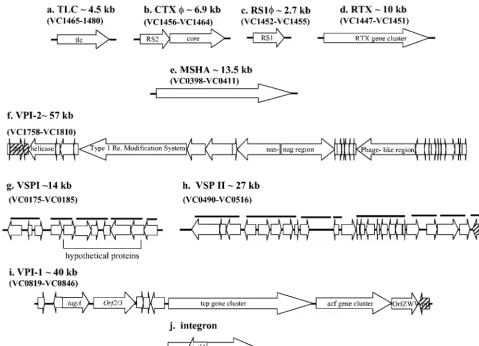
AmongV. choleraeEl Tor isolates, CTXis flanked by an additional filamentous phage RS1that is required for CTX production (16, 22) (Fig. 1). The CTX prophage is also flanked by the toxin-linked cryptic plasmid (TLC), whose role in patho-genesis is unknown (56) (Fig. 1). A number of other gene clusters have also been identified that are found predominantly among epidemicV. choleraeisolates: the RTX toxin gene clus-ter (42), the mannose-sensitive hemolysin agglutination pilin
(MSHA) (32), VPI-2 (31), hemolysin, and PilE pilin (28, 30) (Fig. 1). Recently, comparative genomic studies which used a V. choleraeDNA microarray among 11 epidemic isolates iden-tified two regions, Vibrioseventh pandemic island I (VSP-I), encompassing VC0175 to VC0185, and VSP-II, encompassing VC0490 to VC0497, that were found exclusively among El Tor biotype isolates (18). The role of VSP-I and VSP-II in V. choleraevirulence remains undetermined.
[image:2.603.51.530.67.413.2]To elucidate the steps and significance of virulence gene acquisition in the evolution ofV. choleraeit is essential to know the underlying phylogenetic relationships among strains. In this study we examined a unique collection of 64V. cholerae and 5Vibrio mimicusisolates to determine their evolutionary genetic relationships and multilocus virulence gene profiles to elucidate the steps involved in the emergence of epidemic isolates. Our results show thatV. choleraeserogroup O1 clas-sical and El Tor biotype strains encompass a single epidemic clonal complex and that differences between biotype strains arose through the acquisition of additional virulence regions by El Tor isolates. The emergence of epidemicV. choleraeO139 serogroup strains was not a unique occurrence in the history of cholera, sinceV. choleraeO37 and O8 serogroup isolates phy-logenetically cluster with O1 and O139 serogroup isolates, indicating antigenic switching.
FIG. 1. Schematic representation of nine regions that are associated with pathogenesis in V. cholerae. The positions and directions of transcription of the open reading frames are indicated by the directions of the arrows. The black bold horizontal lines indicate the positions of the PCR primers.
on May 15, 2020 by guest
http://jcm.asm.org/
MATERIALS AND METHODS
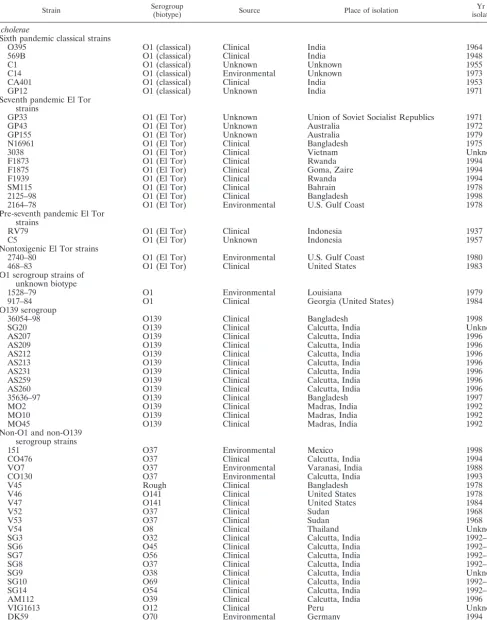
Bacterial isolates.A total of 64V. choleraeisolates were examined in this study (Table 1). The 64V. choleraeisolates belonged to 19 different serogroups, 3 isolates had no serogroup designation, and 23 isolates belonged to serogroup O1, of which 6 isolates were of the classical biotype and 15 isolates were of the El Tor biotype. The O139 serogroup was represented by 13 isolates, the O37 serogroup was represented by 7 isolates, and the O8 and O141 serogroups each were represented by 2 isolates. There were 14 serogroups represented by a singleV. choleraeisolate. TheV. choleraeisolates were recovered from six continents (North and South America, Asia, Europe, Australia, and Africa) over a 60-year period (1937 to 2000) (Table 1). In addition, our study also included fiveV. mimicusisolates, four O115 and one O41 serogroup isolates. Of the 69 strains examined, 51 were clinical isolates and 12 were environmental isolates (Table 1). AllVibriostrains were grown in Luria-Bertani (LB) broth and stored at⫺70°C in LB broth with 20% (vol/vol) glycerol.
DNA isolation.Chromosomal DNA was extracted from eachV. choleraeand V. mimicusisolate by using the G-nome DNA isolation kit from Bio 101 (Vista, Calif.). Briefly, a single colony of each isolate was inoculated into 3 ml of LB broth and incubated overnight at 37°C with shaking at 150 rpm. The bacterial cells were pelleted at 3,000 rpm for 5 min, the supernatant was discarded, and the pellet brought to a final volume of 1.85 ml in cell suspension solution. The cells were lysed and treated with RNase and protease. DNA was extracted with Tris-EDTA buffer and ethanol and resuspended in Tris-EDTA buffer.
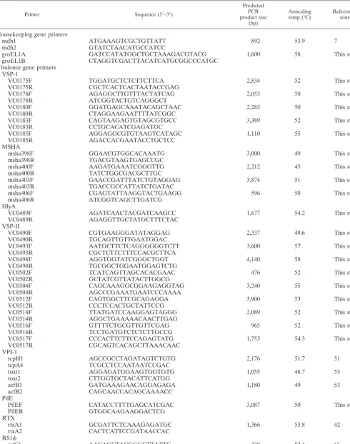
PCR amplification and nucleotide sequencing.PCR primers to amplify the chromosomal housekeeping gene malate dehydrogenase (mdh) were designed from themdhsequence ofV. choleraestrain N16961 (26). The following PCR cycle was used to amplify themdhgene for each isolate: an initial denaturation step at 96°C for 1 min followed by 30 cycles of denaturation at 94°C for 30 s, 30 s of primer annealing at 53.9°C, and 1.5 min of primer extension at 72°C. The primer pair mdh1-mdh2 amplified an 892-bp fragment, representing 84% of the mdhgene. PCR products were purified with the QIAquick PCR purification kit (Qiagen, Hilden, Germany) in accordance with the manufacturer’s instructions. After purification, an aliquot of 10l was used as a sequencing template. The mdhgene sequences were determined in both directions by MWG-Biotech based on the dye deoxy terminator method.
Phylogenetic analyses.Themdhgene sequences were aligned by using the CLUSTALW multiple-sequence alignment program (27). From the mdh se-quence alignments, a 648-bp region was further analyzed by using the Molecular Evolutionary Genetics Analysis (MEGA) suite of programs, version 2.1 (39). Phylogenetic gene trees were constructed by the neighbor-joining method with the Jukes-Cantor distance method (33, 57). Bootstrap values were calculated for 1,000 trees. The proportions of synonymous (silent) substitutions per synony-mous site (Ds) and nonsynonysynony-mous (replacement) substitutions per nonsynony-mous site (Dn) were calculated.
PCR-SSCP.In conjunction withmdhsequencing, an additional 25V. cholerae O1 and O139 serogroup strains were analyzed to confirm sequence identity at this locus within these two serogroups by PCR–single-strand conformational polymorphism analysis (PCR-SSCP), a simple and rapid method to determine point mutations within genes. Two oligonucleotide primers, mdh1 and mdh2 (Table 2), were used to amplify an 892-bp PCR product, which was then re-stricted with HindIII (Roche Molecular Biochemicals, East Sussex, United King-dom) at 37°C to generate two fragments. Then 5l of the restricted DNA was mixed with 5l of denaturation buffer (5 mM EDTA, 0.05% bromophenol blue, and xylene cyanole in formamide), and the mixture was incubated at 95°C for 8 min. The sample was then placed directly in ice for 10 min before being loaded onto a nondenaturing 8% polyacrylamide gel. Samples (8l) were run at 100 V for 2 h. As a control, 4l of undenatured digested DNA (mdhgene) was used. In addition, all epidemicV. choleraeO1 and O139 serogroup isolates were examined at thegroELlocus by PCR-SSCP analysis. Primer pair groEL1A and groEL1B, designed fromV. choleraegenome sequence (26) were used to PCR amplify a 1.6-kb band from 20V. choleraeO1 and O139 serogroup isolates and 2 O37 serogroup isolates. PCR products were digested with BstYI at 60°C to generate four restriction bands. Restricted DNA (10l) was denatured as de-scribed above in 10l of denaturation buffer and electrophoresed at 175 V for 6 h.
After electrophoresis, the 8% polyacrylamide gels were silver stained with a DNA silver staining kit (Pharmacia Biotech). Briefly, the silver staining proce-dure was as follows. The gels were first fixed in 10% acetic acid for approximately 30 min at room temperature and washed with deionized water three times for 2 min. Color impregnation lasted for 20 min at room temperature. The gel was then washed for 5 to 10 s with deionized water, followed by color development for 6 min with a color development solution. The color reaction was stopped, and
the bands were fixed. The gel was air dried for approximately 2 h. SSCP profiles were interpreted visually.
PCR analysis.PCR was used to assay 64V. choleraeand 5V. mimicusisolates for the presence of 12 regions associated withV. choleraevirulence. Of the 12 virulence regions examined, 10 regions were comprised of three or more genes (VSP-I, MSHA pilin, VSP-II, VPI-1, Repeat in toxin [RTX], RS1, CTX, TLC, VPI-2, and class 1 integron) and 2 loci were single gene regions (hlyAandpilE). Of the 10 virulence gene clusters examined, 7 are associated with mobile genetic elements (Fig. 1). A total of 31 primer pairs were used to determine the distri-bution of the 12 regions among the 69Vibrioisolates (Table 2). Five primer pairs were used to assay for the presence of VSP-I, four primer pairs were used to assay for MSHA, nine primer pairs were used to assay for VSP-II, three primer pairs were used to assay for VPI-1, four primer pairs were used to assay for the presence of CTX, and one primer pair (each) was used to assay for the presence of pilE,hlyA, RTX, RS1, TLC, andintl4(Table 2). Gene fragments were amplified from chromosomal DNA isolated from the 64V. choleraestrains and the 5V. mimicusstrains. PCR was performed in a 20-l reaction mixture by using the following cycles: an initial denaturation step at 96°C for 1 min followed by 30 cycles of denaturation at 94°C for 30 s, 30 s of primer annealing at 45 to 58°C, and 1 to 4 min of primer extension at 72°C (Table 2).
Southern blot analysis.To confirm negative PCR results, Southern hybridiza-tion analysis was carried out. DNA from each strain of interest was digested with the restriction enzyme EcoRI (Roche Molecular Biochemicals) and separated by electrophoresis in 0.6% (wt/vol) 1⫻Tris-borate-EDTA agarose. Separated DNA fragments were transferred to a nitrocellulose membrane for Southern hybrid-ization. A single DNA probe was generated for each of the 12 regions by PCR amplification withV. choleraestrain N16961 as a template and labeled with horseradish peroxidase to verify the absence of a particular gene. Southern hybridization was carried out by using the enhanced chemiluminescence direct nucleic acid labeling and detection system according to the manufacturer’s in-structions (Amersham Pharmacia Biotech). In all experiments,V. choleraestrain N16961 was used as a positive control.
RESULTS
Genetic variation at themdh locus among V. cholerae
iso-lates. To determine the evolutionary genetic relationships
among our collection of V. cholerae isolates, we analyzed a 648-bp region of the housekeeping gene malate dehydrogenase (mdh) from 36V. choleraeisolates and 5V. mimicusisolates. Previous studies have shown that comparative nucleotide se-quence analysis of the mdh locus is a reliable indicator of overall genetic relationships between strains (6). Within the 648-bp region from the 36V. choleraestrains examined, there was a total of 44 polymorphic sites, which included two amino acid replacement sites (Table 3; Fig. 2). Of the 44 polymorphic sites, 22 were phylogenetically informative (at least two or more sequences contained the polymorphism) (Table 3). The average pairwise difference for the 36 V. cholerae mdh se-quences was 1.03%, with a maximum pairwise difference of 4.61% observed betweenV. choleraestrain DK71, an environ-mental O66 serogroup strain from Germany, and V. cholerae strain 9581, a clinical O41 serogroup isolate from India. Eight epidemicV. cholerae O1 El Tor and O139 serogroup strains examined had identical mdh sequences which differed from classical biotype strains at a single site. ThreeV. choleraeO37 serogroup strains, V52, V53, and CO130, one O8 serogroup strain, V54, and one rough strain, V45, had mdh sequences identical to the El Tor O1 and O139 serogroupmdhsequence. Among the 23V. choleraenon-O1 and non-O139 isolates ex-amined at themdhlocus, there were a total of 43 polymorphic sites, which resulted in 42 synonymous polymorphic sites and 1 nonsynonymous polymorphic site (Table 3). An additional 25 V. choleraeO1 and O139 serogroup isolates were examined for sequence variation at the mdh locus by PCR-SSCP analysis (Table 1) (Fig. 3). The sensitivity of PCR-SSCP tends to
on May 15, 2020 by guest
http://jcm.asm.org/
TABLE 1. Strains used in this study
Strain Serogroup(biotype) Source Place of isolation isolationYr of
V. cholerae
Sixth pandemic classical strains
O395 O1 (classical) Clinical India 1964
569B O1 (classical) Clinical India 1948
C1 O1 (classical) Unknown Unknown 1955
C14 O1 (classical) Environmental Unknown 1973
CA401 O1 (classical) Clinical India 1953
GP12 O1 (classical) Unknown India 1971
Seventh pandemic El Tor strains
GP33 O1 (El Tor) Unknown Union of Soviet Socialist Republics 1971
GP43 O1 (El Tor) Unknown Australia 1972
GP155 O1 (El Tor) Unknown Australia 1979
N16961 O1 (El Tor) Clinical Bangladesh 1975
3038 O1 (El Tor) Clinical Vietnam Unknown
F1873 O1 (El Tor) Clinical Rwanda 1994
F1875 O1 (El Tor) Clinical Goma, Zaire 1994
F1939 O1 (El Tor) Clinical Rwanda 1994
SM115 O1 (El Tor) Clinical Bahrain 1978
2125–98 O1 (El Tor) Clinical Bangladesh 1998
2164–78 O1 (El Tor) Environmental U.S. Gulf Coast 1978
Pre-seventh pandemic El Tor strains
RV79 O1 (El Tor) Clinical Indonesia 1937
C5 O1 (El Tor) Unknown Indonesia 1957
Nontoxigenic El Tor strains
2740–80 O1 (El Tor) Environmental U.S. Gulf Coast 1980
468–83 O1 (El Tor) Clinical United States 1983
O1 serogroup strains of unknown biotype
1528–79 O1 Environmental Louisiana 1979
917–84 O1 Clinical Georgia (United States) 1984
O139 serogroup
36054–98 O139 Clinical Bangladesh 1998
SG20 O139 Clinical Calcutta, India Unknown
AS207 O139 Clinical Calcutta, India 1996
AS209 O139 Clinical Calcutta, India 1996
AS212 O139 Clinical Calcutta, India 1996
AS213 O139 Clinical Calcutta, India 1996
AS231 O139 Clinical Calcutta, India 1996
AS259 O139 Clinical Calcutta, India 1996
AS260 O139 Clinical Calcutta, India 1996
35636–97 O139 Clinical Bangladesh 1997
MO2 O139 Clinical Madras, India 1992
MO10 O139 Clinical Madras, India 1992
MO45 O139 Clinical Madras, India 1992
Non-O1 and non-O139 serogroup strains
151 O37 Environmental Mexico 1998
CO476 O37 Clinical Calcutta, India 1994
VO7 O37 Environmental Varanasi, India 1988
CO130 O37 Environmental Calcutta, India 1993
V45 Rough Clinical Bangladesh 1978
V46 O141 Clinical United States 1978
V47 O141 Clinical United States 1984
V52 O37 Clinical Sudan 1968
V53 O37 Clinical Sudan 1968
V54 O8 Clinical Thailand Unknown
SG3 O32 Clinical Calcutta, India 1992–1993
SG6 O45 Clinical Calcutta, India 1992–1993
SG7 O56 Clinical Calcutta, India 1992–1993
SG8 O37 Clinical Calcutta, India 1992–1993
SG9 O38 Clinical Calcutta, India Unknown
SG10 O69 Clinical Calcutta, India 1992–1993
SG14 O54 Clinical Calcutta, India 1992–1993
AM112 O39 Clinical Calcutta, India 1996
VIG1613 O12 Clinical Peru Unknown
DK59 O70 Environmental Germany 1994
DK67 O74 Environmental Korea 1994
DK71 O66 Environmental Germany 1994
Continued on following page
on May 15, 2020 by guest
http://jcm.asm.org/
crease with increasing fragment length, therefore the 892-bp amplicon was digested with HindIII to generate shorter frag-ments before PCR-SSCP analysis. Undenatured, digestedmdh DNA of theV. choleraestrain produced two HindIII restricted bands of⬃500 and⬃300 bp. Denatured, digestedmdhDNA produced 11 HindIII-restricted bands for allV. choleraestrains tested. Of the 25 strains analyzed, 23 exhibited PCR-SSCP profile 1, 1 classical strain, CA401, exhibited PCR-SSCP pro-file 2, and strain GP43 exhibited propro-file 3 (Fig. 3). There is a minor difference in the banding pattern of the three profiles, which could have resulted from a single nucleotide substitu-tion. Overall, the mdhsequence and PCR-SSCP analyses in-dicate that the epidemicV. choleraeisolates at themdhlocus are highly homologous.
Genetic variation at themdhlocus betweenV.choleraeand
V. mimicusisolates.Analysis of themdhsequence from the five clinicalV. mimicusisolates identified seven polymorphic sites, six synonymous polymorphic sites, and one nonsynonymous site among these isolates (Table 3). ClinicalV. mimicusO115 serogroup strains PT5, PT48, 9583, and 523-80 all had identical mdh sequences, which differed from strain 531-90, a clinical O41 serogroup isolate recovered in Japan in 1990. Compara-tive nucleotide sequence analysis of themdhlocus betweenV. choleraeand V. mimicusisolates revealed a total of 91 poly-morphic nucleotide sites, of which 81 sites were phylogeneti-cally informative (Table 3). Of the 91 polymorphic sites, 45 were unique toV. mimicusisolates and resulted in two amino acid replacements (Fig. 2). The average pairwise difference for the 36V. choleraeand 5V. mimicus mdhsequences was 3.4%, the maximum difference of 12.03% was between theV. mimi-cus isolates and V. cholerae isolates, which is similar to the divergence between Escherichia coli and Salmonella enterica serovar Typhimurium isolates at themdhlocus.
Genetic variation atgroEL. To elucidate further the
rela-tionships betweenV. choleraeO1 serogroup isolates, we exam-ined 5 classical, 10 El Tor, 5 O139, and 2 O37 isolates by PCR-SSCP analysis at an additional locus,groEL. One of the most widely used techniques to localize mutations is PCR-SSCP, which is capable of detecting almost 100% of mutations. Alteration of the nucleotide sequence of the molecule by as little as a single base can reshape the secondary structure, with consequent changes in electrophoretic mobilities through a gel (52). The 1.6-kb groEL PCR amplicon was digested with BstYI, which resulted in four bands of 705, 435, 342, and 153
bp. Denatured, digestedgroELDNA produced 11 bands rep-resenting profile 1 for all strains examined (Fig. 4). As can be seen from Fig. 4, V. cholerae classical and El Tor biotype strains gave identical banding patterns atgroEL, indicating a lack of polymorphic sites in this gene among these isolates. In addition,V. choleraeO37 serogroup isolates V52 and V53 were also examined by PCR-SSCP at the groEL locus and, as ex-pected, gave an identical banding pattern to the epidemic iso-lates, again confirming a common origin (data not shown). Taken together, the mdh sequence analysis and PCR-SSCP analyses at themdhandgroELloci indicate that theV. cholerae O1 classical and El Tor biotypes and O139 serogroup strains are a highly homologous group of isolates representing a single clonal lineage.
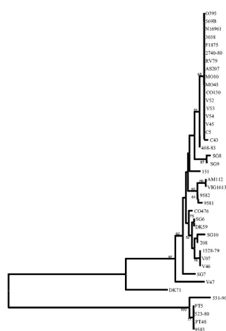
Evolutionary genetic relationships amongV. cholerae
natu-ral isolates. From the 36V. cholerae and 5V. mimicus mdh
[image:5.603.50.540.81.219.2]sequences, we constructed a neighbor-joining tree based on synonymous polymorphic sites, which are sites in a codon pre-dicted to not result in amino acid replacements and are there-fore not under selective pressure (Fig. 5). Themdhgene tree groupsV. choleraeO1 classical and El Tor isolates and O139 serogroup isolates together to form an epidemic clone complex (Fig. 5). Interestingly, several V. cholerae O1 and non-O139 serogroup isolates also clustered with this epidemic clone complex: three toxigenic V. cholerae serogroup O37 strains, V52, V53, isolated in Sudan in 1968, and CO130, isolated in India in 1993, and one toxigenic O8 serogroup strain, V54, recovered in Thailand. In addition, V. choleraestrain V45, a rough isolate clustered with the epidemic clone, as well as a nonagglutinable strain, C43, and a nontoxigenic clinical O1 serogroup strain, 468-83, isolated on the U.S. Gulf Coast in 1983 (Fig. 5). Comparative sequence analysis also demon-strates that strains of the same serogroup may belong to two or more widely divergent lineages (Fig. 5). Thus, for example, of the seven V. cholerae O37 serogroup strains examined, the remaining four strains (SG8, 151, CO476, and VO7), which are nontoxigenic clinical and environmental isolates (SG8, CO476, and VO7 were isolated in India and 151 was isolated in Mex-ico), were found on four separate branches of the mdhgene tree, indicating their diverse evolutionary origins (Fig. 5). A similar picture emerges from the analysis of two O141 strains (V46 and V47) examined; they are also found on divergent branches of themdhtree, suggesting that serogroup designa-tion is not an indicator of overall relatedness but represents TABLE 1—Continued
Strain Serogroup(biotype) Source Place of isolation isolationYr of
SCE4 O8 Environmental India 1997
SCE188 O44 Environmental India 1997
208 O11 Clinical Thailand 1998
C43 NAG Clinical Unknown Unknown
9581 O41 Clinical India 1990
9582 Rough Clinical India 1990
V. mimicus
PT5 O115 Clinical Bangladesh 1985
PT48 O115 Clinical Bangladesh 1985
9583 O115 Clinical United States 1980
523–80 O115 Clinical United States 1980
531–90 O41 Clinical Japan 1990
on May 15, 2020 by guest
http://jcm.asm.org/
TABLE 2. PCR primers used in this study
Primer Sequence (5⬘–3⬘)
Predicted PCR product size
(bp)
Annealing
temp (°C) Reference orsource
Housekeeping gene primers
mdh1 ATGAAAGTCGCTGTTATT 892 53.9 7
mdh2 GTATCTAACATGCCATCC
groEL1A GATCCATATGGCTGCTAAAGACGTACG 1,600 58 This study
groEL1B CTAGGTCGACTTACATCATGCGGCCCATGC
Virulence gene primers VSP-1
VC0175F TGGATGCTCTCTTCTTCA 2,834 52 This study
VC0175R CGCTCACTCACTAATACCGAG
VC0178F AGAGGCTTGTTTACTATCAG 2,053 50 This study
VC0178R ATCGGTACTGTCAGGGCT
VC0180F GGATGAGCAAATACAGCTAAC 2,283 50 This study
VC0180R CTAGGAAGAATTTTATCGGC
VC0183F CAGTAAGAGTGTAGCGTGCC 3,389 52 This study
VC0183R CCTGCACATCGAGATGC
VC0185F AGGAGGCGTGTAAGTCATAGC 1,110 55 This study
VC0185R AGACCACGAATACCTGCTCC
MSHA
msha398F GGAACGTGGCACAAATG 3,000 49 This study
msha398R TGACGTAAGTGAGCCGC
msha400F AAGATGAAATCGGGTTG 2,212 45 This study
msha400R TATCTGGCGACGCTTGC
msha403F GAACCGATTTATCTGTAGGAG 3,874 51 This study
msha403R TGACCGCCATTATCTGATAC
msha406F CGAGTATTAAGGTACTGAAGG 596 50 This study
msha406R ATCGGTCAGCTTGATCG
HlyA
VC0489F AGATCAACTACGATCAAGCC 1,677 54.2 This study
VC0489R AGAGGTTGCTATGCTTTCTAC
VSP-II
VC0490F CGTGAAGGGATATAGGAG 2,337 49.6 This study
VC0490R TGCAGTTGTTGAATGGAC
VC0493F AATGCTTCTCAGGGGGGTCTT 3,600 57 This study
VC0493R CGCTCTTCTTTCCACGCTTCA
VC0498F AGGTGGTATCGGGCTGGT 4,140 58 This study
VC0498R TGCGGCTGGAATGGAGTCTG
VC0502F TCATCAGTTAGCACACGAAC 476 52 This study
VC0502R GCTATCGTTATACTTGGCG
VC0504F CAGCAAAGGCGGAAGAGGTAG 3,240 55 This study
VC0504R AGCCCGAAATGAATCCCAAAA
VC0512F CAGTGGCTTCGCAGAGGA 3,900 53 This study
VC0512R CCCTCCACTGCTATTCCG
VC0514F TTATGATCCAAGGAGTAGGG 2,089 52 This study
VC0514R AGGCTGAAAAACAACTTGAG
VC0516F GTTTTCTGCGTTGTTCGAG 965 52 This study
VC0516R TCCTGATGTCTCTCTTGCCG
VC0517F CCCACTTCTTCCAGAGTATG 1,753 54.3 This study
VC0517R CGCAGTCACAGCTTAAACAAC
VPI-1
tcpH1 AGCCGCCTAGATAGTCTGTG 2,176 51.7 51
tcpA4 TCGCCTCCAATAATCCGAC
toxt1 AGGAGATGGAAGTGGTGTG 1,055 48.7 53
toxt2 CTTGGTGCTACATTCATGG
acfB1 GATGAAAGAACAGGAGAGA 1,180 49 53
acfB2 CAGCAACCACAGCAAAACC
PilE
PilEF CATACCTTTTGAGCATCGAC 3,087 50 This study
PilER GTGGCAAGAAGGACTCG
RTX
rtxA1 GCGATTCTCAAAGAGATGC 1,366 53.8 42
rtxA2 CACTCATTCCGATAACCAC
RS1
rstC1 AACAGCTACGGGCTTATTC 238 52.4 66
rstC2 TGAGTTGCGGATTTAGGC
Continued on following page
on May 15, 2020 by guest
http://jcm.asm.org/
lateral gene transfer of the O-antigen among strains. Of the remaining 17V. choleraenon-O1 and non-O139 isolates exam-ined at themdhlocus, strains SG7, V47, and DK71 formed the most divergent branches. The non-O1 and non-O139 sero-group strains formed separate lineages from the epidemic strains but in general are closely related to one another, hence, the very small branch lengths. Two clinicalV. choleraestrains, AM112, an O39 serogroup isolate from India, and VIG1613, an O12 serogroup isolate from Peru, clustered together, indi-cating identity. In addition, an O45 serogroup strain, SG6, from India and an O70 serogroup strain, DK59, from Germany clustered together, as did strains VO7, V46, and 1528-79 (Fig. 5). These data suggest the occurrence of clones of wide geo-graphic distribution.
We also identified two strains, 9581 and 9582, which were originally designatedV. mimicusbut clustered withV. cholerae non-O1 and non-O139 serogroup isolates on the mdh gene tree (Fig. 5). To determine the species designation of these isolates, we performed two biochemical tests previously used to differentiate V. cholerae and V. mimicus isolates: the Voges-Proskauer and corn oil tests. As expected, strains 9581 and 9582 were positive for both tests, similar to the control V. choleraestrains tested, indicating that these strains are indeed V. cholerae.
Evolutionary genetic relationships betweenV. choleraeand
V. mimicus.As expected, the fiveV. mimicusisolates formed a
separate divergent branch from theV. choleraeisolates on the mdhgene tree. FourV. mimicusisolates, PT5, PT48, 9583, and 523-80, clustered together, and strain 531-90 formed a separate divergent lineage (Fig. 5).
Presence of virulence regions inV. choleraeandV. mimicus.
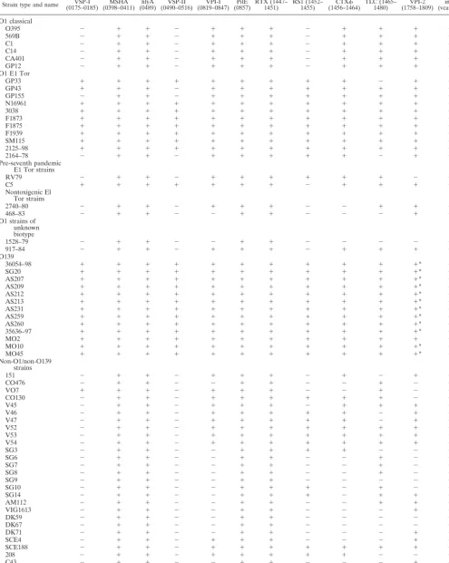
[image:7.603.49.542.81.236.2]In total, 64V. choleraestrains were examined for the presence of 12 regions associated with virulence inV. choleraeby PCR assays with 31 primer pairs (Table 2). Of the six classical biotype strains assayed by PCR, all strains contained the same nine regions, MSHA, hlyA, VPI-1, pilE, RTX, CTX, TLC, VPI-2, andintl4and lacked RS1, VSP-I, and VSP-II (Table 4). The 15 El Tor biotype strains analyzed were divided into three groups based on the year of isolation and the presence of ctxAB(Table 1). Of the 11 toxigenic seventh pandemic strains examined by PCR analysis, 7 strains contained all 12 virulence regions examined. Two toxigenic El Tor strains recovered from Australia in the 1970s, GP155 and GP43, lacked VSP-I and VSP-II and VSP-II, respectively, by PCR and Southern blot analyses (Table 4). In addition, strain GP33 lacked TLC by PCR and Southern blot analyses. PCR analysis with six primer pairs (Table 2) showed that the VSP-II region was larger than previously documented (18) and encompassed an additional 19.4-kb region from VC0498 to VC0516. From our PCR anal-ysis, we estimate that the VSP-II region is an⬃27-kb region encompassing VC0490 to VC0516 (Fig. 1). PCR assays indi-cated that VC0489 marked the 5⬘ flanking region and was TABLE 2—Continued
Primer Sequence (5⬘–3⬘) Predicted PCRproduct size (bp)
Annealing
temp (°C) Reference orsource
CTX
rstA1 ACTCGATACAAACGCTTCTC 1,009 53.7 66
rstA2 AGAATCTGGAAGGTTGAGTG
orfU CGTCACACCAGTTACTTTTCG 1,072 54.5 62
orfU AGAATGTACGCCATCGC
zot1 GGCTTAAACCTTGAACGC 1,036 54.7 24
zot2 AACCCCGTTTCACTTCTAC
ctxA1 AGTCAGGTGGTCTTATGCC 1,037 51.2 This study
ctxB2 TTGCCATACTAATTGCGG
tlc3 GGGAATGTTGAGTTCTCAGTG 1,548 55.5 56
tlc4 GTTGCGAAGTGGATTTTGTG
intl4:3 CCTTCATTGGATCACTCG 597 51.9 This study
intl4:4 GACGGAAAAAGATAGTGCC
TABLE 3. Sequence variation at themdhlocus amongV. choleraeandV. mimicusstrains
No. of strains Fragmentsize (bp) Total no. of sites ds⫾SE dn⫾SE Polymorphic Synonymous Nonsynonymous Informative Singleton
WithinV. cholerae
36 648 44 42 2 22 22 0.038⫾0.008 0.0004⫾0.0003
Within O1/O139 serogroups
13 648 9 8 1 2 7 0.008⫾0.003 0.0006⫾0.0006
Within non-O1/non-O139 serogroups
23 648 43 42 1 21 22 0.049⫾0.010 0.0002⫾0.0002
WithinV. mimicus
5 648 7 6 1 0 7 0.014⫾0.006 0.001⫾0.001
BetweenV. choleraeand
V. mimicus
41 648 91 87 4 81 10 0.144⫾0.017 0.003⫾0.001
on May 15, 2020 by guest
http://jcm.asm.org/
[image:7.603.40.543.570.724.2]found in allV. choleraestrains examined. Similarly, PCR assays with primer pair VC0517F-VCO517R showed that VC0517 marked the 3⬘flanking region of VSP-II and was present in all V. cholerae strains examined. An environmental toxigenic El Tor isolate 2164-78, recovered in the United States in 1978, was shown not to contain VSP-I, VSP-II, and TLC by PCR and Southern blot analyses. A clinical pre-seventh pandemic El Tor strain RV79 isolated in Indonesia in 1937 lacked only three of the virulence regions examined: VSP-I, VSP-II, and VPI-2 (Table 4). The El Tor strain C5 isolated 20 years later in Indonesia contained 11 of the virulence regions with only RS1missing. Two nontoxigenic El Tor isolates, 468-83 and 2740-80, isolated in the United States in the early 1980s both lacked VSP-I, VSP-II, RS1, and CTX; additionally, strain 468-83 did not contain VPI-1 and TLC (Table 4). Two V. cholerae O1 serogroup strains of unknown biotype, 1528-79 and 917-84, were examined. Neither contained VSP-I, VSP-II, nor RS1; strain 1528-79 also lacked VPI-1, CTX, TLC, and VPI-2 (Table 4). The 13V. cholerae O139 serogroup strains examined yielded positive PCR bands for all 12 virulence re-gions. However, as previously shown, there was a partial dele-tion of VPI-2 from 12 of the 13 O139 strains examined (31). These 12 O139 serogroup strains only contained a 20-kb 3⬘ region of VPI-2. Strain MO2 isolated in India in 1992 con-tained the entire VPI-2 region (Table 4) (31).
Twenty-eightV. choleraenon-O1 and non-O139 serogroup strains were assayed by PCR for the presence of 12 virulence regions (Table 4). None of the 28 strains examined by PCR and Southern blot analyses contained VSP-II, and only one strain, VO7, an environmental O37 serogroup isolate from India, contained VSP-I (Fig. 6). Only four regions MSHA,hlyA,pile, and RTX were present in all 28V. choleraeO1 and non-O139 serogroup strains. Theintl4region was absent in strain CO476. The three O37 serogroup strains, V52, V53, and CO130, which form part of the epidemic clone complex, lacked VSP-I and VSP-II; CO130 also lacked VPI-2. Similarly, strain V54 only lacked VSP-I and VSP-II, as did strain V45, which also lacked RS1(Table 4).
Among the five V. mimicus strains investigated, all gave negative PCR results for eight regions assayed: VSP-I, MSHA, hlyA, VSP-II, pilE, RTX, TLC, and intl4. Four V. mimicus strains, PT5, PT48, 9583, and 523-80, contained both VPI-1 and CTX; two strains (PT5 and PT48) also contained RS1.
DISCUSSION
In this study we show that clinical epidemicV. choleraeO1, O139, and O37 serogroup isolates form a highly uniform clone and that the emergence of the sixth and seventh cholera pan-FIG. 2. Polymorphic sites within themdhgene ofV. choleraeandV. mimicusisolates. N16961 represents 3038, F1875, RV79, AS207, MO10, MO45, CO130, V45, V52, V53, and V54. The numbering of the polymorphic sites (vertical format) is from the first position of the sequence segment. The position within the codon for each polymorphic site is shown below the sequences. Asterisks indicate polymorphic sites that gave rise to an amino acid change.
on May 15, 2020 by guest
http://jcm.asm.org/
[image:8.603.51.533.63.404.2]demic strains resulted from the successive acquisition of viru-lence regions.
Genotypic and phenotypic analysis of two pre-seventh pan-demic isolates, RV79 and C5, isolated in Indonesia in 1937 and 1957, respectively, give some interesting insights into a possible scenario for the evolution of epidemic isolates. El Tor strain RV79 is identical to other O1 serogroup strains atmdh and groEL and lacks only 3 of the 12 virulence regions, VSP-I,
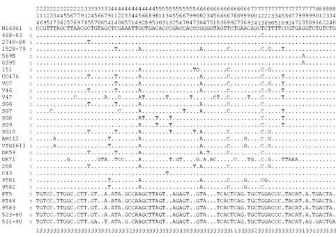
[image:9.603.115.472.76.303.2]VSP-II, and VPI-2, examined in this study. El Tor strain C5, similar to RV79, is identical to other O1 serogroup strains, isolated 20 years later lacks only 1 of the 12 regions examined, RS1. Since classical biotype strains were still circulating in the human population prior to the emergence of the seventh chol-era pandemic El Tor strain, the question arises as to whether El Tor seventh pandemic isolates arose from a classical pro-genitor strain via the acquisition of RS1, VSP-I, and VSP-II FIG. 3. PCR-SSCP profiles ofmdhfragments after HindIII digestion of 18 strains (25 strains profiled, 7 strains not shown). PCR-SSCP profiles are as follows. Profile 1, 2125-98, 2164-78, SM115, F1939, F1873, GP12, C14, C1, RV508, GP33, GP155, 1528-79, 917-84, 36054-98, SG20, and AS209 (AS212, AS213, AS231, AS259, AS260, 35636-97, and MO2 are not shown); profile 2, CA401; profile 3, GP43.
FIG. 4. PCR-SSCP profiles ofgroEL restriction fragments after BstYI digestion from three V. cholerae serogroups separated on a 0.8% nondenaturing polyacrylamide gel. The strains and respective serogroups are as follows: lanes 1 to 8, N16961, 2740-80, F1875, 3038, GP33, 2125-98, 2164-78, C5, O1 serogroup, biotype El Tor; lanes 9 to 12, CA401, O395, 569B, C14, O1 serogroup, biotype classical; lanes 13 to 15, MO10, MO45, 36054-98, O139 serogroup; lane 16, V52, O37 serogroup; lane M, Gibco-BRL 1-kb marker (with sizes in base pairs noted on the left); lane U, undenaturedgroELrestriction fragments from N16961. D1 to D4, single-stranded band pairs of the denatured 705-, 435-, 342-, and 153-bp BstYI
groELrestriction fragments; U1 to U3, renatured double-stranded BstYIgroELrestriction fragments (with sizes in base pairs noted in paren-theses). The smaller 153- and 112-bp fragments were too faint to be presented here.
on May 15, 2020 by guest
http://jcm.asm.org/
[image:9.603.111.476.473.660.2]or, alternatively, whether they arose from an RV79 and C5 progenitor-like isolate. The most parsimonious scenario (one requiring the least number of steps) is that an O1 isolate acquired CTX, VSP-I, VSP-II, and RS1(Fig. 7).
In 1968 there was a large outbreak of cholera in Sudan caused by an O37 serogroup isolate (68). Interestingly, Bik and colleagues (4) determined by IS1004 fingerprinting that this O37 serogroup strain from Sudan is closely related to classical O1 strains and may have acquired the O37 biosynthesis genes via lateral gene transfer. Beltran et al. (2) confirmed the iden-tity of the O37 strain to O1 strains by multilocus enzyme electrophoresis analysis and identified an additional O37 sero-group strain from India that was similar to O1 classical strains.
[image:10.603.129.461.56.541.2]Recently, an analysis of the O-antigen biosynthesis region also identified an O37 serogroup strain from India that had an O1 serogroup core genome (41). In our study, twoV. choleraeO37 serogroup isolates (isolated in Sudan in 1968) had mdh se-quences and PCR-SSCP profiles forgroELidentical to those of the O1 and O139 strains, indicating that these have an O1 serogroup core genome. These strains likely arose by modifi-cation of an O1 strain similar to the emergence of the O139 serogroup clone as previously suggested by Bik et al. (4). Based on multilocus virulence gene profiles of the O37 and O8 sero-groups, it is likely that strains V45, V52, V53, and V54 arose from a classical-like progenitor, since they lack only VSP-I and VSP-II; in addition, V45 lacks RS1, similar to classical FIG. 5. Neighbor-joining tree constructed by the Jukes-Cantor method with the nucleotide sequences ofmdhgene fragments ofV. cholerae
strains. Construction and bootstrapping of the trees were carried out with the MEGA suite of programs. One thousand bootstrap replicates were performed for each analysis, and bootstrap values are given at the nodes.
on May 15, 2020 by guest
http://jcm.asm.org/
TABLE 4. Distribution of 12 regions associated with virulence amongV. choleraenatural isolates as determined by PCR analysisa
Strain type and name (0175–0185)VSP-I (0398–0411)MSHA (0489)hlyA (0490–0516)VSP-II (0819–0847)VPI-I (0857)PilE RTX (1447–1451) RS1 (1452–1455) (1456–1464)CTX TLC (1465–1480) (1758–1809)VPI-2 (vca0291)intl4
O1 classical
O395 ⫺ ⫹ ⫹ ⫺ ⫹ ⫹ ⫹ ⫺ ⫹ ⫹ ⫹ ⫹
569B ⫺ ⫹ ⫹ ⫺ ⫹ ⫹ ⫹ ⫺ ⫹ ⫹ ⫹ ⫹
C1 ⫺ ⫹ ⫹ ⫺ ⫹ ⫹ ⫹ ⫺ ⫹ ⫹ ⫹ ⫹
C14 ⫺ ⫹ ⫹ ⫺ ⫹ ⫹ ⫹ ⫺ ⫹ ⫹ ⫹ ⫹
CA401 ⫺ ⫹ ⫹ ⫺ ⫹ ⫹ ⫹ ⫺ ⫹ ⫹ ⫹ ⫹
GP12 ⫺ ⫹ ⫹ ⫺ ⫹ ⫹ ⫹ ⫺ ⫹ ⫹ ⫹ ⫹
O1 E1 Tor
GP33 ⫹ ⫹ ⫹ ⫹ ⫹ ⫹ ⫹ ⫹ ⫹ ⫺ ⫹ ⫹
GP43 ⫹ ⫹ ⫹ ⫺ ⫹ ⫹ ⫹ ⫹ ⫹ ⫹ ⫹ ⫹
GP155 ⫺ ⫹ ⫹ ⫺ ⫹ ⫹ ⫹ ⫹ ⫹ ⫹ ⫹ ⫹
N16961 ⫹ ⫹ ⫹ ⫹ ⫹ ⫹ ⫹ ⫹ ⫹ ⫹ ⫹ ⫹
3038 ⫹ ⫹ ⫹ ⫹ ⫹ ⫹ ⫹ ⫹ ⫹ ⫹ ⫹ ⫹
F1873 ⫹ ⫹ ⫹ ⫹ ⫹ ⫹ ⫹ ⫹ ⫹ ⫹ ⫹ ⫹
F1875 ⫹ ⫹ ⫹ ⫹ ⫹ ⫹ ⫹ ⫹ ⫹ ⫹ ⫹ ⫹
F1939 ⫹ ⫹ ⫹ ⫹ ⫹ ⫹ ⫹ ⫹ ⫹ ⫹ ⫹ ⫹
SM115 ⫹ ⫹ ⫹ ⫹ ⫹ ⫹ ⫹ ⫹ ⫹ ⫹ ⫹ ⫹
2125–98 ⫹ ⫹ ⫹ ⫹ ⫹ ⫹ ⫹ ⫹ ⫹ ⫹ ⫹ ⫹
2164–78 ⫺ ⫹ ⫹ ⫺ ⫹ ⫹ ⫹ ⫹ ⫹ ⫺ ⫹ ⫹
Pre-seventh pandemic E1 Tor strains
RV79 ⫺ ⫹ ⫹ ⫺ ⫹ ⫹ ⫹ ⫹ ⫹ ⫹ ⫺ ⫹
C5 ⫹ ⫹ ⫹ ⫹ ⫹ ⫹ ⫹ ⫺ ⫹ ⫹ ⫹ ⫹
Nontoxigenic El Tor strains
2740–80 ⫺ ⫹ ⫹ ⫺ ⫹ ⫹ ⫹ ⫺ ⫺ ⫹ ⫹ ⫹
468–83 ⫺ ⫹ ⫹ ⫺ ⫺ ⫹ ⫹ ⫺ ⫺ ⫺ ⫹ ⫹
O1 strains of unknown biotype
1528–79 ⫺ ⫹ ⫹ ⫺ ⫺ ⫹ ⫹ ⫺ ⫺ ⫺ ⫺ ⫹
917–84 ⫺ ⫹ ⫹ ⫺ ⫹ ⫹ ⫹ ⫺ ⫹ ⫹ ⫹ ⫹
O139
36054–98 ⫹ ⫹ ⫹ ⫹ ⫹ ⫹ ⫹ ⫹ ⫹ ⫹ ⫹* ⫹
SG20 ⫹ ⫹ ⫹ ⫹ ⫹ ⫹ ⫹ ⫹ ⫹ ⫹ ⫹* ⫹
AS207 ⫹ ⫹ ⫹ ⫹ ⫹ ⫹ ⫹ ⫹ ⫹ ⫹ ⫹* ⫹
AS209 ⫹ ⫹ ⫹ ⫹ ⫹ ⫹ ⫹ ⫹ ⫹ ⫹ ⫹* ⫹
AS212 ⫹ ⫹ ⫹ ⫹ ⫹ ⫹ ⫹ ⫹ ⫹ ⫹ ⫹* ⫹
AS213 ⫹ ⫹ ⫹ ⫹ ⫹ ⫹ ⫹ ⫹ ⫹ ⫹ ⫹* ⫹
AS231 ⫹ ⫹ ⫹ ⫹ ⫹ ⫹ ⫹ ⫹ ⫹ ⫹ ⫹* ⫹
AS259 ⫹ ⫹ ⫹ ⫹ ⫹ ⫹ ⫹ ⫹ ⫹ ⫹ ⫹* ⫹
AS260 ⫹ ⫹ ⫹ ⫹ ⫹ ⫹ ⫹ ⫹ ⫹ ⫹ ⫹* ⫹
35636–97 ⫹ ⫹ ⫹ ⫹ ⫹ ⫹ ⫹ ⫹ ⫹ ⫹ ⫹* ⫹
MO2 ⫹ ⫹ ⫹ ⫹ ⫹ ⫹ ⫹ ⫹ ⫹ ⫹ ⫹ ⫹
MO10 ⫹ ⫹ ⫹ ⫹ ⫹ ⫹ ⫹ ⫹ ⫹ ⫹ ⫹* ⫹
MO45 ⫹ ⫹ ⫹ ⫹ ⫹ ⫹ ⫹ ⫹ ⫹ ⫹ ⫹* ⫹
Non-O1/non-O139 strains
151 ⫺ ⫹ ⫹ ⫺ ⫹ ⫹ ⫹ ⫺ ⫹ ⫺ ⫹ ⫹
CO476 ⫺ ⫹ ⫹ ⫺ ⫺ ⫹ ⫹ ⫺ ⫺ ⫹ ⫺ ⫺
VO7 ⫹ ⫹ ⫹ ⫺ ⫹ ⫹ ⫹ ⫺ ⫺ ⫹ ⫺ ⫹
CO130 ⫺ ⫹ ⫹ ⫺ ⫹ ⫹ ⫹ ⫹ ⫹ ⫹ ⫺ ⫹
V45 ⫺ ⫹ ⫹ ⫺ ⫹ ⫹ ⫹ ⫺ ⫹ ⫹ ⫹ ⫹
V46 ⫺ ⫹ ⫹ ⫺ ⫹ ⫹ ⫹ ⫹ ⫹ ⫺ ⫹ ⫹
V47 ⫺ ⫹ ⫹ ⫺ ⫹ ⫹ ⫹ ⫹ ⫹ ⫺ ⫹ ⫹
V52 ⫺ ⫹ ⫹ ⫺ ⫹ ⫹ ⫹ ⫹ ⫹ ⫹ ⫹ ⫹
V53 ⫺ ⫹ ⫹ ⫺ ⫹ ⫹ ⫹ ⫹ ⫹ ⫹ ⫹ ⫹
V54 ⫺ ⫹ ⫹ ⫺ ⫹ ⫹ ⫹ ⫹ ⫹ ⫹ ⫹ ⫹
SG3 ⫺ ⫹ ⫹ ⫺ ⫺ ⫹ ⫹ ⫹ ⫹ ⫺ ⫺ ⫹
SG6 ⫺ ⫹ ⫹ ⫺ ⫺ ⫹ ⫹ ⫺ ⫺ ⫹ ⫺ ⫹
SG7 ⫺ ⫹ ⫹ ⫺ ⫺ ⫹ ⫹ ⫺ ⫺ ⫹ ⫺ ⫹
SG8 ⫺ ⫹ ⫹ ⫺ ⫺ ⫹ ⫹ ⫺ ⫺ ⫹ ⫺ ⫹
SG9 ⫺ ⫹ ⫹ ⫺ ⫺ ⫹ ⫹ ⫺ ⫺ ⫺ ⫺ ⫹
SG10 ⫺ ⫹ ⫹ ⫺ ⫺ ⫹ ⫹ ⫹ ⫺ ⫹ ⫺ ⫹
SG14 ⫺ ⫹ ⫹ ⫺ ⫺ ⫹ ⫹ ⫹ ⫺ ⫹ ⫹ ⫹
AM112 ⫺ ⫹ ⫹ ⫺ ⫺ ⫹ ⫹ ⫺ ⫺ ⫹ ⫹ ⫹
VIG1613 ⫺ ⫹ ⫹ ⫺ ⫺ ⫹ ⫹ ⫺ ⫺ ⫺ ⫹ ⫹
DK59 ⫺ ⫹ ⫹ ⫺ ⫺ ⫹ ⫹ ⫺ ⫺ ⫺ ⫺ ⫹
DK67 ⫺ ⫹ ⫹ ⫺ ⫺ ⫹ ⫹ ⫺ ⫺ ⫺ ⫺ ⫹
DK71 ⫺ ⫹ ⫹ ⫺ ⫺ ⫹ ⫹ ⫺ ⫺ ⫺ ⫹ ⫹
SCE4 ⫺ ⫹ ⫹ ⫺ ⫹ ⫹ ⫹ ⫺ ⫺ ⫺ ⫹ ⫹
SCE188 ⫺ ⫹ ⫹ ⫺ ⫹ ⫹ ⫹ ⫹ ⫹ ⫹ ⫹ ⫹
208 ⫺ ⫹ ⫹ ⫺ ⫹ ⫹ ⫹ ⫹ ⫹ ⫺ ⫺ ⫹
C43 ⫺ ⫹ ⫹ ⫺ ⫺ ⫹ ⫹ ⫺ ⫺ ⫺ ⫹ ⫹
9581 ⫺ ⫹ ⫹ ⫺ ⫹ ⫹ ⫹ ⫺ ⫺ ⫺ ⫹ ⫹
9582 ⫺ ⫹ ⫹ ⫺ ⫹ ⫹ ⫹ ⫺ ⫺ ⫺ ⫹ ⫹
aThe numbers in parentheses. refer to the genetic organization, e.g., 0175 is VC0175. Asterisks denote strains that only contain the phage-like region of VP1–2; the
nan-nag region and restriction modification system are absent.⫹, present;⫺, absent.
on May 15, 2020 by guest
http://jcm.asm.org/
strains. Interestingly, a recent study with the infant mouse cholera model has shown that several non-O1 and non-O139 serogroup isolates, including O37 serogroup strains are effi-cient intestinal colonizers (8). Morris et al. (46) also demon-strated that a non-O1 and non-O139 V. cholerae strain was capable of causing severe diarrheal disease in humans.
In contrast to the data for O1 and O139 serogroup strains, which all belong to a single unique epidemic clone, our anal-yses indicate that strains from the same serogroup can belong to divergent lineages and that strains with different serogroup designations can belong to the same lineage, which is expected for regions, such as the O-antigen, that can be acquired by lateral gene transfer (2, 4, 9, 37, 41, 59).
Multilocus virulence gene profile analysis demonstrates the cooccurrence of several virulence regions among V. cholerae isolates (Table 5). For example, with the exception of SG3, CTXwas only found in strains containing VPI-1, which is to
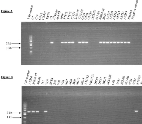
[image:12.603.51.529.75.492.2]be expected, since it encodes the CTXreceptor TCP (64). In addition, RS1 was mainly present in isolates that also con-tained CTX, which again is to be expected, since recent data suggest that both elements require each other for transfer (22). Two strains, SG10 and SG14, however, contained only RS1. This observation may be explained by a recent finding that described an alternative mechanism for RS1 transfer via a novel filamentous phage named KSF-1(23). Consistent with previous studies, we found that all classical strains examined in this study lacked RS1, VSP-I, and VSP-II and that these three regions are all present in El Tor strains (Table 4) (14, 18). Dziejman et al. (18) recently found VSP-I and VSP-II present only in El Tor isolates. In our study, we found VSP-I in an O37 serogroup strain and both VSP-I and VSP-II were absent from several El Tor isolates. Furthermore, Dziejman and colleagues found that VSP-II encompassed open reading frames VC0490 to VC0497; however, we found that VSP-II FIG. 6. PCR amplification of gene VC0180, which is part of VSP-I. (A) Lane 1, molecular size ladder; lanes 2 to 29, PCR amplicon with primer pair VC0180F-VC0180R. (B) Lane 1, molecular size ladder; lanes 2 to 27, PCR amplicon with primer pair VC0180F-VC0180R.
on May 15, 2020 by guest
http://jcm.asm.org/
spanned a larger region encompassing VC0490 to VC0516. Nonetheless, consistent with their results, we found that VSP-II is confined to seventh pandemic strains.
Four of the virulence regions, MSHA,hlyA,pilE, and RTX, were present in allV. choleraeisolates and absent from allV. mimicusstrains examined, indicating that these regions were acquired afterV. choleraeandV. mimicusdiverged from their most recent common ancestor. Among theV. mimicusisolates examined, only three regions, VPI-1, CTX, and TLC, were present. Previous studies of VPI-1 and CTXhave indicated recent interspecies lateral transfer betweenV. choleraeandV. mimicus, suggesting that transfer of virulence factors among isolates is an ongoing process.
Initially, aV. choleraeO1 serogroup strain first acquired the pathogenic island VPI-1, which encodes TCP, an essential col-onization factor and the receptor for CTX. This proposition is supported by the near sequence identity between classical and El Tor biotype strains across most of the VPI-1 region (36). The hypervariability documented at the tcpA gene is likely the result of positive Darwinian selection in this region (8). A second pathogenic island, VPI-2, which encodes genes involved in restriction modification and N-acetyl neuraminic
acid utilization, is found predominantly among O1 and O139 epidemicV. choleraeisolates (31) and was most likely present in an O1 serogroup strain that gave rise to classical and El Tor biotype strains. Following the acquisition of VPI-1 and VPI-2 by an O1 serogroup progenitor strain, classical and El Tor biotype isolates emerged and diverged from one another through the acquisition of VSP-I, VSP-II, and RS1. Studies based on comparative nucleotide sequence analysis of CTX genes indicate that this region was acquired independently in classical and El Tor biotype isolates (7). TheV. cholerae clas-sical biotype was responsible for the sixth cholera pandemic, which began in 1899, and presumably previous cholera pan-demics.V. choleraeEl Tor biotype isolates, which are respon-sible for the ongoing seventh cholera pandemic, which began in 1961, acquired at least three regions in addition to CTX: RS1, which facilitates CTX production, and VSP-I and VSP-II, whose roles inV. choleraevirulence are unknown (18). TheV. cholerae O139 strains that emerged in 1992 were de-rived from an El Tor progenitor by O-antigen switching likely facilitated by bacteriophages as well as the acquisition of a novel CTXand SXT constin (4, 15, 66).
[image:13.603.136.457.68.251.2]Since the beginning of the modern era of cholera pandemics, FIG. 7. Hypothetical evolutionary scenario for the emergence of epidemicV. choleraeO1 and O139 isolates. From the left, the parsimonious evolutionary steps beginning with theV. choleraeprogenitor to the right to the contemporary states are indicated. The model begins with aV. choleraeancestor that most resembles the present dayV. choleraeisolates in metabolic functions. From this ancestral state,V. choleraenatural isolates diverged from one another through mutation. TheV. choleraeO1 serogroup strains appear to have only arisen once and given rise to the highly successful epidemic classical and El Tor biotype isolates through the independent acquisition of CTXby these isolates. TheV. cholerae
El Tor biotype, which is responsible for the present seventh cholera pandemic, acquired additional virulence regions including RS1, VSP-I, and VSP-II. TheV. choleraeO139 serogroup, which arose in 1992, acquired a new O-antigen as well as the SXT element.
TABLE 5. Cooccurrence of virulence regions amongV. cholerae
Serogroup (biotype) Total no. ofstrains No. of strains containing:
VSP-I VSP-II VPI-I PilE RTX RS1 CTX TLC VPI–2
O1 (classical) 6 0 0 6 6 6 0 6 6 6
O1 (El Tor) 15 10 9 14 15 15 12 13 12 14
O1 2 0 0 1 2 2 0 1 1 1
O139 13 13 13 13 13 13 13 13 13 13a
Non-O1/non-O139 28 1 0 14 28 28 11 11 14 16
aThese strains only contain the phage-like region of VP1–2; thenan-nagregion and restriction modification system are absent. MO2 is an exception, it contains the
entire VPI-2.
on May 15, 2020 by guest
http://jcm.asm.org/
[image:13.603.41.542.626.708.2]all epidemicV. choleraeisolates appear to have a highly con-served core genome onto which additional DNA was added via lateral transfer, facilitating pathogenesis. In addition, the se-quence identity atmdhandgroELamongV. choleraeepidemic O1, O139, and O37 isolates suggests that these strains have emerged recently, evolutionarily speaking, which is also indi-cated by the fact that humans are the only known animal hosts forV. cholerae.
ACKNOWLEDGMENTS
We thank Frits Mooi and Matthew Waldor forV. choleraeisolates. The research in E.F.B.’s laboratory is funded by the Higher Educa-tion Authority PRTLI-3 grant and an Enterprise Ireland basic research grant.
REFERENCES
1. Albert, M. J., A. K. Siddique, M. S. Islam, A. S. Faruque, M. Ansaruzzaman, S. M. Faruque, and R. B. Sack.1993. Large outbreak of clinical cholera due toVibrio choleraenon-O1 in Bangladesh. Lancet341:704.
2. Beltran, P., G. Delgado, A. Navarro, F. Trujillo, R. K. Selander, and A. Cravioto.1999. Genetic diversity and population structure ofVibrio cholerae. J. Clin. Microbiol.37:581–590.
3. Berche, P., C. Poyart, E. Abachin, H. Lelievre, J. Vandepitte, A. Dodin, and J. M. Fournier.1994. The novel epidemic strain O139 is closely related to the pandemic strain O1 ofVibrio cholerae. J. Infect. Dis.170:701–704. 4. Bik, E., R. Gouw, and F. Mooi.1996. DNA fingerprinting ofVibrio cholerae
strains with a novel insertion sequence element: a tool to identify epidemic strains. J. Clin. Microbiol.34:1453–1461.
5. Boyd, E. F., A. J. Heilpern, and M. K. Waldor.2000. Molecular analysis of a putative CTXprecursor and evidence for independent acquisition of dis-tinct CTXs by toxigenicVibrio cholerae. J. Bacteriol.182:5530–5538. 6. Boyd, E. F., K. Nelson, F.-S. Wang, T. S. Whittam, and R. K. Selander.1994.
Molecular genetic basis of allelic polymorphism in malate dehydrogenase (mdh) in natural populations ofEscherichia coliandSalmonella enterica. Proc. Natl. Acad. Sci. USA91:1280–1284.
7. Boyd, E. F., K. L. Moyer, L. Shi, and M. K. Waldor.2000. Infectious CTX and theVibriopathogenicity island prophage inVibrio mimicus: evidence for recent horizontal transfer betweenV. mimicusandV. cholerae. Infect. Im-mun.68:1507–1513.
8. Boyd, E. F., and M. K. Waldor.2002. Evolutionary and functional analyses of variants of the toxin-coregulated pilus protein TcpA from toxigenicVibrio choleraenon-O1/non-O139 serogroup isolates. Microbiology148:1655–1666. 9. Byun, R., L. D. Elbourne, R. Lan, and P. R. Reeves.1999. Evolutionary relationships of pathogenic clones ofVibrio choleraeby sequence analysis of four housekeeping genes. Infect. Immun.67:1116–1124.
10. Chakraborty, S., A. K. Mukhopadhyay, R. K. Bhadra, A. N. Ghosh, R. Mitra, T. Shimada, S. Yamasaki, S. M. Faruque, Y. Takeda, R. R. Colwell, and G. B. Nair.2000. Virulence genes in environmental strains ofVibrio cholerae. Appl. Environ. Microbiol.66:4022–4028.
11. Chen, F., G. M. Evins, W. L. Cook, R. Almeida, N. Hargrett-Bean, and K. Wachsmuth.1991. Genetic diversity among toxigenic and nontoxigenic Vibrio choleraeO1 isolated from the Western Hemisphere. Epidemiol. In-fect.107:225–233.
12. Colwell, R. R.1996. Global climate and infectious disease: the cholera paradigm. Science274:2025–2031.
13. Dalsgaard, A., O. Serichantalergs, A. Forslund, W. Lin, J. Mekalanos, E. Mintz, T. Shimada, and J. G. Wells.2001. Clinical and environmental iso-lates ofVibrio choleraeserogroup O141 carry the CTX phage and the genes encoding the toxin-coregulated pili. J. Clin. Microbiol.39:4086–4092. 14. Davis, B. M., K. E. Moyer, E. F. Boyd, and M. K. Waldor.2000. CTX
prophages in classical biotypeVibrio cholerae: functional phage genes but dysfunctional phage genomes. J. Bacteriol.182:6992–6998.
15. Davis, B. M., H. H. Kimsey, W. Chang, and M. K. Waldor.1999. TheVibrio choleraeO139 calcutta CTX⌽is infectious and encodes a novel repressor. J. Bacteriol.181:6779–6787.
16. Davis, B. M., H. H. Kimsey, A. V. Kane, and M. K. Waldor.2002. A satellite phage-encoded antirepressor induces repressor aggregation and cholera toxin gene transfer. EMBO J.21:4240–4249.
17. Dumontier, S., and P. Berche.1998.Vibrio choleraeO22 might be a putative source of exogenous DNA resulting in the emergence of the new strain of Vibrio choleraeO139. FEMS Microbiol. Lett.164:91–98.
18. Dziejman, M., E. Balon, D. Boyd, C. M. Fraser, J. F. Heidelberg, and J. J. Mekalanos.2002. Comparative genomic analysis ofVibrio cholerae: genes that correlate with cholera endemic and pandemic disease. Proc. Natl. Acad. Sci. USA99:1556–1561.
19. Evins, G. M., D. N. Cameron, J. G. Wells, K. D. Greene, T. Popovic, S. Giono-Cerezo, I. K. Wachsmuth, and R. V. Tauxe.1995. The emerging
diversity of the electrophoretic types ofVibrio choleraein the Western Hemi-sphere. J. Infect. Dis.172:173–179.
20. Farfan, M., D. Minana, M. C. Fuste, and J. G. Loren.2000. Genetic rela-tionships between clinical and environmentalVibrio choleraeisolates based on multilocus enzyme electrophoresis. Microbiology146:2613–2626. 21. Faruque, S. M., M. J. Albert, and J. J. Mekalanos.1998. Epidemiology,
genetics, and ecology of toxigenicVibrio cholerae. Microbiol. Mol. Biol. Rev.
62:1301–1314.
22. Faruque, S. M., Asadulghani, M. Kamruzzaman, R. K. Nandi, A. N. Ghosh, G. B. Nair, J. J. Mekalanos, and D. A. Sack.2002. RS1 element ofVibrio choleraecan propagate horizontally as a filamentous phage exploiting the morphogenesis genes of CTXphi. Infect. Immun.70:163–170.
23. Faruque, S. M., D. A. Sack, R. B. Sack, R. R. Colwell, Y. Takeda, and G. B. Nair.2003. Emergence and evolution ofVibrio choleraeO139. Proc. Natl. Acad. Sci. USA100:1304–1309.
24. Fasano, A., B. Baudry, D. W. Pumplin, S. S. Wasserman, B. D. Tall, J. Ketley, and J. B. Kaper.1991.Vibrio choleraeproduces a second enterotoxin, which affects intestinal tight junctions. Proc. Natl. Acad. Sci. USA88:5242– 5246.
25. Ghosh, C., R. K. Nandy, S. K. Dasgupta, G. B. Nair, R. H. Hall, and A. C. Ghose.1997. A search for cholera toxin (CT), toxin coregulated pilus (TCP), the regulatory element ToxR and other virulence factors in non-O1/non-O139Vibrio cholerae. Microb. Pathog.22:199–208.
26. Heidelberg, J. F., J. A. Eisen, W. C. Nelson, R. A. Clayton, M. L. Gwinn, R. J. Dodson, D. H. Haft, E. K. Hickey, J. D. Peterson, L. Umayam, S. R. Gill, K. E. Nelson, T. D. Read, H. Tettelin, D. Richardson, M. D. Ermolaeva, J. Vamathevan, S. Bass, H. Qin, I. Dragoi, P. Sellers, L. McDonald, T. Utter-back, R. D. Fleishmann, W. C. Nierman, and O. White.2000. DNA sequence of both chromosomes of the cholera pathogenVibrio cholerae. Nature12:
477–483.
27. Higgins, D. G., J. D. Thompson, and T. J. Gibson.1996. Using CLUSTAL for multiple sequence alignments. Methods Enzymol.266:383–402. 28. Hobbs, M., and J. S. Mattick.1993. Common components in the assembly of
type 4 fimbriae, DNA transfer systems, filamentous phage and protein-secretion apparatus: a general system for the formation of surface-associated protein complexes. Mol. Microbiol.10:233–243.
29. Huq, A., R. R. Colwell, R. Rahman, A. Ali, M. A. Chowdhury, S. Parveen, D. A. Sack, and E. Russek-Cohen.1990. Detection ofVibrio choleraeO1 in the aquatic environment by fluorescent-monoclonal antibody and culture methods. Appl. Environ. Microbiol.56:2370–2373.
30. Iredell, J. R., and P. A. Manning.1994. The toxin-co-regulated pilus ofVibrio choleraeO1: a model for type 4 pilus biogenesis? Trends Microbiol.2:187– 192.
31. Jermyn, W. S., and E. F. Boyd.2002. Characterization of a novelVibrio pathogenicity island (VPI-2) encoding neuraminidase (nanH) among toxi-genicVibrio choleraeisolates. Microbiology148:3681–3693.
32. Jonson, G., J. Holmgren, and A. M. Svennerholm.1991. Identification of a mannose-binding pilus onVibrio choleraeEl Tor. Microb. Pathog.11:433– 441.
33. Jukes, T. H., and C. R. Cantor.1969. Evolution of protein molecules. Aca-demic Press, Inc., New York, N.Y.
34. Kaper, J. B., J. G. Morris, Jr., and M. M. Levine.1995. Cholera. Clin. Microbiol. Rev.8:48–86.
35. Karaolis, D. K., J. A. Johnson, C. C. Bailey, E. C. Boedeker, J. B. Kaper, and P. R. Reeves.1998. AVibrio choleraepathogenicity island associated with epidemic and pandemic strains. Proc. Natl. Acad. Sci. USA95:3134–3139. 36. Karaolis, D. K., R. Lan, J. B. Kaper, and P. R. Reeves.2001. Comparison of
Vibrio choleraepathogenicity islands in sixth and seventh pandemic strains. Infect. Immun.69:1947–1952.
37. Karaolis, D. K., R. Lan, and P. R. Reeves.1995. The sixth and seventh cholera pandemics are due to independent clones separately derived from environmental, nontoxigenic, non-O1Vibrio cholerae. J. Bacteriol.177:3191– 3198.
38. Kotetishvili, M., O. C. Stine, Y. Chen, A. Kreger, A. Sulakvelidze, S. Sozha-mannan, and J. G. Morris, Jr.2003. Multilocus sequence typing has better discriminatory ability for typingVibrio choleraethan does pulsed-field gel electrophoresis and provides a measure of phylogenetic relatedness. J. Clin. Microbiol.41:2191–2196.
39. Kumar, S., K. Tamura, I. B. Jakobsen, and M. Nei.2001. MEGA2: molec-ular evolutionary genetics analysis software. Bioinformatics17:1244–1245. 40. Li, M., M. Kotetishvili, Y. Chen, and S. Sozhamannan.2003. Comparative
genomic analyses of theVibriopathogenicity island and cholera toxin pro-phage regions in nonepidemic serogroup strains ofVibrio cholerae. Appl. Environ. Microbiol.69:1728–1738.
41. Li, M., T. Shimada, J. G. Morris, Jr., A. Sulakvelidze, and S. Sozhamannan.
2002. Evidence for the emergence of non-O1 and non-O139Vibrio cholerae strains with pathogenic potential by exchange of O-antigen biosynthesis regions. Infect. Immun.70:2441–2453.
42. Lin, W., K. J. Fullner, R. Clayton, J. A. Sexton, M. B. Rogers, K. E. Calia, S. B. Calderwood, C. Fraser, and J. J. Mekalanos.1999. Identification of a Vibrio choleraeRTX toxin gene cluster that is tightly linked to the cholera toxin prophage. Proc. Natl. Acad. Sci. USA96:1071–1076.
on May 15, 2020 by guest
http://jcm.asm.org/
43. Merrell, D. S., S. M. Butler, F. Qadri, N. A. Dolganov, A. Alam, M. B. Cohen, S. B. Calderwood, G. K. Schoolnik, and A. Camilli.2002. Host-induced epidemic spread of the cholera bacterium. Nature417:642–645.
44. Mooi, F. R., and E. M. Bik.1997. The evolution of epidemicVibrio cholerae strains. Trends Microbiol.5:161–165.
45. Morris, J. G., Jr., G. E. Losonsky, J. A. Johnson, C. O. Tacket, J. P. Nataro, P. Panigrahi, and M. M. Levin.1995. Clinical and immunologic character-istics ofVibrio choleraeO139 Bengal infection in North American volunteers. J. Infect. Dis.171:903–908.
46. Morris, J. G., Jr., T. Takeda, B. D. Tall, G. A. Losonsky, S. K. Bhattacharya, B. D. Forrest, B. A. Kay, and M. Nishibuchi.1990. Experimental non-O group 1Vibrio choleraegastroenteritis in humans. J. Clin. Investig.85:697– 705.
47. Mukhopadhyay, A. K., S. Chakraborty, Y. Takeda, G. B. Nair, and D. E. Berg.2001. Characterization of VPI pathogenicity island and CTXphi pro-phage in environmental strains ofVibrio cholerae. J. Bacteriol.183:4737– 4746.
48. Nair, G. B., B. L. Sarkar, S. P. De, M. K. Chakraborti, R. K. Bhadra, and S. C. Pal.1988. Ecology ofVibrio choleraein the freshwater environs of Calcutta, India. Microb. Ecol.15:203–216.
49. Nandi, B., R. K. Nandy, A. C. Vicente, and A. C. Ghose.2000. Molecular characterization of a new variant of toxin-coregulated pilus protein (TcpA) in a toxigenic non-O1/non-O139 strain ofVibrio cholerae. Infect. Immun.
68:948–952.
50. Novais, R. C., A. Coelho, C. A. Salles, and A. C. Vicente.1999. Toxin-co-regulated pilus cluster in non-O1, non-toxigenicVibrio cholerae: evidence of a third allele of pilin gene. FEMS Microbiol. Lett.171:49–55.
51. Ogierman, M. A., E. Voss, C. Meaney, R. Faast, S. R. Attridge, and P. A. Manning.1996. Comparison of the promoter proximal regions of the toxin-co-regulated tcp gene cluster in classical and El Tor strains ofVibrio cholerae O1. Gene170:9–16.
52. Orita, M., H. Iwahana, H. Kanazawa, K. Hayashi, and T. Sekiya.1989. Detection of polymorphisms of human DNA by gel electrophoresis as single-strand conformation polymorphisms. Proc. Natl. Acad. Sci. USA86:2766– 2770.
53. O’Shea, Y. A., and E. F. Boyd.2002. Mobilization of theVibriopathogenicity island betweenVibrio choleraeisolates mediated by CP-T1 generalized trans-duction. FEMS Microbiol. Lett.214:153–157.
54. Popovic, T., C. A. Bopp, O. Olsvik, and K. Wachsmuth.1993. Epidemiologic
application of a standardized ribotype scheme forV. choleraeO1. Clin. Microbiol.31:2474–2482.
55. Rivera, I. N., J. Chun, A. Huq, R. B. Sack, and R. R. Colwell.2001. Geno-types associated with virulence in environmental isolates ofVibrio cholerae. Appl. Environ. Microbiol.67:2421–2429.
56. Rubin, E. J., W. Lin, J. J. Mekalanos, and M. K. Waldor.1998. Replication and integration of aVibrio choleraecryptic plasmid linked to the CTX prophage. Mol. Microbiol.28:1247–1254.
57. Saitou, N., and M. Nei.1987. The neighbor-joining method: a new method for reconstructing phylogenetic trees. Mol. Biol. Evol.4:406–425. 58. Salles, C. A., and H. Momen.1991. Identification ofVibrio cholerae by
enzyme electrophoresis. Trans. R. Soc. Trop. Med. Hyg.85:544–547. 59. Stine, O. C., S. Sozhamannan, Q. Gou, S. Zheng, J. G. Morris, Jr., and J. A.
Johnson.2000. Phylogeny ofVibrio choleraebased onrecAsequence. Infect. Immun.68:7180–7185.
60. Stroeher, U. H., G. Parasivam, B. K. Dredge, and P. A. Manning.1997. NovelVibrio choleraeO139 genes involved in lipopolysaccharide biosynthe-sis. J. Bacteriol.179:2740–2747.
61. Taylor, R. K., V. L. Miller, D. B. Furlong, and J. J. Mekalanos.1987. Use of phoAgene fusions to identify a pilus colonization factor coordinately regu-lated with cholera toxin. Proc. Natl. Acad. Sci. USA84:2833–2837. 62. Trucksis, M., J. E. Galen, J. Michalski, A. Fasano, and J. B. Kaper.1993.
Accessory cholera enterotoxin (Ace), the third toxin of aVibrio cholerae virulence cassette. Proc. Natl. Acad. Sci. USA90:5267–5271.
63. Wachsmuth, I. K., G. M. Evins, P. I. Fields, O. Olsvik, T. Popovic, C. A. Bopp, J. G. Wells, C. Carrillo, and P. A. Blake.1993. The molecular epide-miology of cholera in Latin America. J. Infect. Dis.167:621–626. 64. Waldor, M. K., and J. J. Mekalanos.1996. Lysogenic conversion by a
fila-mentous phage encoding cholera toxin. Science272:1910–1914.
65. Waldor, M. K., and J. J. Mekalanos.1994.Vibrio choleraeO139 specific gene sequences. Lancet343:1366.
66. Waldor, M. K., E. J. Rubin, G. D. Pearson, H. Kimsey, and J. J. Mekalanos.
1997. Regulation, replication, and integration functions of theVibrio cholerae CTX⌽are encoded by region RS2. Mol. Microbiol.24:917–926.
67. Yamasaki, S., S. Garg, G. B. Nair, and Y. Takeda.1999. Distribution of Vibrio cholerae O1 antigen biosynthesis genes among O139 and other non-O1 serogroups ofVibrio cholerae. FEMS Microbiol. Lett.179:115–121. 68. Zinnaka, Y., and C. C. Carpenter.1972. An enterotoxin produced by
non-choleravibrios. Johns Hopkins Med. J.131:403–411.