0095-1137/05/$08.00⫹0 doi:10.1128/JCM.43.6.2881–2885.2005
Multiple Copies of the 16S rRNA Gene in
Nocardia nova
Isolates and
Implications for Sequence-Based Identification Procedures
Patricia S. Conville* and Frank G. Witebsky
Microbiology Service, Department of Laboratory Medicine, Warren G. Magnuson Clinical Center, National Institutes of Health, U.S. Department of Health and Human Services, Bethesda, Maryland
Received 4 August 2004/Returned for modification 5 December 2004/Accepted 14 January 2005
Molecular investigation of twoNocardiapatient isolates showed unusual restriction fragment length
poly-morphism patterns with restriction endonuclease assays (REA) using an amplified portion of the 16S rRNA
gene. Patterns typical ofNocardia novawere obtained with REA of an amplified portion of the 65-kDa heat
shock protein gene. Subsequent sequence analysis of the 16S rRNA gene regions of these isolates showed the presence of ambiguous bases within an expected restriction endonuclease recognition site which were not able to be resolved on repeat testing. Cloning of amplified regions of the 16S rRNA genes and subsequent sequenc-ing of the resultsequenc-ing clones from the two patient isolates showed three different 16S rRNA gene sequences which
corresponded to sequences found inN. nova, a molecular variant ofN. nova, and a previously undescribed
sequence. Hybridization studies using a DNA probe corresponding to an 89-bp conserved region of the 16S
rRNA gene confirmed the presence of at least two copies of the 16S rRNA gene in theN. novatype strain, in
a patient isolate identical to the molecular variant ofN. nova, and in the two other patient isolates. All isolates
were found to belong to the species N. novaas determined by DNA-DNA hybridization. Because minimal
variation has been found in the 16S rRNA gene sequences of different species ofNocardia, those laboratories
employing molecular methods for identification of these species must be aware of the potential identification complications that may be caused by the presence of differing 16S rRNA genes in the same isolate.
Nocardiaspecies have been implicated in a wide variety of infectious diseases in both immunocompetent and immuno-compromised patients. In recent years the number of recog-nized clinically relevant Nocardia species has dramatically increased, and the resulting difficulties in determining an accurate identification for many of these isolates by conven-tional methodologies have been well documented (4, 8, 14). The use of molecular techniques, including restriction endo-nuclease assays (REA) using amplified portions of both the heat shock protein gene and the 16S rRNA gene, has greatly improved the reliability of the identifications obtained for these species (8, 16). To date, however, the method that pro-vides the most definitive identification of mostNocardia iso-lates is sequence analysis of the 16S rRNA gene (14). In many cases, analysis of even a segment of the gene gives sufficient information to allow an accurate identification (6). However, in our experience with 16S rRNA gene sequencing ofNocardia
isolates, we have occasionally observed the occurrence of am-biguous bases which are not able to be resolved, even with repeated sequencing. Occasionally, these ambiguous bases oc-cur within a recognition sequence of a restriction endonu-clease, resulting in restriction fragment length polymorphism (RFLP) patterns that show more bands than expected. We have recently observed this problem with several isolates sus-pected to be related toNocardia nova; RFLP patterns obtained from DpnII digests of an amplified region of the 16S rRNA genes of these isolates corresponded both to those obtained fromN. novaand to an RFLP variant ofN. nova. Sequence analysis confirmed the presence of at least one ambiguous base
occurring within the recognition sequence of the endonucle-ase. We determined that these isolates contained at least two copies of the 16S rRNA gene and that the sequence of the gene at the restriction site was different for the two copies. We here report the detailed molecular analysis of three patient isolates and of theN. novatype strain.
(The possible existence of such isolates was previously reported [Abstr. 104th Gen. Meet. Am. Soc. Microbiol., abstr. U-075, 2004].)
MATERIALS AND METHODS
Organisms.The American Type Culture Collection type strain ofNocardia
nova(ATCC 33726T
) was used as the organism to which all otherNocardia nova-like organisms were compared. Three patient isolates were examined. Iso-late A was obtained from a patient being treated at the Clinical Center of the National Institutes of Health, and isolates B and C were referred to the Univer-sity of Texas Health Center at Tyler for identification and/or susceptibility test-ing. All isolates were grown on Sabouraud dextrose agar, Emmons modification (Hardy Diagnostics, Santa Monica, Calif.); organisms were modified acid-fast stain positive, and colonies showed aerial hyphae. Molecular studies on all isolates were performed on subcultures derived from a single colony.
Restriction endonuclease assay and direct 16S rRNA gene sequencing.DNA
from all isolates was extracted as previously described (8). All organisms were initially identified using PCR of a 999-bp region of the 16S rRNA gene and a 440-bp region of the 65-kDa heat shock protein (HSP) gene and subsequent REA as previously described (8, 16). Briefly, the amplified region of the 16S rRNA gene was digested with the endonucleases HinP1I, DpnII, BstEII, and SphI (New England Biolabs, Beverly, Mass.); the amplified region of the HSP gene was digested with the endonucleases MspI and HinfI (New England Bio-labs). Digests were electrophoresed for 2 h on a 2% MetaPhor agarose gel (Cambrex Bio Science Rockland, Inc., Rockland, Maine). Gels were analyzed using the Bio-Rad Molecular Analyst System (Bio-Rad Laboratories, Hercules, Calif.); resulting RFLP patterns were compared to patterns obtained with the type strains of variousNocardiaspecies known to cause human disease. The sequences of a 1,463-bp region of the 16S rRNA genes of the type strain and patient isolates were determined using procedures previously described (7, 8).
Cloning.PCR was performed using the DNA prepared for use for molecular
identification; the DNA of isolates B and C was extracted as previously described * Corresponding author. Mailing address: 10 Center Drive, MSC
1508, National Institutes of Health, Bethesda, MD 20892-1508. Phone: (301) 496-4433. Fax: (301) 402-1886. E-mail: [email protected].
2881
on May 16, 2020 by guest
http://jcm.asm.org/
(8). Amplification of a 532-bp region of the 16S rRNA gene (corresponding to bases 2 through 533 of the sequence ofNocardia asteroidesATCC 19247T;
GenBank accession number X84850) was performed using the primers 5⬘-CG A-ACG-CTG-GCG-GCG-TGC-TTA-AC-3⬘and 5⬘-ACC-GCC-TAC-AAG-CT C-TTT-ACG-CC-3⬘(Research Genetics, Huntsville, AL), each at a concentra-tion of 0.25M. PCR was performed using puReTaq Ready-To-Go PCR Beads (Amersham Biosciences, Buckinghamshire, United Kingdom) with 2.5l ex-tracted DNA. The DNA was denatured for 5 min at 94°C and then subjected to 40 cycles of amplification (94°C for 60 s, 68°C for 45 s, and 72°C for 60 s) followed by a 10-min extension at 72°C. Cloning was performed using the TA cloning kit (Invitrogen Corporation, Carlsbad, Calif.). Briefly, amplified DNA was ligated into pCR2.1 (Invitrogen Corporation) and transformed into One Shot INV␣F⬘ chemically competentEscherichia coli(Invitrogen Corporation). Transformants were plated on Luria-Bertani agar with ampicillin, X-Gal (5-bromo-4-chloro-3-indolyl--D-galactopyranoside), and IPTG (isopropyl--D -thiogalactopyrano-side) (KD Medical, Columbia, MD), and colonies showing inserts were subcul-tured to Lennox L broth (Quality Biologicals, Gaithersburg, MD) and incubated at 35°C overnight. Plasmids were recovered using the QIAprep spin miniprep kit (QIAGEN Inc., Valencia, Calif.), and inserts were tested for appropriate size by digestion with the restriction endonuclease EcoRI (New England Biolabs). Nine-teen plasmid clones from both isolates B and C were sequenced directly using “reaction 1” sequencing primers with tails containing M13 forward binding sites. One clone of each sequence type was further sequenced with the “reaction 1” sequencing primers containing the M13 forward and reverse binding sites as previously described (8).
Restriction digests of genomic DNA and hybridization studies.Genomic DNA
from all isolates was extracted as previously described (7, 9, 10). DNA was quantitated and digested for 2 h using the restriction endonuclease SphI (New England Biolabs) according to the recommendations of the manufacturer. This endonuclease was chosen because analysis of 16S rRNA gene sequences of all four isolates showed no SphI recognition sites within the region studied. Digests were electrophoresed overnight on a 0.5% SeaKem GTG gel (Cambrex Bio Science Rockland, Inc.) containing 2g ethidium bromide (Amresco, Solon, Ohio) at 20 V. Digests were visualized using the Kodak Image Station 440CF (Eastman Kodak Company, Rochester, NY). DNA fragments were transferred and fixed to a positively charged nylon membrane (Roche Diagnostics, Mann-heim, Germany) (15). A DNA probe was designed which was complementary to an 89-bp conserved region of the nocardial 16S rRNA gene corresponding to bases 1098 through 1186 of the sequence ofN. asteroidesATCC 19247T
(Gen-Bank accession number X84850). The probe sequence was 5⬘ -GAG-ACT-GCC- GGG-GTC-AAC-TCG-GAG-GAA-GGT-GGG-GAC-GAC-GTC-AAG-TCA- TCA-TGC-CCC-TTA-TGT-CCA-GGG-CTT-CAC-ACA-TGC-TAC-AAT-GG-3⬘(Midland Certified Reagent Co., Crawford, Tex.). The probe was labeled at the 3⬘end with digoxigenin-labeled dideoxyuridine triphosphate by using the DIG Oligonucleotide Tailing Kit, 2nd Generation (Roche Diagnostics) accord-ing to the instructions of the manufacturer. Hybridization usaccord-ing 10 pmol of the labeled probe in 7.5 ml DIG Easy Hyb (Roche Diagnostics) with 0.1 mg/ml poly(A) (Roche Diagnostics) was performed overnight in a rotating hybridiza-tion chamber at 55°C. Stringency washes were performed as recommended (Roche Diagnostics). Chemiluminescent detection was performed using the DIG Luminescent Detection Kit (Roche Diagnostics), according to the manufactur-er’s instructions. Luminescent products were visualized and digitized using the Kodak Image Station 440CF.
DNA-DNA hybridization.Genomic DNA from all isolates was extracted as
previously described (7, 9, 10). DNA obtained from the type strain ofN. novawas labeled with [32
P]dCTP by using a nick translation kit (Invitrogen Corporation). The hybridization method for determination of DNA relatedness by absorbance to hydroxyapatite has been described previously (3). All reactions were per-formed in duplicate at 70°C. The relative binding ratio ([percentage of heterol-ogous DNA bound to hydroxyapatite/percentage of homolheterol-ogous DNA bound to hydroxyapatite]⫻100) was calculated by the method of Brenner et al. (2). The percent divergence (calculated to the nearest 0.5%) was determined by assuming that each degree of heteroduplex instability, compared to the melt-ing temperature of the homologous duplex, was caused by 1% unpaired bases (2).
Sequence analysis. Sequences were assembled using SeqMan II software
(DNASTAR, Inc., Madison, Wis.) and aligned using Megalign software (DNA-STAR, Inc.) using the Clustal V algorithm. The presence of SphI recognition sites within the 16S rRNA gene sequence was evaluated using MapDraw soft-ware (DNASTAR).
RESULTS
REA. HinP1I digests of the amplified region of the 16S rRNA gene showed identical RFLP patterns for the type strain ofN. nova and the three patient isolates (bands of approxi-mately 420, 350, and 225 bp) (data not shown). DpnII digests of the amplified region of the 16S rRNA gene showed three different RFLP patterns (Fig. 1), one for the N. nova type strain, one for theN. novamolecular variant, and a third for isolates B and C. TheN. novatype strain and isolate A (des-ignated “N. nova variant”) each showed three bands of 640, 200, and 95 bp and 700, 200, and 95 bp, respectively. Isolates B and C showed identical patterns of four bands (700, 640, 200, and 95 bp) that appeared to be a combination of the patterns observed withN. novaand theN.novavariant. MspI and HinfI digests of the amplified region of the HSP gene showed iden-tical patterns for all isolates, corresponding to the pattern expected forN. nova(16) (data not shown).
16S rRNA gene sequencing. BLAST searches of the
se-quences obtained for isolates A, B, and C all showed closest similarity to N. nova. Alignment of 1,369-bp sequences (se-quences were trimmed to the size of the shortest sequence) of isolate A and theN. novatype strain showed two base discrep-ancies (⬎99.9% similarity) (Table 1); one of these base dis-crepancies occurred within a recognition sequence of the en-donuclease DpnII. The 1,369-bp sequences of isolates B and C each showed two base discrepancies from the sequence of the type strain ofN. nova(⬎99.9% similarity); each of these base discrepancies were ambiguous bases which were not able to be resolved (Fig. 2). All of the discrepancies for all isolates occurred within the regions corresponding to bases 102 to
FIG. 1. DpnII digests of a 999-bp amplified region of the 16S rRNA gene. Lane 1, base pair marker; lane 2,N. novaATCC 33726T; lane 3, isolate A (N. novavariant); lane 4, isolate B; lane 5, isolate C; lane 6, base pair marker.
on May 16, 2020 by guest
http://jcm.asm.org/
154 ofN. asteroidesATCC 19247T(GenBank accession
num-ber X84850). No SphI recognition sites were detected within the 16S rRNA gene sequences of any of the isolates.
Clones.Analysis of the sequences of the 19 clones of isolate
B showed two different sequence patterns (Table 1). The se-quences of 10 of the clones were identical to the sequence obtained for the type strain ofN. nova; the sequences of 9 of the clones were identical to the sequence obtained for isolate A, theN. nova variant. For isolate C, two different base se-quence patterns were also found with the 19 clones sese-quenced (Table 1). The sequences of 14 of the clones were identical to the sequence obtained for the type strain ofN. nova, and the sequences of the five remaining clones showed two base dis-crepancies from theN. novatype strain. These two discrepan-cies were in adjacent bases, both corresponding to the recog-nition site of DpnII (Table 1). Base discrepancies noted for the cloned plasmids of isolates B and C correspond to the ambig-uous bases noted in the direct sequences of isolates B and C (Table 1; Fig. 2).
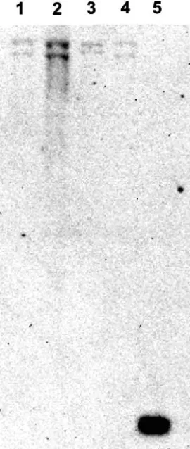
Hybridization assay with digested genomic DNA. The
la-beled probe hybridized with two regions of the SphI-digested genomic DNAs of all isolates (Fig. 3).
DNA-DNA hybridization.DNA hybridization results for
iso-lates A, B, and C with theN. novatype strain showed that all three patient isolates belonged to the speciesN. nova. Com-pared to the type strain ofN. nova, isolates A, B, and C showed relative binding ratios (percent divergences) of 95 (2.0), 100 (2.0), and 86 (1.0), respectively.
DISCUSSION
The presence of multiple copies of the 16S rRNA gene has been documented for numerous bacterial species; recent re-ports have shown that within some species, the multiple copies show distinct sequence differences (1, 5, 11–13, 17). We are unaware of any previous documentation of the presence of more than one copy of the 16S rRNA gene in members of the genus Nocardia. The three patient isolates included in this study were proven to be members of the speciesN. nova by DNA-DNA hybridization. These three isolates and theN. nova
type strain all contained at least two copies of the 16S rRNA gene. For the N. nova type strain and isolate A (an isolate determined to be theN. novavariant), the within-organism 16S rRNA gene copies are identical to each other as determined by the distinct RFLP patterns obtained by REA and by the lack of ambiguous bases in the gene sequences. TheN. novavariant has previously been considered to be related to the type strain ofN. novabased on the similarities of the 16S rRNA sequences (8); theN. nova variant, however, showed a different RFLP pattern from the type strain ofN. novawhen REA was per-formed using a DpnII digest of the amplified region of the 16S rRNA gene. The sequences of the 16S rRNA genes of the
N. novatype strain and isolate A differed by two bases; one of these base differences occurred within the recognition
[image:3.585.300.538.68.489.2]se-FIG. 2. Sequence trace data for isolates B and C, showing direct sequencing with ambiguous bases and sequence data for individual clones. The region corresponds to the gene region between bases 152 and 156 of the sequence ofN. asteroidesATCC 19247T(GenBank accession no. X84850).
TABLE 1. Base discrepancies among theN. novatype strain and patient isolates B and C
Isolate Base at position
a
:
102 103 104 152 153 154 155 156
N. novaATCC 33726T G C A G
G A T C
Isolate A (N. novavariant) G T A G A A T C
Isolate B
Direct sequence G Y A G R A T C
Clone 5 G T A G A A T C
Clone 24 G C A G G A T C
Isolate C
Direct sequence G C A G R R T C
Clone 1 G C A G G A T C
Clone 6 G C A G A G T C
aY, either C or T; R, either A or G. Boldface indicates DpnII recognition
sites; underlined bases are ambiguous bases. Compared toN. asteroidesATCC 19247T(GenBank accession no. X84850), bases at positions 105 through 151 are
identical.
on May 16, 2020 by guest
http://jcm.asm.org/
[image:3.585.44.284.88.240.2]quence of DpnII, resulting in the different RFLP patterns obtained for the two isolates.
The patient isolates B and C were also shown to possess at least two copies of the 16S rRNA gene. Cloning experiments showed that the two genes in each of the isolates were different from each other. For each isolate, the sequence of one of the genes was identical to that of the N. nova type strain. For isolate B, the sequence of the second gene was identical to that of isolate A, theN. novavariant. For isolate C, the sequence of the second gene was unlike either the sequence of theN. nova
type strain or that of theN. novavariant.
We are confident that the sequence differences that we note here represent real base differences between the two 16S rRNA gene copies and not mixed cultures or polymerase tran-scription errors. All molecular work was initiated from sub-cultures of single colonies. The 16S rRNA gene sequences obtained for the multiple gene copies were reproduced by analyzing multiple clones from each isolate, and multiple se-quences from each clone were studied.
We were unable to unequivocally determine if there are more than two copies of the 16S rRNA gene present in these isolates. DNA hybridization with a probe complementary to a conserved region of the 16S rRNA gene showed two distinct regions of hybridization for each isolate. The SphI
endonucle-ase does not have a restriction site within any of the 16S rRNA genes we sequenced, so the double bands shown in Fig. 2 in-dicate that each of the isolates we studied contains at least two separate 16S rRNA genes. Although it seem improbable, it is remotely possible that the hybridized area of the blot may contain more than one band of nearly identical size that was not clearly resolved. Preliminary work with other patient iso-lates not included in this study suggests that some Nocardia
isolates may possess more than two copies of the 16S rRNA gene (data not shown). Further study using digestion with ad-ditional restriction endonucleases may allow more definitive determination of the total number of 16S rRNA copies with theN. novaisolates studied here.
Cilia et al. reported that with theEnterobacteriaceae, which are known to have multiple 16S rRNA copies, the sequences of the multiple within-organism copies tend to be different within the most variable regions of the gene (5), and they noted that these regions represent the locations of most mutations. This is also the case with the isolates presented here. The DpnII rec-ognition site is located within the “variable region” of the 16S rRNA gene ofNocardia(8); it is this region which tends to vary from species to species and which is particularly useful in the identification of variousNocardiaspecies by sequence analysis. The presence of multiple copies of the 16S rRNA gene has some specific implications for the widespread use of molecular methods for the identification ofNocardiaspecies. If both gene copies of a particular isolate are of the same sequence, iden-tification of the isolate should be straightforward by any meth-od, as long as that isolate belongs to a previously described species. The presence of multiple copies with base differences within a restriction endonuclease recognition site may result in uninterpretable RFLP patterns when REA testing of the 16S rRNA gene is performed; frequently these isolates show RFLP patterns with extra bands resulting from amplification and di-gestion of both gene regions. In such cases, when more than one gene is present, the sum of the sizes of all bands may exceed the size of the original amplified product. It is impor-tant to be aware of the fact that RFLP patterns will not appear unusual for isolates that have more than one copy of the 16S rRNA gene unless those genes have base changes within a restriction endonuclease recognition site. If sequencing is per-formed to identify isolates with multiple gene copies, ambigu-ous bases will be seen in the tracings where there are base discrepancies (Fig. 2). Care should be taken to analyze these sequences carefully and to attempt resolution of any ambigu-ous bases by repeat testing, if necessary. This is especially im-portant when attempting to identify isolates such asNocardia
which have been shown to have particularly small 16S rRNA gene sequence differences among species (7). Cilia et al. noted that when analyzing phylogenies of closely related species, at-tention should be paid to the method used for obtaining se-quence data, as sese-quences derived from clones may be more definitive than sequence data derived from direct sequencing (5).
[image:4.585.96.231.70.389.2]The presence of multiple 16S rRNA gene copies within a particular organism could be of particular importance if, as we suspect is possible, the sequence of one or more of the copies is unlike the sequence of any known organism. Under these circumstances, only DNA-DNA hybridization would be able to provide definitive identification of the isolate.
FIG. 3. DNA hybridization of SphI digests of genomic DNA with a probe complementary to an 89-bp region of the 16S rRNA gene. Lane 1,N.novaATCC 33726T; lane 2, isolate A (N. novavariant); lane 3, isolate B; lane 4, isolate C; lane 5, positive control.
on May 16, 2020 by guest
http://jcm.asm.org/
Because of the difficulties encountered in conventional iden-tification ofNocardiaspecies and the recent reliance on mo-lecular methods for accurate species determination, the aware-ness of the presence of multiple copies of the 16S rRNA gene in at least some isolates ofNocardiaspecies should enhance the reliability of identifications obtained using these proce-dures. Laboratorians must be diligent when analyzing and us-ing 16S rRNA sequences for the identification of Nocardia
species and should carefully analyze any ambiguities or dis-crepancies detected.
ACKNOWLEDGMENTS
We thank Richard Wallace, Barbara Brown-Elliott, and Rebecca Wilson of the University of Texas Health Care Center at Tyler for supplying the isolates resemblingN. novathat were used in this study. We thank Stephen Fischer and Li Li of the Microbiology Service, Department of Laboratory Medicine, Clinical Center, NIH, for tech-nical advice concerning probe hybridization procedures. We thank Patrick Murray, Microbiology Service, Department of Laboratory Medicine, Clinical Center, NIH, for critically reviewing the manu-script.
REFERENCES
1.Bercovier, H., O. Kafri, and S. Sela.1986. Mycobacteria possess a
surpris-ingly small number of ribosomal RNA genes in relation to the size of their genome. Biochem. Biophys. Res. Commun.136:1136–1141.
2.Brenner, D. J., F. W. Hickman-Brenner, J. V. Lee, A. G. Steigerwalt, G. R.
Fanning, D. G. Hollis, J. J. Farmer III, R. E. Weaver, S. W. Joseph, and R.
Seidler.1983.Vibrio furnissii(formerly aerogenic biogroup ofVibrio
fluvia-lis), a new species isolated from human feces and the environment. J. Clin. Microbiol.18:816–824.
3.Brenner, D. J., A. C. McWhorter, J. K. Leete Knutson, and A. G. Steigerwalt.
1982.Escherichia vulneris: a new species of Enterobacteriaceaeassociated with human wounds. J. Clin. Microbiol.15:1133–1140.
4.Brown, J. M., and M. M. McNeil.2003.Nocardia,Rhodococcus,Gordonia,
Actinomadura,Streptomyces, and other aerobic actinomycetes, p. 502–531.In P. R. Murray et al. (ed.), Manual of clinical microbiology, 8th ed., vol. 1. ASM Press, Washington, D.C.
5.Cilia, V., B. Lafay, and R. Christen.1996. Sequence heterogeneities among
16S ribosomal RNA sequences, and their effect on phylogenetic analyses at the species level. Mol. Biol. Evol.13:451–461.
6.Cloud, J. L., P. S. Conville, A. Croft, D. Harmsen, F. G. Witebsky, and K. C.
Carroll.2004. Evaluation of partial 16S ribosomal DNA sequencing for
identification ofNocardiaspecies by using the MicroSeq 500 system with an expanded database. J. Clin. Microbiol.42:578–584.
7.Conville, P. S., J. M. Brown, A. G. Steigerwalt, J. W. Lee, D. E. Byrer, V. L.
Anderson, S. E. Dorman, S. M. Holland, B. Cahill, K. C. Carroll, and F. G.
Witebsky.2003.Nocardia veteranaas a pathogen in North American
pa-tients. J. Clin. Microbiol.41:2560–2568.
8.Conville, P. S., S. H. Fischer, C. P. Cartwright, and F. G. Witebsky.2000.
Identification ofNocardiaspecies by restriction endonuclease analysis of an amplified portion of the 16S rRNA gene. J. Clin. Microbiol.38:158–164.
9.Lasker, B. A., J. M. Brown, and M. M. McNeil.1992. Identification and
epidemiological typing of clinical and environmental isolates of the genus Rhodococcus with use of a digoxigenin-labeled rDNA gene probe. Clin. Infect. Dis.15:223–233.
10.Loeffelholz, M. J., and D. R. Scholl.1989. Method for improved extraction of
DNA fromNocardia asteroides. J. Clin. Microbiol.27:1880–1881.
11.Menendez, M. C., M. J. Garcia, M. C. Navarro, J. A. Gonzalez-y-Merchand,
S. Rivera-Gutierrez, L. Garcia-Sanchez, and R. A. Cox.2002.
Characteriza-tion of an rRNA operon (rrnB) ofMycobacterium fortuitumand other my-cobacterial species: implications for the classification of mycobacteria. J. Bacteriol.184:1078–1088.
12.Ninet, B., M. Monod, S. Emler, J. Pawlowski, C. Metral, P. Rohner, R.
Auckenthaler, and B. Hirschel.1996. Two different 16S rRNA genes in a
mycobacterial strain. J. Clin. Microbiol.34:2531–2536.
13.Reischl, U., K. Feldmann, L. Naumann, B. J. M. Gaugler, B. Ninet, B.
Hirschel, and S. Emler.1998. 16S rRNA sequence diversity in
Mycobacte-rium celatumstrains caused by presence of two different copies of 16S rRNA gene. J. Clin. Microbiol.36:1761–1764.
14.Roth, A., S. Andrees, R. M. Kroppenstedt, D. Harmsen, and H. Mauch.2003.
Phylogeny of the genusNocardiabased on reassessed 16S rRNA gene se-quences reveals underspeciation and division of strains classified asNocardia asteroidesinto three established species and two unnamed taxons. J. Clin. Microbiol.41:851–856.
15.Sambrook, J., E. M. Fritsch, and T. Maniatis.1989. Molecular cloning: a
laboratory manual, 2nd ed. Cold Spring Harbor Laboratory, Cold Spring Harbor, N.Y.
16.Steingrube, V. A., R. W. Wilson, B. A. Brown, K. C. Jost, Jr., Z. Blacklock,
J. L. Gibson, and R. J. Wallace, Jr.1997. Rapid identification of clinically
significant species and taxa of aerobic actinomycetes, including Actinoma-dura,Gordona,Nocardia,Rhodococcus,Streptomyces, andTsukamurella iso-lates, by DNA amplification and restriction endonuclease analysis. J. Clin. Microbiol.35:817–822.
17.Wang, Y., Z. Zhang, and N. Ramanan.1997. The Actinomycete
Thermo-bispora Thermo-bisporacontains two distinct types of transcriptionally active 16S rRNA genes. J. Bacteriol.179:3270–3276.