Development of a Concept Demonstrator for QRS
Complex Detection using Combined Algorithms
Ida Laila binti Ahmad, Masnani binti Mohamed, Norul Ain binti Ab Ghani
Dept. of Electronic EngineeringUniversiti Tun Hussein Onn Malaysia (UTHM) Batu Pahat, Johor
[email protected], [email protected], [email protected]
Abstract—This paper describes a study on combined algorithms used for classification of QRS Complex in ECG signals. The proposed algorithm/detector uses Hillbert transform on a Wavelet base for the pre-processing stage. Both Hillbert transform and Wavelet base known to be superior in reducing unwanted noise resembles in ECG signal such as baseline wander and muscle noise. In addition, the Pan-Tompkins algorithm was employed as the QRS peak detection. A testing against MIT-BIH Arrhythmias Database results in a reliable detection error rate (DER) of 98.7%. It can be concluded that, the proposed method offers significant noise reduction in pre-processing stage and still produce a reliable results even with the contaminated ECG signal.
Keywords-component; ECG, QRS Complex, Hillbert transform, Wavelet, Pan-Tompkins algorithm
I. INTRODUCTION
Electrocardiogram (ECG) information is vital in determining the conditions of the heart of a patient. ECG signal is represented by the bioelectrical signals or activity by heart due to cyclical contractions and relaxations of human heart muscles. The QRS complex is the most prominent feature in the ECG signal and corresponds to the ventricular excitation [1]. QRS detection is difficult, not only because of the physiological variability of the QRS complexes, but also because of the various types of noise that can be present in the ECG signal. Noise sources include muscle noise; artifacts due to electrode motion, power-line interference, and baseline wander or tread noise [2]. The baseline is the isoelectric line from the heart. It is a horizontal line that would be observed on an ECG signal, if the heart had no electrical activity. In contrast, the baseline has a wavy path due to the patient’s movements and breathing. Due to the variation in the baseline, the detection of QRS complexes will be more difficult [3].
Many researches have been conducted particularly in suggesting various pre-processing techniques for QRS detection. An extensive review on these various approaches can be found in [4]. Another range of approaches use wavelet methods where the ECG signal is represented in the time-scale domain, permitting the representation of the temporal characteristics of the signal at different resolutions [5, 6]. Other
QRS complex detection methods include neural networks, adaptive filters and hidden Markov models [4].
In this paper, two automatic QRS detection algorithms namely the Pan-Tompkins algorithm and the Wavelet algorithm were used. In addition to that, the Hillbert transform was applied to the wavelet algorithm to improve the noise rejection capability on ECG signal. This paper is organized as follows: In Section II a brief description of the algorithm used; Pan and Tompkins (PT), Wavelet algorithm and Hillbert transform. Section III is dedicated to explain the method of combining the two algorithms. In Section IV presents the experimental results of applied methodology, and, finally, in Section V we conclude our study.
II. RELATED CONCEPTS
A. Wavelet Algorithm
The Wavelet transform, as proposed by many can be applied as a means to characterize the ECG signal and QRS complex [7]. Generally, this method offers many enhancements to the ECG signal in terms of signal to noise ration (SNR), and to reduce the complexity of the proposed algorithm [8]. Noise rejection capability of Wavelet transform has been proven to be profound in reducing baseline wander and high frequency noise whereby a good SNR value can be obtained from non stationary ECG signal sample. The authors in [5], [10-12] mentioned analysis on denoising techniques using Wavelet transform. This technique segregates the signals into several components that appear at different scale.
Various Wavelets operators such as Dyadic Wavelet transform, Discrete Wavelet transform and Complex Wavelet transform works in the similar manner which comprises signal decomposition, followed by analyzing the signal and compressing the given ECG signal. As stated by [7], Wavelet transform approach offers computational simplicity and faster output return since it considers a fixed point processor implementation.
functions are dilations and translations of the sine function. Comparably, the wavelet basis functions are dilations (characterized by a parametera) and translations (characterized by a parameter b) of a prototype function ψ(x) with bounded support, called the mother wavelet [1]. The transformation
W(a, b) of functionx(t) is, thus, defined as:
dt a b t t x a x b a W
) ( 1 ) )( , ( In our experiments, we used the wavelet QRS detector described in [6]. Mostly, all QRS detection based on signal derivative and digital filters uses band pass filters with cut-off frequencies at about 10Hz and 25Hz, in order to attenuate other signal and artifacts.
B. Pan-Tompkins Algorithm
The Pan and Tompkins (PT) QRS detection algorithm identifies the QRS complexes based upon digital analyses of the slope, amplitude, and width of the ECG data. The algorithm implements a special digital band-pass filter. It can reduce false detection caused by the various types of signal contamination present in the ECG signal. This filtering permits the use of low thresholds, thereby increases the detection sensitivity. The algorithm automatically adjusts the thresholds and parameters periodically to adapt to changes in QRS morphology and heart rate. Details of the algorithm can be found in [2]. In a nutshell, it consists of the following processing steps as stated in [11]. Figure 1 shows the processing steps as in block diagram.
band-pass filtering
differentiation
squaring
moving window integration
fiducial mark determination
thresholds adjustment
average RR interval and rate limits adjustment
T wave identification
Figure 1. Pan-Tompkins Algorithm Processing Steps [3]
C. Hillbert Transform
From [14-15], the Hillbert transform is known to be helpful in determining R peak to detect the QRS complex. Then, the RR interval is calculated to determine the beat detection. The natural variations of the RR intervals had been taken into consideration due to the respiratory activity. This method is able to minimize the unwanted effects of large peaked of T and P waves. Given a linear functionx(t), the Hillbert transform is defined as:
x t d
t x H t
x() [ ()] 1 ( ) 1
Furthermore, signal x(t)is a linear function of x(t). It can be obtained by applying convolution with (t)1as shown in the following relationship:
() 1*x(t)
t t x
Rewriting (3) by applying Fourier Transform will result in following equation, where;
{ ()} 1 1 F{x(t)}
t F t x F It is known that;
e j f x
t
F 1 1 j2fxdx sgn
Where; 0 1 0 0 0 1 sgn f f f f Therefore, (4) can be re-expressed as;
[image:2.595.59.267.569.647.2]The motivation of using the Hilbert transform is due to its capacity of distinguishing high positive frequencies from high negative frequencies. Then, the real and imaginary parts of the resulting complex wavelets will be close to a Hilbert transform pair [16]. In this paper we utilize the properties of Hilbert transform on a wavelet base in order to develop a pass band filter between 5-40 Hz with the goal to get a resulting signal emphasizing on the R wave peaks.
III. METHODOLOGY
A. Proposed Detector
The following arrangement has been adapted for the proposed detector. The ECG signal or later used as originalin the equation. It can be seen that the properties of Hillbert transform on a wavelet based was used for the QRS detection. The resultant pass band filter in the range of 5-40Hz was able to produce a reliable detection mechanism which emphasizing the R peak to make the QRS detection easier.
Figure 2. Proposed ECG Detector
By having the wavelet function as a pre-processing mechanism, baseline wandering can be suppressed and a more explicit and stationary ECG can be obtained. The pass band filter in a range 5-40Hz designed by applying Hillbert transform properties was used to further suppress the muscular noise and maximize the QRS complex respectively. Finally, by employing Pan Tompkins algorithm, the QRS complex detection was done. The signal S1is basically given as:
0
1
1 S(t ) ( )d
S
Next, is to calculate the Hilbert transform absolute value of the signal and then it is obtained the function S2as depicted in the following:
t d S
S
( ) 1
1
2
By substitution and considering the associative property of the convolution, the equation can be rewrite as (11).
S2 nt1 *original*(t) original*nt1 *(t)
S2 original*H{(t)} Below, the wavelet base pair is expressed in frequency domain.
0 )
(
0 )
( )
( ))} ( ( {
w w j
w w j w t
H
F n
Thus, the equation (13) is understandable to be a convolution function between the ECG signal and wavelet base.
S2 original*n(t)
B. Development Platform
The algorithm was developed using MATLAB R2012b with the usage of Wavelet Toolbox. The proposed algorithm or detector was later being tested with the commercially available database MIT-BIH Arrhythmias Database [17]. Episodes of ventricular flutter (in record 207) were excluded from the analysis. A false negative (FN) occurs when the algorithm fails to detect a true beat (actual QRS) in the corresponding annotation file of the MIT-BIH record and a false positive (FP) represents a false beat detection. Sensitivity (Se) [18], positive prediction (+P) [18], and detection error rate (DER) was calculated using equation (14) to (16) respectively:
(%) %
FN TP
TP y
Sensitivit
Positive Prediction %
FP TP
TP
%
# (%)
ex ofQRSCompl Total
FN FP
DER
[image:3.595.54.251.287.332.2]IV. RESULTS ANDANALYSIS
Fig. 3 below shows the detected R peaks from a sample of ECG signal.
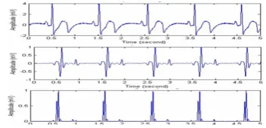
Figure 3. The original ECG Signal, the proposed pre-processing output resultant signal S1 and the output before the QRS detection S2, respectively
[image:3.595.353.558.393.525.2] [image:3.595.345.539.592.685.2]Further classification has been done by the Pan Tompkins algorithm and the resultant QRS peak detection is shown by Fig. 4 below.
Figure 4. QRS Complexes marked on the ECG signal
[image:4.595.63.258.105.178.2]The subsequent table I demonstrates the performance of the proposed combined algorithm. It can be seen that the algorithm provides a reliable QRS detection performance when tested against MIT-BIH Database.
TABLE I. QRS DETECTION PERFORMANCE ON THE MIT-BIH DATABASE
FP FN DER Se +P
185 203 0.38 96.3 97.83
The high-quality performance of the proposed detector can be explained by applying the Hilbert transform algorithm, baseline wander and noise are removed from the ECG signal and the R peaks are easily recognized.
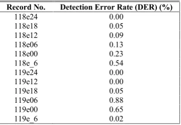
The noise effects were quantified by using the noise stress test procedures recommended by the ANSVAAMI EC38-1998 standard [18]. The MIT-BIH Noise Stress Test Database includes 12 sample records contaminated with electrode motion artifacts and significant amount of baseline wander and muscular noise.
TABLE II. NOISE EFFECTS PERFORMANCE ON THE MIT-BIH NOISE STRESS TEST DATABASE
Record No. Detection Error Rate (DER) (%)
118e24 0.00
118e18 0.05
118e12 0.09
118e06 0.13
118e00 0.23
118e_6 0.54
119e24 0.00
119e12 0.00
119e18 0.05
119e06 0.88
119e00 0.65
119e_6 0.02
From this study, it is found that a good pre-processing technique can basically help to improve the QRS peak detection. By employing Hillbert transform on a Wavelet base, the proposed detector gives reliable results such as in table I. The QRS detection rate was proven to be 98.7% which considers being in acceptable performance. Moreover, the testing done against the MIT-BIH Database yields comparable values against results discussed in prior works. Record of 207 was excluded due to ventricular flutter and record 105 contains
contaminated signal. Even at the noisy signal, the proposed detector still gives a reliable result. Furthermore, the noise stress test was carried out to verify noise tolerance of the proposed detector and the DER results are given in table II.
V. CONCLUSION
In conclusion, the usefulness of Hillbert transform on a Wavelet base was proved to be an effective pre-processing approach. In this study, the proposed pre-processing stage was later engaged with Pan-Tompkins algorithm for QRS detection purpose. Using the MIT-BIH arrhythmia database, the algorithm developed performed effectively even in the presence of significant noise. Further enhancement and validation on the proposed algorithm can also be tested for premature ventricular contraction (PVC) and to determine the correlation with the sudden cardiac arrest (SCA) cases.
ACKNOWLEDGMENT
This project is funded by Short Term Grant Phase 1/2011 (Vot.no: 0847) from Office for Research, Innovation, Commercialization and Consultancy Management (ORICC), Universiti Tun Hussein Onn Malaysia (UTHM).
REFERENCES
[1] Carsten Meyer, Jose Fernandez Gavela, and Matthew Harris, “Combining Algorithms in Automatic Detection of QRS Complexes in ECG Signals”, IEEE Transactions on Information Technology in Biomedicine, Vol. 10, No.3, July 2006.
[2] J. Pan and W. Tompkins, “Real Time Algorithm Detection for QRS”, IEEE Trans. Eng. Biomed Eng, 32(3), 1985, pp.230-236.
[3] N Debbabi, S El Asmi, H Arfa, “Correction of ECG Baseline Wander Application to the Pan & Tompkins QRS Detection Algorithm,” IEEE I/V Communications and Mobile Networks (ISVC), September 30-Oct 2, 2010.
[4] B. U. K¨ohler, C. Henning and R. Orgelmeister, “The Principles of Software QRS Detection”, IEEE Eng. Med. Biol.Mag., 21:42-57, Jan/Feb 2002.
[5] J.P. Martinez, S Olmos and P. Laguna, “Evaluation of a Wavelet Based ECG Waveform Detector on the QT Database,” Computers in Cardiology, 81-84, 2000.
[6] C. Li, C. Zheng and C. Tai, “Detection of ECG Characteristic Points Using Wavelet Transforms,” IEEE Trans. Biomed. Eng., 42:21-28, 1995.
[7] 7. A. Schuck Jr.', J. 0. Wisbeck', “QRS Detector Pre-processing Using the Complex Wavelet Transform,” Proceedings of the 25th Annual International Conference of the IEEE EMBS Cancun, Mexico September 17-21, 2003.
[8] Miad Faezipour,Ttarun M. Tiwari, Adnan Saeed, Mehrdad Nourani and Lakshman S. Tamil, “Wavelet Based Denoising and Beat Detection of ECG Signal,” IEEE/NIH Life Science Systems and Applications Workshop (LiSSA 2009).
[9] Martinez JP, Almeida R, Olmos S, Rocha AP, Laguna P., “A Wavelet-Based ECG Delineator: Evaluation on Standard Databases,” IEEE Trans on Biomed Eng.,51:570–581, 2004.
[10] P.M.Agante, J.P. Marques de Sa, “ECG Noise Filtering Using Wavelets with Soft Tresholding Methods,” Computers in Cardiology, pp.535-538, Sep 1999.
[image:4.595.74.254.471.595.2][12] H.A.N. Dinh, D.K.Kumar, N.D.Pah and P.Burton, “Wavelets for QRS Detection,” Proceedings of the 23rd IEEE EMBS International Conference, pp.1883-1887, Oct.2001.
[13] Natalia M. Arzeno, Chi-Sang Poon, and Zhi-De Deng, “Quantitative Analysis of QRS Detection Algorithms Based on the First Derivative of the ECG,” Proceedings of the 28th IEEE EMBS Annual International Conference New York City, USA, Aug 30-Sept 3, 2006.
[14] D. S. Bemitez, P. A. Gaydecki, A. Zaidi, and A. P. Fitzpatrick, “A New QRS Detection Algorithm Based on the Hilbert Transform,” Xl Computers in Cardiology, Vol. Zi, pp. 379-382, 2000.
[15] Francisco Ivan de Oliviera and Paulo Cesar Cortez, “A QRS Detection Based on Hillbert Transform and Wavelet Bases,” in IEEE Workshop on Machine Learning for Signal Processing, pp. 481-489, 2004.
[16] J. Weiss; "The Hilbert Transform of Wavelets are Wavelets", Applied Mathematics Group, Arlington, MA.
[17] MIT-BIH Database Distribution, Massachusetts Institute of Technology, 77 Massachusetts Avenue, Cambridge, MA 02139, 1998.
![Figure 1.Pan-Tompkins Algorithm Processing Steps [3]](https://thumb-us.123doks.com/thumbv2/123dok_us/8776022.901486/2.595.59.267.569.647/figure-pan-tompkins-algorithm-processing-steps.webp)