Size regulates particulate adjuvant induced
IL-10 and IL-12p70 production
Seán McCluskey
B.A (mod) Molecular Medicine
A Thesis submitted to
Trinity College Dublin
for the degree of Doctor of Philosophy
Supervisor: Prof. Ed Lavelle
Adjuvant Research Group
School of Biochemistry and Immunology
Trinity College Dublin
i
Declaration of authorship
This report represents the sole work of the author except where stated and has not been
submitted in whole or in part to any other university or institution as a part of a degree or other
qualification.
ii
Acknowledgements
Firstly, I would like to thank Prof. Ed Lavelle for the invaluable help, guidance, optimism and
his easy-going nature making this PhD project very enjoyable and interesting. It’s been a
pleasure to work in the lab these past four years where I have learned so much. Hopefully a
few papers will be coming.
For everyone in the lab both past and present who have taught, advised or helped me over the
last four years, this project would not have been possible. A massive thank you to Craig, Filipa,
Katie, Hannah, Stephanie, Alex, Aqel, Joanna, Aoife and Mimmi. A very big thank you to
Natalia who is always giving helpful advice, tips and ideas on new experiments. Not forgetting
the past members of the lab either, Ciaran, Chris, Claire, Dulce, Graham, Áine, Clíona, Lorraine,
Liz and Ewa.
Thanks to all the current and past TBSI staff members, who I’ve known since my undergraduate
degree and are always willing to stop for a chat. I would especially like to thank Gavin
McManus for all his help, suggestions and long hours spent trying to optimise my confocal
experiments. Also, a big thanks to Derek Nolan for his advice on all the fluidity experiments,
without those ideas we wouldn’t have got many of the results in this thesis.
To all my friends and family. I would like to thank my friend Barry, who encouraged me to
pick science over accounting way back, he still does not let me forget it. Finally, to my
girlfriend Clare who I met in our first day of Chemistry labs in Trinity and still going strong.
You have put up with my late hours or any rants when experiments didn’t go according to plan
yet would still meet me late in the day and make me laugh. Thank you for all your support you
have given me over the years and the holidays you have taken me on to give me a much-needed
iii
Abbreviations
APCs Antigen presenting cells
ATP adenosine triphosphate
AS Adjuvant system
Adju-Phos aluminium phosphate
Alum aluminium hydroxide
ASC apoptosis-associated speck-like protein containing a CARD
APS ammonium persulphate
BMDCs
BCG
Bone marrow-derived dendritic cells
Bacille Calmette–Guérin
BSA Bovine Serum Albumin
Bregs Regulatory B cells
BCR B cell receptors
CLRs C-type lectin receptors
CREB cAMP response element-binding protein
cDCs conventional DCs
CXCR C-X-C chemokine receptor
DNA deoxyribonucleic acid
cDNA Complementary DNA
CARD caspase recruitment domain
cAMP cyclic adenosine monophosphate
CpG cytidine-phosphate-guanosine
CTL Cytotoxic T lymphocyte
cGAS Cyclic GMP-AMP Synthase
ChIP-Seq Chromatin Immunoprecipitation Sequencing
DCs Dendritic cells
DAMPs Damage associated molecular patterns
DC-SIGN DC-specific intercellular adhesion molecule-3-grabbing non-integrin
DMEM Dulbecco’s Modified Eagle Medium
ECL enhanced chemiluminescence reagent
ERK extracellular signal-regulated kinase
EDTA Ethylenediaminetetraacetic acid
iv
ELISA
ECM
GF
enzyme-linked immunosorbent assay
extracellular matrix
Growth Factor
MSKs Mitogen-and Stress-activated protein Kinases
MSU Monosodium urate
MDP Muramyl dipeptide
MAPKs Mitogen-activated protein kinases
Mo-DCs Monocyte derived inflammatory DCs
MHC Major histocompatibility complex
mTOR Mammalian target of rapamycin
M-CSF Macrophage colony-stimulating factor M-CSF
MyD88 Myeloid differentiation primary response gene 88
MβCD Methyl-beta-cyclodextrin
MPL 3-O-desacyl-4’-monophosphoryl lipid A
nm Nanometre
NLRs Nod-like receptors
NAD NACHT-associated domain
NLRC4 NLR family CARD domain-containing 4
NLRP3 NLR family pyrin domain-containing 3
NOD Nucleotide oligomerisation domain
NF-κB Nuclear factor kappa-light-chain-enhancer of activated B cells
OPD o-Phenylenediamine dihydrochloride
OVA Ovalbumin
OTII OVA transgenic
O/W Oil-in-water
PI Propidium iodide
PI3K Phosphatidylinositol 3-kinases
PLA Poly (lactic acid)
PLG Poly (D L-lactide)-co-glycolide
PLGA Poly (lactic-co-glycolic acid)
PRRs Pattern recognition receptors
PMA Phorbol 12-myristate 13-acetate
v
poly I:C Polyriboinosinic acid-polyribocytidylic acid
PAMPs Pathogen-associated molecular patterns
PFA Paraformaldehyde
pDCs
PLGA
Plasmacytoid DCs
poly(lactic-co-glycolic acid)
PYD Pyrin domain
PS Polystyrene
PBS Phosphate-buffered saline
QS21 Quillaja saponaria fraction 21
RIG Retinoic acid-inducible gene
RLRs Retinoic acid-inducible gene RIG-like receptors
RORγt
ROS
RAR-related orphan receptor gamma
reactive oxygen species
RNA Ribonucleic acid
RPMI Roswell Park Memorial Institute
SARM Sterile-alpha and Armadillo motif containing protein
Syk Spleen tyrosine kinase
SDS Sodium dodecyl sulphate
SDS-PAGE SDS-polyacrylamide gel electrophoresis
STAT Signal transducer and activator of transcription
TRAM TRIF-related adaptor molecule
TLRs Toll-like receptors
TRIF TIR-domain-containing adapter-inducing IFN-β
Tfh Follicular T cell
TGF-β Transforming growth factor beta
TCR T cell receptor
TEMED Tetramethylethylenediamine
TMB Tetramethyl benzidine
TH Helper T cell
TIR Toll/Interleukin-1 receptor
Treg Regulatory T cell
T-bet T-box transcription factor
vi
TRAF
VLP
WT
W/O
µm
TNF receptor-associated factor
Virus like particles
Wild type
Water-in-oil
vii
Abstract
Until recently, vaccines were largely comprised of whole cell killed and attenuated pathogens.
However, over recent decades, there has been a shift towards subunit vaccine development,
using purified antigens which although much safer are less immunogenic, requiring adjuvants
to generate protective immunity. There is an urgent need to develop improved vaccines that
generate potent Th1 and CD8+ responses for diseases including TB, malaria, HIV and for
cancer. However, adjuvants such as alum and oil in water emulsions are strong inducers of
humoral immunity but less effective at promoting cellular immunity.
The hypothesis underlying this project was that this limited capacity to drive cellular immunity
was related to adjuvant induced production of the anti-inflammatory cytokine IL-10 and
inhibition of the Th1 polarising cytokine IL-12p70 by dendritic cells (DCs) and macrophages.
One key parameter dictating the effectiveness of particle based adjuvants is size but how size
influences the induction of T cell polarising cytokines has not been elucidated. Using
biodegradable poly (lactide-co-glycolide) (PLGA) particles between 100 nm and 30 µm in size,
it was shown that 500 nm-2 µm particles potently enhanced IL-10 production by DCs and these
DCs could inhibit T cell proliferation. These particles also potently enhanced IL-10 production
in the draining lymph nodes following intramuscular injection. Moreover, deficiency in IL-10
resulted in enhanced Th1 responses after intra-muscular immunisation with OVA and 500 nm
PLGA particles.
Particles of this specific size were shown to selectively interact with lipid rafts on the DC
membrane and activate the kinase Syk, leading to p38 and CREB phosphorylation and
enhanced transcription and secretion of IL-10. Remarkably it was found that this mechanism
is more broadly employed by DCs in response to physical stresses. Specifically, 500 nm PLGA
particles, osmotic stress, oxidative stress or disruption of actin polymerisation resulted in DCs
becoming rounded, decreasing their membrane fluidity and enhancing their capacity for IL-10
production. Conversely, increasing membrane fluidity using local anaesthetics or causing cell
swelling inhibited IL-10 production. Overall, a novel mechanism by which particulate
adjuvants and mechanical signals enhance IL-10 production by DCs is proposed, highlighting
the therapeutic potential of tailored biodegradable particles to block inflammation and promote
viii
Table of Contents
Declaration of authorship...i
Acknowledgements…...ii
Abbreviations…...iii
Abstract…...vii
Chapter 1. Introduction ... 1
Vaccines ... 2
Innate Immunity ... 3
PRRs, PAMPs and DAMPs ... 4
MAPKs: p38 and ERK... 10
Phosphatidylinositol 3-kinase ... 11
Dendritic cells and antigen presentation ... 12
IL-10 and suppression of immune responses ... 15
IL-10 regulation ... 18
IL-10 transcription factors ... 20
Role of Syk and kinase mediators ... 22
Role of the plasma membrane and actin in cell signalling ... 26
Regulation of cell-mediated immunity by the IL-12 family ... 33
T-Cell Responses and Adaptive Immunity ... 35
T helper cells ... 36
Th1 cells ... 36
Th2 cells ... 37
T regulatory cells ... 37
CD8+ T cells ... 38
B cells and the humoral immune response ... 39
Adjuvants ... 41
Novel and next generation adjuvants ... 46
Immunostimulatory adjuvants ... 46
Combination vaccine adjuvants ... 48
Particulate vaccine adjuvants ... 50
Aluminium adjuvants ... 56
ix
Particle size: from nano to micron scale ... 64
Aims and Objectives ... 67
Chapter 2. Materials and Methods... 68
Materials ... 69
General cell culture materials ... 69
Cell culture treatments ... 70
Real-time PCR materials... 72
Particulate adjuvants ... 73
Western blot materials ... 73
Enzyme-linked immunosorbent assay (ELISA) materials ... 76
Flow cytometry materials ... 79
Confocal microscopy materials ... 81
Methods ... 82
Mice ... 82
Cell culture ... 82
Culture of J558 granulocyte-macrophage colony-stimulating factor (GM-CSF)-expressing cell line ... 83
Production of M-CSF conditioned medium from L929 cells ... 83
Isolation and culture of BMDCs ... 84
Generation of Bone Marrow Derived Macrophages ... 84
Isolation and culture of human monocytes ... 85
Isolation and culture of splenocytes and draining lymph nodes ... 85
Isolation and culture of muscle tissue ... 86
Flow cytometry ... 86
Real-time PCR ... 90
Western Blot ... 91
Enzyme-linked immunosorbent assay (ELISA) ... 92
Confocal Microscopy ... 94
x
Chapter 3. Investigating the modulation of IL-10 and IL-12 production by particulate vaccine adjuvants ... 96
Introduction ... 97
Results ... 99
Different sized PS particles elicit distinct cytokine profiles in Dendritic cells……. ... 99
PLGA particle-induced cytokine modulation is size dependent ... 102
PLGA particles selectively inhibit IL-12p35 expression in a size-dependent manner… ... 107
The ability of PLGA particles to promote IL-10 production is not restricted to BMDCs.. ... 109
Alum enhances IL-10 expression and secretion by DCs and macrophages .... 111
PLGA particles alone do not promote the upregulation of costimulatory molecules on the surface of BMDCs or induce IL-6 secretion. ... 114
PLGA particle mediated cytokine modulation in BMDC is not due to toxicity in vitro…… ... 116
PLGA particles can modulate the potential of DCs to drive T cell proliferation and cytokine production in a size dependent manner. ... 119
The capacity of PLGA particles to enhance DC IL-10 production peaks around the 500 nm size range ... 128
Size dependent PLGA particle-induced cytokine modulation in DCs is also seen in combination with alternative TLR ligands. ... 132
The size-dependent ability of PLGA particles and alum to enhance IL-10 translates to human monocytes ... 136
Intramuscular injection of PLGA particles promotes innate and adaptive immune cell recruitment into the injection site ... 139
Injection of 500 nm PLGA particles and alum can promote IL-10 production and inhibit Th1 associated responses in vivo ... 143
Discussion ... 154
Chapter 4. Investigating the mechanism underlying particulate adjuvant modulation of IL-10 and IL-12 production ... 169
Introduction ... 170
Results ... 172
xi
PLGA particle mediated enhancement of IL-1β is NLRP3 and cathepsin S
dependent ... 175
Dissecting the mechanism by which PLGA particles promote IL-10 ... 178
The PI3K and ERK signalling pathways partially regulate PLGA particle induced IL-10 secretion in BMDCs ... 180
The size of PLGA particles influences the shape and size of DCs. ... 187
500 nm PLGA particles require cholesterol to induce IL-10 and enhance lipid raft formation. ... 191
Disrupting actin polymerisation drives increased IL-10 and partially reverses PLGA particle modulation of IL-12p35. ... 199
500 nm PLGA particles selectively exhibit a sustained interaction with the DC plasma membrane ... 207
Dendritic cell membrane fluidity is a key regulator of IL-10 production. ... 212
The enhancement of IL-10 secretion by PLGA particles in BMDCs requires Syk……. ... 223
The p38 signalling pathway is required for the enhancement of IL-10 production by PLGA particles ... 231
Alum induced IL-10 production in BMDCs also requires Syk and p38 signalling… ... 236
Phosphorylation of the transcription factor CREB is enhanced in cells incubated with 500 nm PLGA particles ... 238
Discussion ... 242
Chapter 5. General Discussion... 259
Future plans and experiments ... 272
1
Chapter 1
2
Vaccines
Infectious diseases remain the second leading cause of death worldwide after cardiovascular
disease, but the leading cause of death in infants and children (1). Vaccines continue to be the
safest, cheapest and most dependable strategy to counter infectious diseases. As of 2017, there
are over 70 licensed vaccines (FDA approved), which prevent the pathogenesis associated with
and reduce the incidence of around 30 viral and bacterial infections. It has been one of the most
successful and efficient forms of protection against infectious diseases for well over 100 years.
Through vaccination, smallpox has been completely eradicated worldwide while Polio has
nearly been abolished. Childhood vaccination has also reduced mortality and morbidity
associated with infectious diseases such as measles, mumps, rubella, diphtheria, tetanus and
Haemophilus influenzae type b in adults and children (2). Vaccines have also been successful
in targeting chronic diseases that can lead to cancers, with the hepatitis B and human
papillomavirus (HPV) vaccines reducing the incidence rates of liver and cervical cancer
respectively (3).
The fundamental idea underlying vaccination is to mimic natural infection and induce specific
long-lasting protection against infection or disease while avoiding unwanted side effects and
risk of developing the disease (4, 5). Vaccines act by initiating the innate immune response and
activating Dendritic cells (DCs) and other antigen presenting cells (APCs) to induce protective
adaptive immune response. Upon exposure to antigens, the APCs capture, process and present
the resulting antigen epitope to T cells (6). Upon re-exposure to a particular pathogen, rapid
activation of host memory T and B cells triggers an effective immune response, preventing
disease.
Traditionally, early vaccines were developed using live-attenuated pathogens or whole killed
pathogens with reduced pathogenicity to induce protective immunity, successfully eradicating
disease (Smallpox) or dramatically reducing the number of cases worldwide (Polio) (7).
Nevertheless, the need for new vaccines remains high as there are several diseases for which
optimal vaccines do not exist (HIV, TB, and Malaria) and there is a desire to replace existing
vaccines with less reactogenic formulations. The use of live attenuated or inactivated pathogens
is not suitable for all diseases due to safety concerns regarding toxicity, the potential for
reversion to virulence and limited efficacy in some cases (8, 9). For example, one virus strain
in the attenuated oral polio virus vaccine has been associated with rare Poliomyelitis outbreaks,
3
recommended for use in patients with impaired immunity. The RotaShield vaccine against
rotavirus was withdrawn from the market due to unwanted side effects (11). In other cases, the
inactivated pertussis vaccine was shown to induce strong reactogenicity at the injection site
and a potential to induce high fever (12, 13). Furthermore, some vaccines do not provide
lifelong protection (Tetanus, Diphtheria, and Pertussis), only generate partial protection (BCG
for leprosy and TB), require booster vaccinations (pertussis, MMR and Polio) or are only
effective against certain strains (annual influenza vaccine, pneumococcal and meningococcal
vaccines).
In order to further impact global health, there is a need to develop effective vaccines against
Human immunodeficiency virus (HIV), TB, and Malaria as well as cancers and autoimmune
diseases. While the use of live attenuated vaccines has been highly effective they are not
applicable to these diseases due to safety concerns (e.g. highly mutagenic HIV virus) (14). This
has led to the development of the so-called subunit vaccines composed of a single or few
well-characterised and highly purified antigens. Subunit vaccines can be based on highly purified
recombinant proteins, polysaccharides or protein-polysaccharide conjugates rather than whole
microorganisms.
While these subunit vaccines containing well defined purified antigens will improve safety
profiles compared to live attenuated and whole killed pathogen vaccines, they are also less
immunogenic due to the removal of microbial motifs such as TLR ligands. For example, live
attenuated vaccines provide both the antigen and the PAMPs to activate APCs, subunit
vaccines are typically unable to activate APCs on their own and induce protective immunity.
Therefore, they require adjuvants to induce adequate immune responses. To date, there are only
a few adjuvants in clinical use with limited abilities to induce a broad and robust Th1 response
needed for many complex pathogens such as TB, malaria and HIV.
Innate Immunity
The immune system is categorised into two arms: innate and adaptive. The innate immune
system acts as the first line of defence and provides the host with a rapid response for detecting
infection by viral, microbial and fungal pathogens. The innate response is mediated and
coordinated by cells including antigen presenting cells (APCs) particularly macrophages and
DCs, neutrophils, natural killer cells and secondary soluble components secreted by these cells
4
respond to pathogens. APCs can phagocytose and kill pathogens through detection of
conserved molecular patterns. DCs are a very important cell type at coordinating protective
immunity in response to infection or immunisation by linking the innate and adaptive immune
response.
In contrast, the adaptive immune system involves a slower antigen-specific response mediated
by B and T lymphocytes promoting the generation of memory cells which upon reinfection can
mount a quicker and more effective immune response. It had long been believed that the innate
immune response functions as a temporal defence system against infection until the adaptive
immune response can be elicited. However, recent studies have demonstrated that innate
immunity is essential for the effective induction of adaptive immunity. The ever-increasing
understanding of how the innate and adaptive immune system function has been exploited to
accelerate, enhance and manipulate the induction of vaccine-specific adaptive immune
responses.
PRRs, PAMPs and DAMPs
Recognition of infection is triggered through binding of specific pathogen-associated
molecular patterns (PAMPs) to pathogen recognition receptors (PRRs) on innate immune cells
to initiate an immune response (15, 16). These PAMPS are a set of evolutionarily conserved
motifs expressed either on the surface or intracellularly by pathogens and are crucial for their
survival, for example Lipopolysaccharide (LPS) a component found on the cell wall of
gram-negative bacteria. PRRs are the first line of defence in immunity and regulate the inflammatory
response according to the type of threat they encounter (17). PRRs are expressed primarily by
cells at the front line of the innate immune system such as Macrophages and DCs. In addition
to PAMPs, PRRs can also respond to damage associated molecular patterns (DAMPs),
5
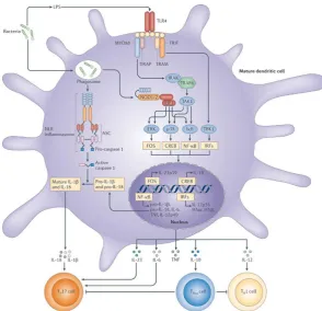
Figure 1.1: DCs bridge the innate and adaptive immune responses
Immature DCs act as sentinels and survey the periphery where they efficiently phagocytose and process
antigens into peptides. These antigenic peptides are then presented in the context of either MHC class
I (MHCI) or MHC class II (MHCII) molecules to naïve T cells. Upon activation, DCs lose their
endocytic ability but acquire an enhanced potential for antigen processing and presentation, facilitating
the provision of signal 1 to T cells through peptide display on major histocompatibility complex (MHC)
molecules and gain chemokine receptor expression (CCR7 and CXCR4) to enable migration to T-cell
rich zones in draining lymph nodes. Activated DCs also express enhanced levels of co-stimulatory
molecules (e.g. CD80 and CD86 which interact with CD28 on T cells or CD40 which interacts with
CD40L on T cells) (Signal 2) and produce cytokines to determine the fate of the naïve T cells (signal
3) (19, 20). DC subsets can specialise in driving either CD4+ T cell or CD8+ T cell adaptive immune
responses. PRRs (e.g. TLR4, NOD1/2 and inflammasome NLRs) can detect PAMPS and DAMPS
within the environment to mediate cytokine release. TLR4 and NOD1/2 engagement activates
signalling pathways (p38, ERK etc.) to modulate proinflammatory and anti-inflammatory cytokine
release influencing T cell polarisation. NLRP3 activation is coordinated through multiple pathways. In
the priming step, PRRs induce NF-κB-dependent induction of components of NLRP3 itself and
pro-IL-1β. The second signal allows NLRP3 to oligomerise and form the inflammasome to process pro-IL-1β
and pro-IL-18 into their active forms. The inflammasome can sense various danger signals that emerge
during infections, tissue damage or metabolic imbalances to trigger formation of the inflammasome.
This includes extracellular ATP, microbial toxins, monosodium urate crystals, alum and other
6
PRRs are expressed on either the cell surface or in intracellular compartments of immune cells.
PRRs consist of four main families: Toll-like receptors (TLRs) (Fig 1.2), retinoic
acid-inducible gene RIG-like receptors (RLRs), Nucleotide-binding and oligomerisation domain
NOD-like receptors (NLRs) and C-type lectin receptors (CLRs) (22-24) (Fig 1.4 and 1.6).
Moreover, there are also various cytoplasmic DNA receptors that respond to cytoplasmic DNA
after microbial infection, which belong to different protein families, including
cyclic-di-GMP-AMP synthase (cGAS) and DNA-dependent activator of interferon-regulatory factors (DAI)
(25, 26).
TLR activation is a hallmark of innate immune responses. The TLRs are the most studied
family of PRRs, sensing a diverse range of PAMPs (27). The binding of PAMPs and/or DAMPs
to PRRs activates intracellular signalling cascades, leading to the release of inflammatory and
anti-inflammatory mediators including cytokines, chemokines, and type 1 interferons (IFNs)
inducing innate immune response as well as the development of acquired immunity against a
particular pathogen (28) (Fig 1.1 and 1.9). TLRs are located either on the surface or intracellular
endosomal compartments (Fig 1.2). The intracellular localisation of TLRs is thought to be
critical for ligand recognition as well as for preventing TLRs from encountering self-nucleic
acids, which could potentially cause autoimmunity. For example, Endogenous TLR9 is not
exposed to mammalian DNA and can only be activated by viral DNA ingested and acidified
within endosomes (29).
One class of PRRs located on the cell surface, namely, C-type lectins (i.e., Dectin-1, 2 and
Mincle), has the capacity to signal in a TLR-independent manner (30). They mainly recognise
carbohydrate moieties on pathogens, including mannose, fucose and glucan structures. For
example, fungal components (from Candida albicans and Aspergillus fumigatus) such as
β-glucans are recognised by the C-type lectin dectin-1 to mediate Th1 and Th17 immune
responses leading to the activation and recruitment of neutrophils and macrophages (31, 32).
Another example is the dendritic cell-specific intercellular adhesion molecule-3-grabbing
non-integrin (DC-SIGN), which can recognise a wide range of pathogens, including HIV, Measles,
Mycobacterium tuberculosis and Helicobacter pylori (24, 33).
NLRs and RLRs maintain immune surveillance in the cytoplasm. Of these, NLRs are a large
group of cytoplasmic sensors that recognise not only microbial products but also endogenous
7
twenty cytosolic receptors in mammals, divided into different groups based on the N-terminal
activation domains involved in signal transduction (NLRA, NLRB, NLRC and NLRP) (35).
Some NLRs have the ability to form an inflammasome, a tightly regulated multi protein
complex mediating the maturation of the inactive precursor forms of interleukin (IL)-1β and
IL-18 into active proinflammatory cytokines (36). The complex consists of a central NOD (or
NACHT) domain, required for oligomerisation, a C-terminal series of leucine-rich repeats
(LRRs) involved in agonist sensing or ligand binding and the variable N-terminal protein–
protein interaction domain defined by the caspase recruitment domain (CARD) (NLRC), pyrin
domain (PYD) (NLRP), acidic transactivating domain (NLRA), or baculovirus inhibitor repeat
(BIR) (NLRB) (34, 37). For example, the NLR family member, NACHT, LRR, PYD domain
containing protein 3 (NLRP3) can sense a wide variety of molecules and compounds, including
monosodium urate (MSU), adenosine triphosphate (ATP) microbial toxins (nigericin and
α-hemolysin), silica and aluminium adjuvants to mediate assembly of the inflammasome (38).
Subsequently, NLRP3 oligomerise and the N terminal associates with the adaptor
apoptosis-associated speck-like protein containing CARD (ASC) via its pyrin domains. ASC in turn
recruit’s pro-caspase-1 through CARD-CARD interactions. Pro-caspase-1 clustering on the
oligomerised NLRP3 results in cleavage of procaspase-1 into caspase-1 to catalyse the
processing of pro-IL-18 and pro-IL-1β into IL-18 and IL-1β (35, 39) (Fig 1.1).
Furthermore, a range of NLRs function independently of inflammasomes, including NOD1,
NOD2 and others (Fig 1.1 and 1.2). NOD1 senses diaminopimelic acid (DAP) expressed on
most Gram-negative and specific Gram-positive bacteria while NOD2 recognises muramyl
dipeptide (MDP), a ubiquitous component of all bacterial peptidoglycans to mediate
proinflammatory cytokine production (34, 40). Additionally, NOD2 can interact with
ATG16L1 and stimulate the formation of the autophagosome (41).
Finally, the RIG-I like receptors (RLRs) are a class of PRRs implicated in the recognition of
and response to most RNA viruses. Retinoic acid inducible gene-1 (RIG-I) and melanoma
differentiation-associated gene 5 (MDA5) are intracellular sensors of dsRNA (from ds RNA
viruses or dsRNA generated during replication of ssRNA viruses) leading to type I IFN gene
8
Following ligand binding, TLRs dimerise and recruit TIR domain-containing adaptor
molecules and subsequently propagate intracellular signal transduction. These adaptor
molecules include; myeloid differentiation primary response gene 88 (MyD88), TIR-domain
containing adapter-inducing IFN-β (TRIF), TRIF-related adaptor molecule (TRAM) and
MyD88 adaptor-like (Mal) (Fig 1.2) (44).
There is also evidence that specific adaptor molecules can negatively regulate TLR related
immune responses. Sterile-alpha and Armadillo motif containing protein (SARM) can
negatively regulate TRIF-dependent Toll-like receptor signalling (45). Likewise, the recently
described B-cell adaptor for PI3K (BCAP) negatively regulates proinflammatory cytokine
secretion and links TLR signalling to the PI3K signalling cascade (46). Most TLRs directly
interact with MyD88 to trigger downstream signalling pathway, although TLR2 and TLR4
require the bridging adaptor Mal to associate with MyD88. TLR4 (and TLR3) also engage a
MyD88-independent, TRIF-dependent pathway which requires the bridging adaptor TRAM to
associate with TRIF (Fig 1.2).
Downstream signalling from TLR4 is driven by recruitment of the adaptor proteins, MyD88
and TRIF (44, 47). The early response relies on the MyD88-dependent pathway, leading to
downstream activation of nuclear factor kappa-light-chain-enhancer of activated B cells
(NF-κB) and MAPKs (p38, ERK, JNK), which induce proinflammatory and anti-inflammatory
responses (28) (Fig 1.1 and 1.2). The late response relies on engagement of the TRIF pathway
following endocytic engagement of TLRs and subsequent activation of IFN regulatory factor
3 (IRF3) and ‘late’ NF-κB transcription factors triggering cytokine (IFN-β) and chemokine
expression (IP-10) (48, 49) (Fig 1.1 and 1.2). Thus, MyD88- and TRIF-dependent pathways
have overlapping functions to regulate both proinflammatory and adaptive immune responses
(50).
Vaccine-induced immunity is heavily reliant on the activation of innate immune mechanisms,
using either PAMPs or DAMPs to engage PRRs and subsequently condition the adaptive
immune response. The PAMPs could for example be TLR agonists from the microbial cell
surface. Additionally, adjuvants within the formulation could release endogenous DAMPs at
9
Figure 1.2 Mammalian Toll like receptors and downstream signalling pathways
TLRs are pattern recognition receptors with the ability to distinguish conserved pathogen-associated
molecular patterns from self. TLR2 heterodimerises with TLR1 or TLR6 and recognises triacylated or
diacylated forms of lipoproteins, respectively. TLR4 recognises LPS from enteric bacteria and a number
of viral and bacterial proteins (e.g. Escherichia coli LPS and fusion proteins fromRespiratory syncytial
virus). TLR4 moves from the plasma membrane to the endosomes in order to switch signalling from
MYD88 to TRIF. The only known protein ligand for human TLRs is bacterial flagellin, which is
recognised by TLR5 (51). Intracellular receptors located in the endosomal compartments can recognise
viral and intracellular bacterial nucleic acids, including unmethylated cytidine-phosphate-guanosine
(CpG) motifs (TLR9), ssRNA (TLR7/8) and dsRNA (TLR3). Engagement of a PAMP with its
corresponding TLR results in the recruitment of two main signalling adaptor molecules, MyD88 and
TRIF. All TLRs use MyD88 except TLR3, which signals through TRIF. TLR4 is the only TLR known
to use MyD88 and TRIF molecules. TLR2 and TLR4 also require MAL for recruitment of MyD88.
Upon receptor ligation, adaptor molecules stimulate downstream pathways involving interaction
between IL1R-associated kinases (IRAKs) and the TNF receptor-associated factors (TRAFs) leading
to downstream phosphorylation and activation of MAPKs (p38, ERK1/2 and JNK) and other signalling
molecules. This leads to translocation of transcription factors (CREB, AP1, NF-κB and IRF3/7),
ultimately resulting in the expression of genes required for cell activation and immunity. Taken from
10
MAPKs: p38 and ERK
Mitogen-activated protein kinases (MAPKs) are a conserved serine-threonine family of kinases
that mediate intracellular signalling events, promoting cellular responses to a wide range of
extracellular cues including growth factors (GF), hormones, cytokines and stress. The MAPK
cascade has fundamental roles in many cellular processes including, cytokine production,
proliferation, survival and differentiation acting as positive and negative regulators at multiple
levels within the signalling cascade (52-54) (Fig 1.1 and 1.2).
TLR stimulation with PAMPs such as LPS activates several highly regulated MAPK signalling
pathways crucial for synthesis of proinflammatory and anti-inflammatory cytokines (55). Any
dysregulated signalling from PRRs leads to excessive proinflammatory cytokine production
and host tissue damage in disease states including chronic inflammatory conditions,
autoimmune disease and cancers (56-58).
The MAPK cascade consists of at least three protein kinases in series that leads to the activation
of a multifunctional MAPK. MAPK pathways consist of a MAPK kinase kinase (MKKK) that
phosphorylates and activates a MAPK kinase (MKK) that then activates the functional terminal
MAPK by dual phosphorylation. Upstream of the kinase module are members of the Ras and
Rho families of GTPases that relay signals from receptor complexes to the module (52, 53, 59,
60).
Among the most important kinases activated are the p38 mitogen-activated protein kinases.
There are four members of the p38 MAPK family (p38α, p38β, p38γ and p38δ), with the p38α
isoform the most abundant and best-characterised isoform (61). The p38 family plays a role in
angiogenesis and cell proliferation and is critical for normal immune and inflammatory
responses. The p38 MAPK pathway is a key regulator of pro-inflammatory cytokine
biosynthesis at the transcriptional and translational level (61) (Fig 1.1 and 1.2). The
extracellular signal-regulated kinase (ERK) family, consists of several isoforms: ERK1 to
ERK8. In the ERK signalling pathway, ERK1 or ERK2 are especially important, regulating
cytokine production, cell cycle, differentiation and apoptosis (58, 62, 63). The c-Jun N-terminal
kinase (JNK) is also an important member of the MAPKs. JNKs have been shown to have
important roles in a number of cell processes, including apoptosis, the development of multiple
11
Activated MAPKs (ERK1/2 and p38) phosphorylate and activate Mitogen-and Stress-activated
protein Kinases (MSKs) and various substrate proteins including transcription factors. For
example, MSK1 and MSK2 can phosphorylate transcription factors such as cAMP response
element-binding protein (CREB) and AP-1 (e.g. c-Fos and c-Jun) which subsequently bind to
the IL-10 promoter to induce transcription (64, 65) (Fig 1.1 and 1.2).
ERK1/2 and p38 MAPKs are critical for both pro- and anti-inflammatory immune responses.
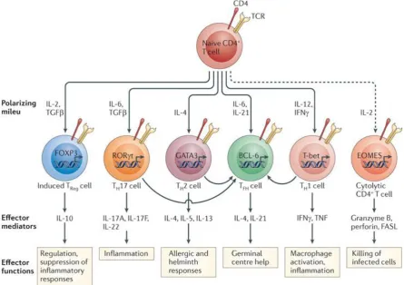
They mediate DC production of cytokines which determine the polarisation of naive CD4+ T
cells into Th1 (66, 67), Th2 (68), Th17 (69, 70), or regulatory T cell subsets (71, 72). For
example, p38 has been shown to be essential for both IL-12 and IL-10 induction. In contrast,
ERK is crucial for IL-10 induction, but mediates a negative feedback of IL-12 inhibition
(through inhibition of the IL-12p40 subunit) (66, 73-77). Both p38 and ERK cooperate and
cross talk with each other during TLR-induced 10 production. ERK- and p38-dependent
IL-10 production has also been shown to be inhibited by IFN-γ (78).
Phosphatidylinositol 3-kinase
Phosphatidylinositol 3-kinase (PI3K) consists of a family of lipid kinases that play crucial roles
in metabolism, rearrangement of the cytoskeleton, growth, proliferation, survival, apoptosis
and immune cell effector functions (79-81). PI3Ks are divided into three classes (I, II, III) based
on their structures and substrate specificities. Class I PI3Ks are further divided into class IA
(p110α, β and γ) and class IB (p110γ) (81). Class I PI3Ks are heterodimers composed of a
regulatory subunit (p85) and a catalytic subunit (p110) involved in receptor-mediated
signalling pathways in the immune system (82).
TLR-induced activation of the PI3K pathway can differentially regulate IL-10 and IL-12
production in both human and mouse macrophages (83, 84). The PI3K pathway has an
important role in the differentiation and activation of DCs, particularly the regulation of
proinflammatory cytokines (IL-12, IL-6, and TNF-α) and partially regulating IL-10 secretion
(85).
Class I PI3K signals to the serine/threonine kinase Akt, also termed PKB (protein kinase B)
and can control pro-inflammatory cytokine production (81, 86, 87). Akt can also activate the
12
two separate pathways. Signalling through the PI3K/AKT/mTOR (mTORC1) pathway can
enhance IL-10 production, leading to IL-12 inhibition in an IL-10 dependent manner (88).
Furthermore, the PI3K/Akt/mTOR pathway has extensive roles in innate immune cells,
including neutrophils, mast cells, monocytes, macrophages and myeloid as well as
plasmacytoid dendritic cells (mDCs and pDCs, respectively) (89). Activation of PI3K/AKT
can also inhibit GSK-3β, a multifunctional enzyme regulating cytokine production and other
cellular functions. GSK3 enhances IL-12p70, IL-6, and TNF-α secretion, and inhibits IL-10 in
E. coli–stimulated human monocyte-derived DCs (85). TLR4 mediated GSK3β activation has
shown to suppress CREB mediated IL-10 production in response to Leishmania
donovani infection (90). In contrast, IFN-γ can also trigger GSK-3β activation by antagonising
the PI3K/AKT pathway. This leads to inhibition of TLR-induced IL-10 production through
inhibition of the transcription factors, CREB and AP-1 (78, 90, 91) (Fig 1.4).
Dendritic cells and antigen presentation
DCs were first described by Steinman and Cohn in 1973 as cell population with a distinctive
morphology (clearly distinct from macrophages) with a unique ability to activate and prime
naïve T cells. Thus, they are the primary instigators of adaptive immunity, and function as the
bridge between innate and adaptive immunity (92, 93). DCs are not the only immune cell
capable of presenting antigens; other APCs including Macrophages and B cells also play
significant roles.
DCs represent a crucial class of APCs which possess multiple PRRs capable of recognising
various PAMPs and DAMPs and subsequently activate a specific adaptive immune response.
DCs thus play important roles in triggering immune response against pathogens or initiating
tolerance or autoimmunity against self-antigens during the steady state. Therefore, DCs are
essential mediators for bridging and shaping the innate and adaptive immune response (94, 95).
DCs can adopt two main states, either an immature tolerogenic state which samples the
peripheral environment for antigens or a mature state that induces an effector or memory T cell
response. A switch of DCs from tolerance maintenance to immune stimulation, referred to as
maturation, may be initiated by PAMPS, DAMPs, cytokines or CD40/CD40L signalling (20).
DCs phagocytose and process antigen into peptides which are loaded onto MHC molecules and
13
determines whether the peptide is displayed on MHC class I or II. Extracellular pathogens will
be endocytosed, degraded in lysosomes by pH dependent proteases and the peptides are
delivered and loaded onto the MHC II complex and transported to the cell surface. In contrast,
endogenous antigens (intracellular) such as viral proteins undergo proteasomal degradation in
the cytosol and are transported to the endoplasmic reticulum where they are loaded onto MHC
class I molecules (96). Finally, a third pathway exists called cross presentation. This
mechanism allows the loading and presentation of exogenous peptides on MHC class I
molecules (97).
In addition to the antigen presenting capabilities of DCs, a range of cytokines secreted by DCs
influence T-cell differentiation (Fig 1.3b and 1.9). DCs producing IL-12 promote Th1
expansion in response to microbial stimuli such as Toxoplasma gondii or to LPS (98) while
secretion of the cytokines IL-23 and IL-1β promotes Th17 differentiation (99, 100). In the case
of Th2 differentiation, IL-10 and IL-33 play a crucial role (68, 101, 102). IL-4 is also important
for Th2 differentiation but DCs are not believed to be the main source (103). Pathogens linked
to a Th2 response can cause secretion of IL-4 (IL-5 and IL-13) rapidly by a variety of other
innate cells (104, 105), but whether IL-4 has a direct effect on DC function remains unclear.
DCs represent a heterogeneous group of cells that differ in their origin, anatomic location,
phenotype, surface markers and function but all are capable of activating naïve T cells (106)
(Fig 1.3a and 1.3b). For instance, PRRs can be differentially expressed on DC subsets which
influences their capacity to respond to a particular PAMP or DAMP. Murine and human DCs
in steady state are predominantly classified as either plasmacytoid DCs (pDCs) or conventional
(cDCs) differing in their function and phenotype such as surface markers (107) (Fig 1.3a and
1.3b).
pDCs represent a small subset of DCs and express a restricted set of receptors and low levels
of major histocompatibility complex class II (MHCII), CD11c and costimulatory molecules in
the steady state. They selectively express high levels of TLR7 and TLR9 to recognise viral
nucleic acids or host-derived nucleic acids, leading to the production of large amounts of type
I IFNs (108, 109) (Fig 1.3a). This is especially crucial for an effective antiviral response. They
also link the innate and adaptive immune response by controlling the function of cDCs, T cells,
14
On the other hand, cDCs (also known as classical or myeloid) main function is to initiate and
drive the adaptive response by capturing antigen in peripheral tissue and migrate to T cell zones
of secondary lymphoid tissues. They have a short half-life (approximately 3-5 days) and are
continuously replaced from bone marrow precursors, a process dependent on the cytokine
Fms-like tyrosine kinase-3 ligand (Flt3L) whereas monocytes require macrophage
colony-stimulating factor (M-CSF) (111). cDCs are found in lymphoid tissues including the thymus,
spleen, lymph nodes, and bone marrow, but are also widely distributed among non-lymphoid
tissues such as the skin, liver, kidney, intestinal tract and lung allowing them to constantly
acquire tissue and blood antigens (112, 113).
In mice, cDCs are defined by expression of the transcription factor Zbtb46 and can be further
separated into two main subsets based on expression of the transcription factors BATF3 or
IRF4, whereas pDCs express the transcription factor E2-2 (114, 115). Batf3 expressing cDCs
express CD8α in the spleen and CD103 or CD24 in the periphery, whereas IRF4+ cDCs express
CD11b and are negative for CD8α. In contrast to CD8a and CD103 DCs, the population
currently defined as CD11b+ cDCs is heterogeneous and remains less well characterised (116).
CD11b+ cDCs are the most abundant cDCs in lymphoid organs except for the thymus and can
also be found in nonlymphoid tissue (117).
The CD8+ and CD103+ cDCs specialise in mediating cross-presentation of exogenous antigens
on MHC-I molecules to CD8+ T-cells, presenting glycolipid antigens in a CD1d manner and
IL-12 secretion to promote Th1 responses (116). For instance, stimulating TLR receptors on
the CD8a+ and CD103+ cDC lineage induces IL-12p70 production (98). In contrast, CD11b+
DCs are more efficient at driving CD4+ T cell responses, potentially because of their higher
expression of MHC-II presentation machinery (118) and may be the dominant subtype
involved in driving Th2 (119) or Th17 responses (120). CD11b+ DCs can also be characterised
by their production of cytokines, such as IL-6, IL-23 and proinflammatory chemokines (116).
Interestingly, both subtypes differ in their ability to acquire and phagocytose particular types
of antigen. CD11b+ DCs preferentially acquire and transport soluble antigen, whereas
CD103+ DCs are more efficient at dealing with particulate material in the lung of mice (121,
122).
DCs can also arise from monocytes in lymphoid and nonlymphoid organs, these monocyte
15
These DCs can be generated in-vitro from bone marrow cells using GM-CSF (Section 2.2.5)
expressing high levels of CD11c and major histocompatibility complex class II (MHC II),
capable of migrating to draining lymph nodes to activate T cells. They play an essential role in
clearing pathogens by secreting large amounts of TNF-α and inducible nitric oxide synthase
(iNOS) (19, 123). Mature Mo-DCs migrate to draining lymph nodes and activate CD4+ and
CD8+ T cells. In terms of antigen presentation function, Mo-DCs are as active as cDCs,
including cross presentation of proteins on MHCI in vivo (124). Although, Nussenzweig et al
demonstrated that Mo-DCs were less efficient in presenting phagocytosed antigen than cDCs
(125). Therefore, it remains unclear whether Mo-DCs significantly contribute to presenting
phagocytosed antigen to CD8+ or CD4+ T cells. It has been suggested that their primary
function is to act as innate immune effector cells that destroy phagocytosed antigen (126).
DCs are known to play a central role in vaccination due to their ability to induce and regulate
T cell responses. Therefore, understanding how DCs respond to established and novel vaccine
formulations is essential for designing vaccines with improved clinical efficacy and
understanding how established vaccines work.
IL-10 and suppression of immune responses
Interleukin-10 (IL-10) is a pivotal anti-inflammatory cytokine that protects the host from
excessive tissue damage during host defence to pathogens and acts as one of the key molecules
critically involved in the development and maintenance of immune tolerance and homeostasis.
IL-10 was first called cytokine synthesis factor (CSIF). It was identified as a factor produced
by Th2 cells with the ability to inhibit Th1 responses (127). Since this discovery, the list of
cells which can produce IL-10 and information on the functions of this cytokine has greatly
expanded. Even in recent years new roles for IL-10 have been reported. For example, IL-10
secreted by Tregs during the resolution phase of infection has been linked to the promotion of
memory CD8+ T cells (128). In contrast, IL-10 restricts the magnitude of the effector and
memory Th1 responses (and memory CD8 responses) during the early stages of an acute viral
16
Figure 1.3: Dendritic cell lineages have specialised functions
A) DCs form a heterogeneous population that have been classified into various groups
according to their origin, location, patterns of migration and expression of PRRs.
B) Progenitors in the bone marrow, called macrophage and DC precursors (MDP), gives
rise to pre-DCs and monocytes. pDCs differentiate in the bone marrow, while Pre-cDCs
can also move into the blood and migrate to lymphoid and non-lymphoid organs, where
they terminally differentiate into cDCs controlled by different transcription factors,
including the CD8α+/CD103+ and CD11b+ subset. Different DC subsets carry out
specific and specialised functions depending on environmental cues. They can respond
differently to pathogen challenge through the secretion of T cell polarising cytokines.
Taken from (111).
A.
17
The initial immune response to pathogens involves the rapid release of pro-inflammatory
cytokines that trigger host defences. Over activation or excess inflammation can lead to host
damage and autoimmunity. As a result, the immune system has evolved to trigger
anti-inflammatory mechanisms simultaneously to limit pro-anti-inflammatory cytokine production and
restore homeostasis after infection or damage. IL-10 is a crucial multifunctional
anti-inflammatory cytokine, primarily acting to limit excessive immune responses and resolve
inflammation, thereby limiting chronic and acute inflammation and preventing autoimmunity
(130, 131). For example, human genetic deficiencies and polymorphisms in IL-10 or its
receptor can strengthen the inflammatory response to gut microbes but also lead to
development of inflammatory bowel disease (ulcerative colitis and Crohn’s disease) (132-135).
IL-10 deficient mice are much more prone to autoimmune diseases such as experimental
autoimmune encephalomyelitis (EAE) as the immune response shifts to an amplified Th1
phenotype (136). IL-10 producing B cells were found to be required to prevent disease onset
(137, 138). Viruses have evolved to manipulate the anti-inflammatory properties of IL-10 to
limit host immune responses, leading to persistent infection (139, 140). Similarly, IL-10
producing DCs and T regulatory cells are upregulated during Schistosoma mansoni and
Mycobacterium tuberculosis infection, inhibiting the effector T-cell response and reducing
parasite control (141, 142).
Its importance is highlighted by the fact that the majority of innate and adaptive immune cells
are capable of secreting IL-10 and responding to its secretion by expressing the IL-10 receptor
(IL-10R) (143, 144). IL-10 can be secreted by macrophages, monocytes, DCs, mast cells,
eosinophils and B cells (143).Additionally, various subsets of CD4+ and CD8+ T cells are a
significant source of IL-10, especially Th2 and Treg cells, but it can also be produced by Th1,
Th17 and CD8+ T cells (130, 143). Even non-immune cells are capable of producing IL-10
including keratinocytes, epithelial cells and tumour cells to evade the immune system (130,
145). APCs are one of the most important sources of IL-10 (146) (Fig 1.1).IL-10 has many
suppressive mechanisms, including inhibiting DC maturation and function by restricting
antigen presentation and downregulating MHC II and costimulatory molecule expression
(147).
IL-10 can provide an autocrine feedback to limit or resolve inflammatory gene expression by
macrophages and DCs activated by TLR agonists such as LPS, thus limiting the production of
18
RANTES, IL-8, IP-10, and MIP-2) (148, 149). IL-10 can inhibit phagocytosis and microbial
killing by limiting the production of reactive oxygen and nitrogen intermediates in response to
IFN-γ (150). IL-10 also acts directly on IL-10R expressing T cells to suppress their functions
(144), by inhibiting proliferation and production of IL-2, IFN-γ, IL-4, IL-5 and TNF-α (151,
152) and can promote anergy and tolerance by Treg cells (153, 154). In B cells, IL-10 prevents
apoptosis, enhances proliferation and modulates immunoglobulin (Ig) class switching (155).
IL-10 has been suggested to drive Th2 cell differentiation through IRF4 activation in DCs (68).
Studies using IL-10-/- DC demonstrated a shift in the balance between Th2 and Th1 response
in a mouse model of allergic dermatitis (156). Novel therapies focusing on the induction or
inhibition of IL-10 have been used to treat chronic inflammatory bowel disease, rheumatoid
arthritis, psoriasis, asthma, systemic lupus erythematosus, multiple sclerosis, transplant
rejection and cancer (155, 157, 158).
IL-10 regulation
IL-10 regulation has several layers, including changes in the chromatin structure, enhancement
or silencing of IL-10 transcription and post-transcriptional regulatory mechanisms (159).
Distinct mechanisms control the production of IL-10 in different cell types. IL-10 production
in APCs is triggered by engagement of TLRs, intracellular PRRs including RIG-I and
Nod2-like family receptors, C-type lectin signalling, or ligation of CD40 and Fc receptors (157) (Fig
1.4).
Once IL-10 is secreted, it binds to a heterodimeric receptor complex, consisting of IL-10R1
and IL-10R2 subunits on the cell surface, activating the Janus kinase (JAK1) and signal
transducer and activator of transcription (STAT3) (JAK-STAT) pathway. STAT3 then
activates IL-10 responsive genes such as suppressor of cytokine signalling 3 (SOCS3) to inhibit
19
Figure 1.4: IL-10 regulation in APCs
A) IL-10 expression is induced by TLR (TLR2, 3, 4, 9) or non-TLR (e.g. dectin-1 or DC-SIGN)
signalling in macrophages and DCs. Following engagement of TLRs and adaptor molecules (MyD88 and TRIF), ERK1/2, p38 and Nf-κB pathways and downstream signalling molecules (MSK1/2) result in transcription factor (e.g. CREB) binding to the IL-10 promoter and subsequent IL-10 production. In myeloid DCs. Non TLR signalling through c type lectin receptors such as Dendritic Cell-Specific Intercellular adhesion molecule-3-Grabbing Non-integrin (DC-SIGN) and RAF1 kinase augments TLR2 induced IL-10 production (prolongs and increases IL-10 transcription after TLR2 ligation) (161). Dectin-1 ligation results in recruitment and activation of Syk and ERK and IL-10 secretion. Macrophages also use NOD2 signalling to enhance IL-10 production by cross talking with TLR2 (162). Taken from (143).
B) Positive and negative feedback loops also regulate IL-10 in APCs. The p38 and ERK pathways
20
IL-10 transcription factors
There are a number of transcription factors that regulate IL-10 expression (Fig. 1.5). The
promoter region in human and murine IL-10 share a high degree of homology. The number of
transcription factors involved in different cell types reflects the complexity of IL-10 regulation.
The importance of different promoter elements varies depending on the cell type and stimuli.
The transcription factors known to regulate IL-10 in APCs are CREB (165), c-maf (166),
C/EBPβ (167), activating transcription factor 1 (ATF1) (143), specific protein (SP1) (168) and
NF-κB (143, 169) (Fig 1.5).
CREB can regulate IL-10 transcription following p38 signalling after engagement of TLR4
and Mal (170, 171). Factors leading to elevated levels of secondary messenger cAMP activate
cAMP-dependent protein kinase A (PKA) that in turn will phosphorylate CREB at Ser-133.
Likewise, increased Ca2+ signalling activates a Ca2+/calmodulin-dependent protein kinase
(CaMK) family member and phosphorylates CREB (172). Similarly, TLR interaction can
trigger Ser-133 phosphorylation of CREB, and in association with agents that elevate cAMP,
lead to increased CREB-mediated transcription. CREB has been associated with TLR
dependent and independent IL-10 production in macrophages and DCs (165, 173, 174).
The main inflammatory transcription factors activated by TLRs are the NF-κB family.
However, along with upregulation of pro-inflammatory cytokines including IL-6, IL-12, IL-1β
and TNF-α, the NF-κB1 (p50) homodimer can enhance expression of IL-10 by binding to a κB
site upstream of the IL-10 promoter (169). More recently, the transcription factor interferon
regulatory factor 4 (IRF4) whose expression is regulated by NF-κB and TLR4 has been linked
to upregulation of IL-10 in dendritic cells (68). Interestingly, IRF4 has also shown to inhibit
IL-12 production and thereby control Th1 response (175, 176). While IRF4 has important roles
in development of both innate and adaptive immune cells including DCs (177, 178), recent
studies have only started to show how IRF4 can regulate the function of DCs including
determining the cytokine secretion profile. Furthermore, the transcription factor DC-specific
transcript (DC-SCRIPT; also known as ZNF366) which is specifically expressed in DCs has
been shown to regulate IL-10 and subsequently IL-12p70 levels in human DCs (179, 180).
Post transcriptional regulation is also important by stabilising the IL-10 transcript. Like many
21
contains clusters of adenosine and uridine rich elements (ARE) located on the 3’ untranslated
region (UTR). These AREs are very important for determining RNA stability as they are targets
for tristetraprolin (TTP), proteins that can induce rapid degradation of mRNA following
binding to ARE regions (182). Furthermore, p38 activation has been reported to stabilise
IL-10 mRNA in BMDMs by inhibiting the action of TTP, suggesting that p38 is involved in both
IL-10 transcription and post transcriptional regulation to promote IL-10 secretion (183).
IL-10 mRNA stability can be positively or negatively regulated by small non-coding RNA
sequences called microRNA. Several well-established microRNA, including miR98, miR-27,
miR-106 and let7 negatively regulate IL-10 (184-187). While miR-4661 can upregulate IL-10
production by antagonising TTP (188).
Figure 1.5: Signalling molecules and transcription factors controlling IL-10 regulation by
innate and adaptive immune cells
A host of signalling molecules (outer circle) and transcription factors (inner circle) have been
found to regulate the expression of IL-10 both in APCs and T helper cells. DCs and
macrophages rely on signalling molecules such as ERK, p38, Syk and NF-κB to activate
transcription factors such as CREB, MAF, SP1, ATF1, NF-κB and C/EBpβ. T helper cells
mainly rely on ERK and STAT signalling molecule to activate several transcription factors.
22
Role of Syk and kinase mediators
Spleen tyrosine kinase (Syk), a non-receptor protein tyrosine kinase expressed in numerous
cell types, particularly in immune cells has been associated with activation of both pro and
anti-inflammatory responses (143, 189, 190).
Syk signalling was initially believed to be restricted to classical immunoreceptors such as B
cell receptors (BCR), T cell receptors (TCR) and Fc Receptors (FcR) of the adaptive immune
system (191). Recently, Syk has been shown to be required for several innate cell immune
functions and certain non-immune functions, such as cytokine regulation, detection of fungi
and tissue damage, cell migration, pathogen clearance, cell differentiation and proliferation,
vascular development and bone metabolism (191-193).
Certain receptors (e.g. FcγRIIA) incorporate signalling adaptors containing a short,
tyrosine-containing peptide sequence known as the immunoreceptor tyrosine-based activation motif
(ITAM) on the cytoplasmic tail chain of the receptor. Others receptors (BCR and TCR)
associate with transmembrane signalling adaptors containing ITAMs (191). Receptor ligation
leads to signal transduction, initiated by phosphorylation of two ITAM tyrosine residues by Src
family kinases (Fig 1.6). The phosphorylated ITAMs recruit and activate Syk leading to
activation of downstream kinases such as PI3K (by binding to its p85a subunit) and NF-κB.
C-Type lectin receptors (CLRs) are a large family of proteins containing a
carbohydrate-recognition domain that can recognise carbohydrate-based PAMPs (e.g. fungal β-glucan) and
non-carbohydrate ligands, such as lipids and proteins. CLRs are primarily expressed on
myeloid cells that mediate several immune responses, including phagocytosis, respiratory
burst, autophagy, Th1 and Th17 polarising cytokines and anti-inflammatory cytokines (e.g.
IL-10) (24, 33, 194). For instance, the CLR Dectin-1 recognises β-glucan expressed on the cell
wall to mediate protection against Candida albicans and other fungi species. In addition to
fungi, CLRs have a role in the defence against bacterial pathogens, including mycobacteria
which are recognised by several CLRs such as Dectin-1, Mincle, CLECSF8 and Dectin-2 (24,
195). However, their role in vivo remains unclear as most CLRs appear redundant during
infection, which may be due to compensation by other CLRs. CLRs have also been linked to
controlling homeostasis, autoimmunity, allergy and involved in the recognition of dead cells
23
Interestingly, some classes of receptors that do not contain conventional ITAMs, including
CLRs (e.g. CLEC7A, also known as dectin-1) (196) and integrins (197, 198) can also activate
Syk on innate immune cells (Fig 1.4). Syk is able to bind to hemi-ITAM motifs in dimerised
C-type lectins (196, 199) (Fig 1.4 and 1.6). For instance, engagement of the fungal β-glucan
receptor dectin-1 can promote synthesis of IL-2, IL-23 and IL-10 through recruitment of Syk
kinase and CARD9 and activation of the transcription factor CREB (64, 200, 201).
Additionally, Syk-coupled PRRs that signal through the ITAM-containing adaptor protein
FcRγ, CLEC6A (also known as Dectin-2) and CLEC4E (also known as MINCLE) were
recently identified (202). Although the exact roles are unclear, receptor activation induces
CARD9 and NF-κB-mediated pro-inflammatory responses, promoting a strong Th17 adaptive
immune response (191). Furthermore, FcR ligation in the presence of TLR signals in
macrophages can also lead to ERK activation and augment IL-10 production (203).
Syk also has the ability to associate with receptors in an ITAM-independent manner (191). In
response to stimulation by TLR ligands, Syk is phosphorylated and associates with TLRs,
including TLR4 and TLR9 (204, 205). TLR4 ligation results in phosphorylation of tyrosine
residues and subsequent recruitment of Syk to initiate responses through activation of various
downstream signalling molecules (206, 207). Phosphorylated Syk associates with TLR4 and
MyD88 in human monocytes after LPS treatment (206). Syk knockout BMDMs have been
shown to secrete enhanced concentrations of proinflammatory cytokines (TNF-α, 6 and
IL-1β) in response to TLR agonists, implying that the TLR signal might be indirectly and
negatively regulated through Syk (208). In contrast, blocking Syk activity with the
pharmacological agent piceatannol reduces IL-10, IL-12p40 and proinflammatory (TNF-α and
IL-6) cytokine production in LPS stimulated human and mouse monocytic cells (206, 209).
Syk has also been shown to negatively regulate TLR4-mediated production of IFNβ and IL-10
and promotes inflammatory responses in DCs through PI3K-Akt and NF-κB signalling
pathways (210). Therefore, the role of Syk in TLR-associated innate immunity remains unclear.
Interestingly, Syk in B cells has now been linked to proliferation, as well as cytokine and
antibody production independent of the BCR (211).
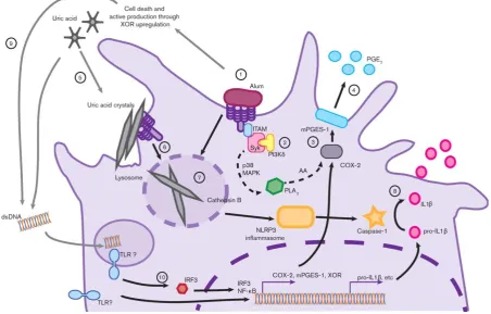
Recent studies have reported that Syk is also involved in pathogen-induced NLRP3
inflammasome activation and IL-1β secretion (212, 213) (Fig 1.6b). This response has been
seen for anti-fungal host defence (214), allergic sensitisation (215), alum, MSU and hemozoin
24
crystals (present in the joints of gout patients) (218). In neutrophils, LPS engagement of TLR4
recruits Syk and mediates inflammasome assembly and caspase-1 activation, explaining why
the LPS signal alone is sufficient to stimulate IL-1β secretion by neutrophils (219). This could
explain why neutrophils only need one signal while DCs and macrophages need two signals to
activate the inflammasome.
Neutrophils can produce large amounts of IL-10 after microbial stimulation due to
co-activation of MyD88 and Syk pathways (220). Simultaneous co-activation of TLR-MyD88 and
CLR-Syk dependent pathways led to prolonged phosphorylation of p38 MAPK and PI3 kinases
in neutrophils. Genetic deletion of Syk can attenuate antibacterial host defence in mice (221).
Mycobacterium tuberculosis can activate Syk through a number of CLRs although how
important this response is for host defence is unclear (202). The increased understanding of
how Syk can modulate the immune response has led to the emergence of Syk inhibitors as
promising therapeutic agents. For example, an oral SYK inhibitor Fostamatinib was tested in
a phase III clinical trial in patients with rheumatoid arthritis (222).
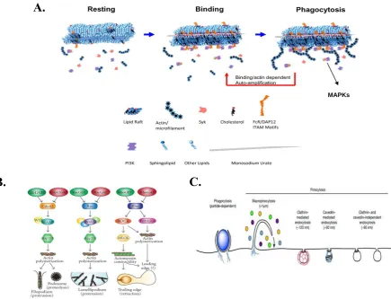
Recently, it has been suggested that the adjuvanticity of alum (and MSU crystals) could be
related to its engagement with cholesterol and other lipids on the plasma membrane, leading to
lipid sorting and forming of lipid rafts. Furthermore, this induces receptor independent
activation through the aggregation of immunoreceptor signalling motif (ITAM)-containing
25
Figure 1.6: Activation of Syk, downstream signalling molecules and cellular effects
A) Ligation of receptors leads to recruitment of Syk (ζ-chain associated protein kinase of
Zap70) to the plasma membrane through binding of tandem SH2 domains on Syk (or
Zap70) to phosphorylated tyrosine residues on the receptor. The tyrosine residues can
be on ITAMs, either present on the cytoplasmic tail of the receptor itself or present in
receptor associated transmembrane adaptors. Additionally, two hemITAMS on two
different associated receptors can also activate Syk. Further, Syk can be activated in an
ITAM independent manner. Taken from (191).
B) ITAM phosphorylation by SRC kinases leads to recruitment and activation of Syk,
leading to downstream signalling and activation of other kinase mediators, including
PI3K (triggers p85a subunit), NLRP3 inflammasome, p38, ERK, AKT, NF-κB.
Binding of Syk associated binding partners activates downstream signalling pathways,