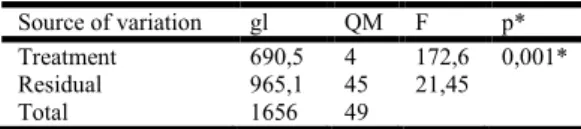
Full Length Research Article
INFLUENCE OF DIFFERENT LASER IRRADIATION ENERGY ON CERAMIC BOND STRENGTH
*
Danilo de S. Andrade, Fernanda A. Feitosa, Rodrigo M. de Araújo, Pablo L. Benitez, César R.
Pucci
São Paulo State University UNESP, Institute of Science and Technology, São Jose dos Campos,
São Paulo, Brazil 12245-000
ARTICLE INFO ABSTRACT
Objective: This study aimed to verify the influence of differentsNd:YAG laser energy parameters
on the bond strength between lithium disilicate ceramic and resin cement. Methods: One hundred
Lithium disilicate ceramic specimens with truncated cones shape were prepared and divided into 5 groups (n=10): 1- Control(without laser irradiation); 2- 80Nd – laser irradiation with Nd:YAG laser at 80mJ energy intensity for 1 min; 3- 100Nd - irradiation with Nd:YAG laser with an intensity of 100 mJ for 1 min; 4- 120Nd - Nd:YAG laser irradiation at 120mJ energy intensity parameters for 1 min; 5- 140Nd - Nd: YAG laser irradiation at 140mJ energy intensity parameters for 1 min. The laser irradiation on the specimen was performed in lower base of thetruncated cones shape. After all lasers treatments the groups were etched with hydrofluoric acid at 10% for 1 minute and silanized. Half of the treated ceramic specimens were cemented with resin cement (Variolink II-Ivoclar-Vivadent) to the other half resultingin hourglasses-shaped specimens.The bonded specimens were cycled thermomechanicallyand stressed to failure under tension.Data were analyzed using ANOVA and Tukey tests (α=0.05).
Results: One-way ANOVAindicated that the factor “laser energy”,statistically significant differences were observed among groups (p= 0.0), The 80Nd group (21.22 ± 6.00) showed greater results means than the other groups (Control-12.37 ± 3.46; 100Nd-15.15 ± 5.89; 120Nd-14.61 ± 2.53; 140Nd-10.12 ± 4.23)(Results in MPa). The other groups, including the control, were not statistically different.
Conclusions: Laser energy intensity parameter significantly affected tensile strength of the resin cement to the lithium disilicate ceramic.
Copyright© 2017,Danilo de S. Andrade et al.This is an open access article distributed under the Creative Commons Attribution License, which permits unrestricted use, distribution, and reproduction in any medium, provided the original work is properly cited.
INTRODUCTION
Using an adhesive cementation strategy for treatments involving pure ceramics, generated the need of bond improvement between the resin cement and the ceramic substrate. In the last decade several studies have been
conducted to bring solutions to this problem (Carvalho et al.,
2011; Cavalcanti et al., 2009; Demir et al., 2012; Kirmali et
al., 2014). Among the treatments evidenced are sandblasting
with aluminum oxide particles (5.6), the silane drying with hot air (1.7) and the irradiation on surfaces with high power laser such as Er:YAG laser (erbium-doped: yttrium aluminum garnet) and Nd:YAG laser (neodymium-doped: yttrium aluminum garnet).Some studies have indicated that the use of Nd:YAG laser increase the roughness on the surface and bond strength improvement in adhesive resin bonding (Cavalcanti
et al., 2009; Demir et al., 2012; Foxton et al., 2011; Guarda
et al., 2013; Kirmali et al., 2014; Nagai, 2005).
Corresponding author: Danilo de S. Andrade,
São Paulo State University UNESP, Institute of Science and Technology, São Jose dos Campos, São Paulo, Brazil 12245-000.
Studies has shown that treatment of the ceramic surface with neodymium laser results in a higher bond strength between ceramic and resin cement when compared to conventional treatment with 10% hydrofluoric acid for 1 minute and
sandblasting with aluminum oxide particles (Akın et al., 2011;
Nagai et al., 2005).However, there are studies that reported
the influence of the use of Nd: YAG laser on the bond strength between pure ceramic and resin cement, doesn’t exist consensus on which parameters is indicated for the types of ceramics available in the market, so that its clinical use is still
restricted (Akın et al., 2011; Usumez et al., 2013). A gap
regarding this aspect relates to the fact that there are few studies in the treatment on vitreous ceramic surface of lithium di-silicate, one of the most used in current clinical with the disproportion in the number of articles about ceramics based
on zirconia (Akın et al., 2011; Cavalcanti et al., 2009; Demir
et al., 2012; Foxton et al., 2011; Ozcan et al., 2008; Usumez
et al., 2013). Due to the growing demonstration of the
advantages of Nd: YAG laser irradiation as a way of increasing the bond strength between resin cement and
ISSN: 2230-9926
International Journal of Development Research
Vol. 07, Issue, 06, pp.13080-13084, June,2017
DEVELOPMENT RESEARCH
Article History: Received 24th March, 2017
Received in revised form 19th April, 2017
Accepted 07th May, 2017
Published online 16th June, 2017
Key Words:
ceramic, together with the fact that there are few articles in the current literature relating lithium di-silicate ceramics with High power laser, this study have as objective to test different parameters of irradiation of the Nd: YAG laser on ceramic surface of lithium di-silicate and after cementation a simulation and mechanical tensile test, to verify the influence of the parameters tested in the bond strength between the substrates.
MATERIALS AND METHODS
Ethical aspects
This article does not contain any studies with human participants or animals performed by any of the authors.
Specimen preparation
One hundred lithium disilicate truncated cones (IPS e.max Press; Ivoclar Vivadent) were fabricated from low contraction wax (Renfert Geo; Renfert GmbH), using a 4-mm thick metal device with a 2-mm diameter wide base and a 4-mm diameter wide top surface. The specimens were heat-pressed using the
lost-wax technique according to the manufacturer´s
instructions (Esquivel-Upshaw et al., 2004; Valenti et al.,
2009).
The 100 ceramic specimens were randomly divided into 5 equal groups. All specimens were cleaned in ultrasonic bath with 96% isopropanol for 3 min. And then submitted to the proposed treatments:
Control. Specimens were etched with HF (60 sec), rinsed
with water spray for 60 sec, silanized with Monobond Plus (Ivoclar Vivadent) for 60 sec and air-dried.
80Nd. Specimens were irradiated using a Nd:YAG laser
(Pulse Master 600 IQ; American Dental Technologies) with 80 mJ energy, using a pulse repetition rate set at 15 pps, using a 320 µm diameter laser optical fiber placed in contact with the specimen surface for 1 min with water spray cooling (5 sec). After irradiation, as surfaces were cleaned on ultrasonic bath with distilled water for 3 min. Following a surface was conditioned for 1 min. With 10% hydrofluoric acid, and silane actively for 60s followed by air-jet drying.
100Nd. The process is similar to those described in the
second group.Specimens were irradiated using a Nd:YAG laser with 100 mJ energy intensity.
120Nd. The process is similar to those described in the
second group.Specimens were irradiated using a Nd:YAG laser with 120 mJ energy intensity.
140Nd. The process is similar to those described in the
second group.Specimens were irradiated using a Nd:YAG laser with 140 mJ energy intensity.
Bonding procedures
After surfaces treatment, the specimens were joined by applying the Variolink II dual resin cement (IvoclarVivadent, Liechtenstein), handled in a ratio of 1:1 for 10s, following the manufacturer's recommendations resulting in 10 specimens per group (n=10). Each set was positioned in adapted eyeliner to the cementation with the bonding surface perpendicular to the application of a static vertical load of 630g.
After positioning of the ceramic / cement / ceramic set, a photopolymerization with LED light curing (Radii cal - SDI) was light-cured for 40s, starting at the proximal edges of each side of the set, obtaining 50 sets (n/2) (Figure 1).
Thermomechanical aging
The bonded specimens were subjected to thermomechanical challenge that consisted of 24,000 mechanical cycles (30 N load at 4 Hz) and 1,000 thermal cycles (30 sec dwell-time in each water bath with temperatures maintained at 5°C, 37°C and 55°C, respectively). Aging was conducted simultaneously in a thermomechanical cycler (Model ER-37000; ERIOS, São Paulo, Brazil).
Tensile bond strength
Bond strength was evaluated in a universal testing machine (model DI-1000; EMICSão José dos Pinhais, Brazil) by attaching a specimen to a custom-made device (Figure 2), using a 10 kg load cell at a cross-head speed of 1 mm/min. Loading was performed in tension until failure. Results of tensile testing (in MPa) were statistically analyzed using one-factor analysis of variance (ANOVA) and Tukey after affirming that the normality and equal variance assumptions of the data were not violated.
The null hypotheses tested were.
H0: The alteration of the irradiation parameters of the Nd:
YAG laser does not interfere in the bond strength between ceramic and resin cement.
H1: Nd: YAG laser irradiation followed by conditioning with 10% hydrofluoric acid gives lower tensile strength results than treatment with only 10% hydrofluoric acid.
RESULTS
Analysis of fractured interface
The failures due to the mechanical tensile test were predominantly adhesive, and the cohesive failures in ceramic (one in the 80Nd group and one in the 120Nd group) were disregarded from the statistical test.
Tensile bond strength
The analysis of the 1-factor ANOVA test showed significant differences between the different groups studied (p value <5%). The 1-factor ANOVA test results are showed in Table 1.
Table 1. Variance Analysis Results - ANOVA 1 factor.
Source of variation gl QM F p* Treatment 690,5 4 172,6 0,001* Residual 965,1 45 21,45
Total 1656 49
*p<5%
Table 2. Tukey's test for the comparison between the different irradiation parameters of the Nd: YAG laser on the ceramic
surface
Groups Means (Mpa) Groups Homogeneous
140Nd 10,12 A
Control 12,37 A
120Nd 14,62 A
100Nd 15,15 A
80Nd 21,22 B
Figure 1 - Final view of ceramic specimens cemented with resin cement
Figure 2 - Adaptation of the ceramic specimens in the metallic model for mechanical testing
According to the Tukey test result for the different irradiation parameters of the Nd: YAG laser on the ceramic surface it can be observed that the 80Nd group was statistically superior to the other groups, while all other groups were similar to each other.
Analysis of Treated Ceramic Surfaces
Two samples referring to each of the groups were submitted to Scanning Electron Microscopy (SEM) (Figures 3-7) for qualitative analysis of the surfaces.The photomicrographs show that there was no change in the morphological pattern of the ceramics for any group, but in the groups 100Nd and 120Nd it is possible to verify the appearance of irregularities that appear to arise from the pulses of laser irradiation.
Figure 3 - Control group with a 2000x magnification, showing the conventional microstructure of the ceramic used, with needle type lithium disilicate crystals with the appearance of agglutination
with each other and in the vitreous matrix in which they are embedded.
Figure 4 - Group 80Nd with 2000x magnification, showing structural normality, but with a higher exposure of lithium
disilicate crystals
Figure 5. Group 100Nd with increase of 2000x. Is possible to detect striking surface irregularities due to the laser emission
pulses
Figure 6 - Group 120Nd with increase of 2000x. Surface irregularities due to laser emission pulses are even more evidente
Figure 7. Group 140Nd with increase of 2000x. The image suggests the presence of deep irregularities and cracks
DISCUSSION
The results obtained in current study demonstrate an increase in bond strength values between lithium disilicate vitreous ceramic and resin cement after irradiation of ceramic surface with Nd: YAG laser in the parameters of 80mJ of energy intensity and 15Hz of frequency with Constant air cooling followed by conditioning with 10% hydrofluoric acid for 1 minute.Therefore, the null hypothesis was not accepted, since the alteration of the irradiation parameters of the Nd: YAG laser interfered in the bond strength between ceramic and resin cement, and the irradiation with Nd: YAG laser followed by the conditioning with hydrofluoric acid to 10 % gave results of tensile strength higher than treatment with only 10% hydrofluoric acid.
The lithium disilicate ceramic IPS E.max Press has indication for anterior and posterior regions, with the possibility of using the same material for infrastructure and cover.The choice of this ceramics for the preparation of the specimens was based on its extensive clinical use, which has been showing positive results regarding the duration rate in the buccal medium, low
index of fractures besides good color maintenance (Etman et
al., 2010; Taskonak, 2006;Toman et al., 2015). Studies have
shown that the area of the adhesive surface to be tested is
directly proportional to the type of failure obtained (Sano et
al., 1994; Sano et al., 1994). Larger surfaces usually result in
a prevalence of cohesive failures, while cross-sections of less
than 2mm result in most adhesive failures (Sano et al., 1994;
Sano et al., 1994; Ghassemieh et al., 2008). According to
the literature, the smaller the surface of the specimen, the smaller the area available for failure, resulting in better bond
strength results (Escribano et al., 2003). The larger the test
area, the lower the adhesive strength, and up to 3mm the
results are more favorable for macrotracking tests (Kara et
al., 2012). In order to obtain results closer to the reality of the
adhesive resistance between the materials and based on the parameters reported above, in this study was used a model of test specimen of small dimensions, with cementation area of 2mm in diameter, made in single body and Prepared for a macrotrace test with the smallest possible dimensions without the disadvantages of samples obtained from various sections of a test piece (sticks), but with the advantage of the test in a small area. The faults obtained from the macrotrace test were mostly adhesive (there was only 1 cohesive failure in ceramic in the 80Nd group and 1 in the 120Nd group).
This result corroborates with that described by Sano et al.,
1994 since the size of the surface possibly influenced the type of failure obtained. The results concerning cohesive failures were removed from the statistical analysis of union resistance since these do not adequately represent their groups and maintaining them could induce a false result.In the present study 10% hydrofluoric acid was used for 1 minute (control group), however the means found were lower than those in the 80Nd, 100Nd and 120Nd groups. A previous study showed that the use of 10% HF created clear pores and exposure of crystals of disilicate due to the marked dissolution of the
vitreous matrix (Prochnow et al., 2016).These data indicate a
probable increase in the surface roughness of the ceramic, which acts as an element favorable to the bond strength. SEM photomicrographs suggest areas of agglutination between crystals of lithium di-silicate and vitreous matrix in the control group (Figure 3), while in the 80Nd, 100Nd and 120Nd groups it is possible to visualize a larger exposure of the surface crystals, which may have interfered with In the quality of the union (Figures 4-6).
The best results of bond strength were found in the irradiated group with 80mJ of Energy intensity and 15Hz of frequency). According to Tukey's test, the 80Nd group was the only one that differed statistically from the others. These results differ
from those found by Akyil et al., 2011, where comparing
different surface treatments in feldspathic ceramics found that surface treatment with hydrofluoric acid was more effective when compared to the use of Nd: YAG and Er: YAG lasers and Even when applied to the surface after irradiation with
both lasers (Akyıl et al., 2011). SEM photomicrograph for the
140Nd group shows areas that suggest cracks and faults in the ceramic surface (Figure 7). The slits also appear in the study of
Liu et al., 2015, when using the Nd: YAG laser in the
parameters of 100mJ and 1 and 2W for the irradiation of the zirconia surface. However, as mentioned previously, the surfaces of zirconia and lithium disilicate have very different structures, which makes comparison impossible. In this study, the cementation of two identical ceramic substrates was used with the purpose of not include another substrate that could provide, for example, cohesive failure in the non-ceramic substrate, compromising the work goal and masking the obtained results. Although the results obtained by this research regarding the bond strength of the vitreous ceramics of lithium di-silicate to resin cement can not be directly extrapolated to a clinical situation, we can suggest them for new strategies of surface treatment of this type of ceramic a So that indirect restorations with longer longevity can be performed.
Conclusion
The results of this study allow us to conclude that irradiation with Nd: YAG laser in the parameters of 80mJ of energy intensity and 15 Hz of frequency with constant cooling air followed by conditioning with 10% hydrofluoric acid for 1 minute is the most suitable for Vitreous ceramic of lithium disilicate used among the treatments tested, being superior to the treatment with only 10% hydrofluoric acid for 1 minute and to the other parameters tested.
REFERENCES
Various Laser Treatments. Photomedicine and Laser Surgery. 2011;29(12):797-802.
Akyıl MŞ, Yılmaz A, Bayındır F, Duymuş ZY. Microtensile bond strength of resin cement to a feldspathic ceramic. Photomed Laser Surg.2011Mar;29(3):197-203.
Carvalho RF, Martins ME, de Queiroz JR, Leite FP, Ozcan M. Influence of silane heat treatment on bond strength of resin cement to a feldspathic ceramic. Dent Mater J.2011;30(3):392-7.
Cavalcanti AN, Foxton RM, Watson TF, Oliveira MT, Giannini M, Marchi GM. Bond Strength of Resin Cements to a Zirconia Ceramic with Different Surface Treatments. Operative dentistry. 2009;34(3):280-7. Demir N, Subaşi MG, Ozturk AN. Surface roughness and
morphologic changes of zirconia following different
surface treatments. Photomed Laser
Surg. 2012 Jun;30(6):339-45.
Escribano NI, Del-Nero MO, de la Macorra JC. Inverse relationship between tensile bond strength and dimensions of bonded area. J Biomed Mater Res B Appl Biomater. 2003 Jul 15; 66(1): 419-24.
Esquivel-Upshaw JF, Anusavice KJ, Young H, Jones J, Gibbs C. Clinical performance of a lithiadisilicate-based core
ceramic for three-unit posterior FPDs. Int J
Prosthodont. 2004 Jul-Aug;17(4):469-75.
Etman MK, Woolford MJ. Three-year clinical evaluation of two ceramic crown systems: a preliminary study. J Prosthet Dent. 2010 Feb;103(2):80-90.
Foxton RM, Cavalcanti NA, Nakajima M, Pilecki P, Sheriff M, Melo L, Watson TF. Durability or resin cement bond
to aluminum oxide and
zircôniaceramics after air abrasion and laser treatment. J
Prosthodont. 2011 Feb;20(2):84-92.
Ghassemieh E. Evaluation of sources of uncertainties in microtensile bond strength of dental adhesive system for
different specimen geometries.Dent Mater. 2008
Apr;24(4):536-47.
Guarda GB, Correr AB, Gonçalves LS, Costa AR, Borges GA, Sinhoreti MA, Correr-Sobrinho L. Effects of surface treatments, thermocycling, and cyclic loading on the bond strength of a resin cement bonded to a lithium disilicate glass ceramic. Oper Dent. 2013 Mar-Apr;38(2):208-17. Kara HB, Dilber E, Koc O, Ozturk AN, Bulbul M. Effect of
different surface treatments on roughness of IPS Empress 2 ceramic. Lasers Med Sci. 2012 Mar;27(2):267-72.
Kirmali O, Akin H, Kapdan A. Evaluation of the surface roughness of zirconia ceramics after different surface treatments. Acta Odontologica Scandinavica. 2014; 72: 432–439.
Liu L, Liu S, Song X, Zhu Q, Zhang W. Effect of Nd:YAG laser irradiation on surface properties and bond strength of zirconia ceramics. Lasers Med Sci. 2015; 30:627-34. Nagai T, Kawamoto Y, Kakehashi Y, Matsumara H. Adhesive
bonding of a lithium disilicate ceramic material with resin-based luting agents. J Oral Rehabil. 2005 Aug;32(8):598-605.
Ozcan M, Nijhuis H, Valandro LF. Effect of various surface conditioning methods on the adhesion of dual-cure resin cement with MDP functional monomer to zirconia after thermal aging. Dent Mater J. 2008 Jan;27(1):99-104.
Prochnow C, Venturini AB, Grasel, Bottino MC, ValandroLf.
Effect of etching with distinct hydrofluoric acid concentration on the flexural strenght of a lithium disilicates based glass ceramic. J Biomed Res B Appl Biomater. 2016 Feb 5. doi: 10.1002/jbm.b.33619.
Sano H, Ciucchi B, Matthews WG, Pashley DH.
Tensile properties of mineralized and demineralized
human and bovine dentin.J Dent
Res. 1994 Jun;73(6):1205-11.
Sano H, Shono T, Sonoda H, Takatsu T, Ciucchi B, Carvalho R, Pashley DH. Relationship between surface area for adhesion and tensile bond strength--evaluation of a micro-tensile bond test.Dent Mater. 1994 Jul;10(4):236-40. Taskonak B, Sertgöz A. Two-year clinical evaluation of
lithia-disilicate-based all-ceramic crowns and fixed partial dentures. Dent Mater. 2006 Nov;22(11):1008-13.
Toman M, Toksavul S. Clinical evaluation of 121 lithium disilicate all-ceramic crowns up to 9 years. Quintessence Int. 2015 Mar;46(3):189-97.
Usumez A, Hamdemirci N, Koroglu BY, Simsek I, Parlar O, Sari T. Bond strength of resin cement to zirconia ceramic with different surface treatments. Lasers Med Sci.2013 Jan;28(1):259-66.
Valenti M, Valenti A. Retrospective survival analysis of 261 lithium dissilicate crowns in a private general practice. Quintessence Int. 2009 Jul-Aug;40(7):573-9.