R E S E A R C H A R T I C L E
Open Access
Mining the deep Red-Sea brine pool
microbial community for anticancer
therapeutics
Luke Esau
1, Guishan Zhang
2,3, Sunil Sagar
1, Ulrich Stingl
3,4, Vladimir B. Bajic
1and Mandeep Kaur
1,5*Abstract
Background:Microbial species in the brine pools of the Red Sea and the brine pool-seawater interfaces are exposed to high temperature, high salinity, low oxygen levels and high concentrations of heavy metals. As adaptations to these harsh conditions require a large suite of secondary metabolites, these microbes have a huge potential as a source of novel anticancer molecules.
Methods:A total of 60 ethyl-acetate extracts of newly isolated strains from extreme environments of the Red-Sea were isolated and tested against several human cancer cell lines for potential cytotoxic and apoptotic activities.
Results:Isolates from the Erba brine-pool accounted for 50% of active bacterial extracts capable of inducing 30% or greater inhibition of cell growth. Among the 60 extracts screened, seven showed selectivity towards triple negative BT20 cells compared to normal fibroblasts.
Conclusion:In this study, we identified several extracts able to induce caspase-dependent apoptosis in various cancer cell lines. Further investigations and isolation of the active compounds of these Red Sea brine pool microbes may offer a chemotherapeutic potential for cancers with limited treatment options.
Keywords:Deep Red-Sea, Triple negative breast cancer, Brine pools, Anticancer
Background
The hallmarks of cancer, which categorises the survival and proliferative mechanisms of cancer cells, was first described by Hanahan and Weinberg [1]. Although these aspects of cancer are well studied, and even with the advances in personalized medicine for patient tailored treatment, there still exists cancer forms for which limited treatment options are available. Breast cancer, the most prevalent cancer in women globally, has an estimated 1.67 million cases diagnosed in 2012 [2]. Women of Arab [3] and African descent [4] present breast cancer at an earlier age, have more aggressive tumour types with a high prevalence of triple negative breast cancer. Penta-negative tumours (i.e., negative for
the estrogen and progesterone receptors, EGFR, HER2, and cytokeratin 5/6) have also been reported in Saudi women [5]. In our previous seminal investigations [6, 7], we reported the anticancer potential of extracts obtained from brine-pool microbes. Most of these extracts were active against estrogen receptor positive (ER+) breast cancer cells. Since the triple-negative type of breast cancer is a more aggressive form and as currently no therapies are available, we aimed to identify new sources of anticancer compounds that can pave the way to de-velop novel therapies for triple negative breast cancer.
Approximately 60 % of current anticancer therapeutics are derived from natural products, including, for ex-ample, marine-derived compounds such as cytarabine, trabectedin, eribulin, and dolastatins [8]. A review by Agrawal et al. [9] described nonribosomal peptides iso-lated from marine microbes having anticancer activity while in a research article Neelam et al. [10] discovered a marine halo-alkaliphilic bacteria species possessing anti breast cancer activity. This evidence supports the
© The Author(s). 2019Open AccessThis article is distributed under the terms of the Creative Commons Attribution 4.0 International License (http://creativecommons.org/licenses/by/4.0/), which permits unrestricted use, distribution, and reproduction in any medium, provided you give appropriate credit to the original author(s) and the source, provide a link to the Creative Commons license, and indicate if changes were made. The Creative Commons Public Domain Dedication waiver (http://creativecommons.org/publicdomain/zero/1.0/) applies to the data made available in this article, unless otherwise stated. * Correspondence:[email protected]
1King Abdullah University of Science and Technology (KAUST), Computational Bioscience Research Center (CBRC), Thuwal, Jeddah 23955-6900, Saudi Arabia
5School of Molecular and Cell Biology, University of the Witwatersrand, Johannesburg 2050, South Africa
pursuit of mining marine environments for the discovery of new anticancer agents. Twenty-five (mostly) anaerobic deep-sea brine pools with extremely high salt concentra-tions have been reported in the Red Sea. Multi-extremophilic microbes that inhabit these environments are not only adapted to high salinity (4–26%), but also to elevated temperature, low oxygen concentrations, and high concentrations of heavy metals [11–14]. These extreme marine environmental conditions favour the production of secondary metabolites and thus potentially unique and potent natural compounds. Extremophilic marine bacterial species from these environments, there-fore, present a unique opportunity for discovering novel anticancer compounds [6, 7] that address the ever-changing need for improved chemotherapeutic drugs.
This study reports on anticancer activity of extracts from bacterial isolates from different habitats around brine pools within the Red Sea. We screened a total of 60 extracts against seven cell lines representing colorec-tal carcinoma, fibrosarcoma, breast carcinoma, cervical carcinoma, neuroblastoma, and normal cell lines. After initial screening, active extracts (> 30% growth inhib-ition) were selected for testing in selected cell lines to investigate if apoptotic activities were moderated by caspases (executors of apoptotic cell death).
Methods Field sampling
The inoculum for microbial isolations were collected during a cruise between Oct 16 and Nov 3 2011 as described in Sagar et al. [7].
Source of bacterial isolates
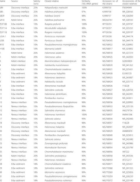
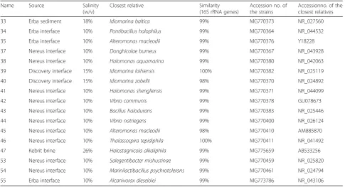
Sixty bacterial strains were isolated from different habitats in or around the brine pools (Table 1). While the deep-sea brine pool habitats differ in their physico-chemical characteristics [15], several studies showed that all of these environments harbour high microbial diver-sity and biomass.
PCR amplification
DNA extraction, PCR amplification, and bioinformatics analyses of 16S rRNA genes from biomass of bacterial strains was performed according to Sagar et al. [7]. Se-quences were deposited in Genbank, and accession numbers are listed in Table1.
Bacterial biomass
The inocula were streaked using three different media types as described by (Sagar et al. 2013 [6, 7]). These solidified media types were supplemented with either 10% or 15% or 20% or 26% NaCl (w/v) before autoclav-ing to mimic salt concentrations in their original habitat. All incubations were done in a Binder incubator (Type
BD53, Binder, Tuttlingen, Germany). Strains reported here were isolated under oxic conditions (air, 21% O2)
and atmospheric pressure (1 atm) described elsewhere [16,17]. To collect biomass, all strains were grown for 2 to 3 weeks with constant agitation at 30 °C in 5.0 l of Marine Broth (Difco) supplemented with the respective concentration of NaCl. Bacterial biomass was harvested and ethyl acetate extracts were prepared according to Sagar et al. [6,7].
Cell culturing
BJ (Fibroblast), HCT (Colorectal adenocarcinoma), HT-1080 (Fibrosarcoma), MCF-7 (Breast Adenocarcinoma), IMR-32 (Neuroblastoma), BT20 (Breast Adenocarcin-oma), and HeLa (Cervical carcinoma) were obtained from the American Type Cell Culture Collection (ATCC, Manassas, VA). The cells were cultured in DMEM (Dulbecco's Modified Eagle's Medium) contain-ing 10% FCS (Foetal calf serum), and streptomycin (100μg/mL) and penicillin (100 U/mL) in a 37 °C incu-bator supplying 5% CO2.
MTT assay
2.5 × 10³ cells were seeded per well in 384-well culture plates and treated with 200μg/mL marine bacterial extracts for 48 h. Growth inhibitory effects of extracts were estimated by an MTT (3-(4, 5-Dimethylthiazol-2-yl)-2, 5-diphenyltetrazolium bromide) assay as previously described [7]. A microtiter plate reader (BMG Labtech-PHERAstar FS, Germany) was used to measure the OD (optical density) at 595 nm and the results were analyzed using Microsoft Office Excel©.
APOPercentage assay
Cells were seeded in quadruplicates in 96 well plates at a density of 5 × 10³cells per well in 90μL of media. After 24 h, 200μg/mL extracts were added to the cells for 48 h with, while 30 min treatment with 10 mM H2O2 was
used as a positive control. The cells were lifted and stained with the APOPercentage dye (Biocolor, UK), and analysed as described previously [18].
Caspase-3/7 activity assay
Table 1Identification of microbial stains. Taxonomic identification based on 16S rRNA gene analysis and the source of inoculum for 60 microbial strains
Name Source Salinity
(w/v)
Closest relative Similarity
(16S rRNA genes)
Accession no. of the strains
Accessionno. of the closest relatives
SB9 Discovery interface 25% Haloprofundus marisrubri 100% KJ999759 FN594944
SB3 Discovery interface 25% Haloferax prahovense 97% KJ999758 NR_028165
SB29 Discovery interface 25% Haloferax larsenii 98% KJ999757 NR_028209
SA10 Kebrit brine 26% Haloferax prahovense 99% MG563761 NR_028165
ZGT108 Erba interface 10% Ruegeria profundi 100% KP726355 NR_029197
ZGT114 Erba interface 10% Microbulbifer salipaludis 98% KP726357 NR_025232
ZGT118 Erba interface 10% Ruegeria marisrubri 100% KP726356 NR_029197
SJ5A-1 Erba interface 10% Ponticoccus marisrubri 97% KP726358 NR_044174
SJ5B Erba interface 10% Ponticoccus litoralis 99% MG764545 NR_044174
XI10 Erba interface 10% Pseudoalteromonas mariniglutinosa 99% MG768922 NR_028992
H106 Erba interface 10% Idiomarina zobellii 99% MG768917 NR_024892
1 Kebrit brine 26% Halomonas axialensis 99% MG768918 NR_027219
2 Kebrit interface 20% Halomonas salina 99% MG768925 NR_042050
3 Kebrit interface 20% Marinimicrobium haloxylanilyticum 99% MG768919 GQ920839
4 Kebrit interface 20% Halobacillus kuroshimensis 99% MG768920 NR_041262
5 Erba interface 10% Chromohalobacter israelensis 99% MG768921 NR_025431
6 Erba sediment 18% Alteromonas halophila 99% MG768928 EU583725
7 Erba sediment 18% Halomonas taeanensis 98% MG768923 NR_043087
8 Erba sediment 18% Halobacillus locisalis 99% MG768924 NR_025715
9 Erba interface 10% Alteromonas macleodii 99% MG768926 Y18228
10 Erba interface 10% Salinivibrio costicola. 99% MG768927 NR_028703
11 Erba interface 10% Halomonas denitrificans 99% MG768930 NR_042491
12 Erba interface 10% Pontibacillus marinus 99% MG768932 NR_043011
13 Nereus interface 10% Pseudoalteromonas mariniglutinosa 98% MG768936 NR_028992
14 Nereus interface 10% Pseudoalteromonas flavipulchra 99% MG768933 NR_025126
15 Nereus interface 10% Salinivibrio sharmensis 99% MG768935 AM279734
16 Nereus interface 10% Halomonas hamiltonii 100% MG768937 AM941396
17 Nereus interface 10% Salinicola salarius 99% MG768934 NR_042490
18 Discovery interface 15% Alteromonas macleodii 98% MG768957 Y18228
19 Discovery interface 15% Halomonas halophila 99% MG768941 NR_042697
20 Discovery interface 15% Alteromonas macleodii 97% MG768929 AM885870
21 Discovery interface 15% Pontibacillus chungwhensis 98% MG768940 NR_025812
22 Nereus interface 10% Salinicola salarius 98% MG768944 NR_042490
23 Nereus interface 10% Zunongwangia profunda 99% MG768951 NR_043986
24 Nereus interface 10% Marinobacter flavimaris 99% MG768954 NR_025799
25 Nereus interface 10% Chromohalobacter marismortui 99% MG768956 X87222
26 Kebrit interface 20% Salinivibrio proteolyticus 99% MG768958 NR_043536
27 Nereus interface 10% Halomonas meridiana 99% MG768959 AF212217
28 Erba sediment 18% Chromohalobacter israelensis 98% MG768971 NR_025431
29 Erba sediment 18% Salinivibrio siamensis 99% MG770368 NR_041552
30 Erba sediment 18% Idiomarina seosinensis 99% MG770369 NR_025826
31 Erba sediment 18% Pseudoalteromonas carrageenovora 99% MG770359 NR_026220
Statistical analysis
The samples (untreated vs. treated) were compared by using Student’s t-test and statistical significance was noted atp< 0.05. A Z-score of≥0.6 indicated robustness of assays [15].
Results
Taxonomic classification of microbes isolated from the Red Sea
Most bacteria isolated from the four brine pools Erba, Discovery, Kebrit, and Nereus were closely related to known and well described halophilic species within ProteobacteriaandArchaea(Table1).
Anticancer activities of isolates in a panel of cell lines
A total of 60 extracts isolated from bacterial cultures from the four brine pools Erba, Discovery, Kebrait, and Nereus were screened for anticancer activity by deter-mining cell growth inhibition through MTT assay (Table 2). Extracts induced varying levels of growth in-hibition and were classified into five groups - empty cir-cle (< 30%), quarter-moon (> 30 and < 40%), half-moon (> 40 and < 60%), three-quarter-moon (> 60 and < 80%) and full-moon (> 80%). The skin fibroblast cell line BJ was used as a normal cell line control for screening anti-cancer activity of the extracts. BJ cell growth was inhib-ited by approximately 50% of extracts while the growth of neuroblastoma cell line IMR-32 remained largely insensitive to the treatment. Interestingly, the growth of
the triple negative breast cancer cell line BT20 was sensitive to the majority of the extracts.
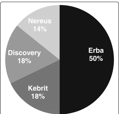
Further analysis revealed that the majority of bacterial isolates inducing active growth inhibition (above 30%) in this study set were isolated from the Erba brine pool (Fig. 1). Figure 1 represents the distribution of active bacterial extracts inducing greater than 30% growth in-hibition found across the brine-pools.
Apoptosis as mode of anticancer activity
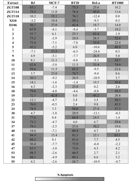
We assessed phosphatidylserine exposure in cancer cells treated with selected microbial extracts using APOPer-centage assay and monitored change in caspase-3/7 activity to determine if extracts induced apoptosis. For this purpose, the extracts inducing > 30% growth inhib-ition were selected for apoptosis screening (Table 3). Due to the availability of limited amount of microbial extracts, we performed apoptosis and caspase-3/7 activ-ity assays only on extracts active in breast cancer, cer-vical cancer, and fibrosarcoma cell lines. Again, BJ cells were used as a control to identify extracts with selective anticancer activity. Extracts induced apoptosis in all cell lines. However four (ZGT118, XI10, 7 and 13), six (ZGT118, XI10, 13, 16, 30 and 55), one (9), and three (10, 15 and 22) extracts selectively induced apoptosis in MCF-7, BT20, HeLa and H T-1080 cells compared to BJ cells, respectively. The extract number 7 was only active against MCF-7 cells, whereas extracts 16 and 55 were selectively active against BT20 cells when the apoptosis-Table 1Identification of microbial stains. Taxonomic identification based on 16S rRNA gene analysis and the source of inoculum for 60 microbial strains(Continued)
Name Source Salinity
(w/v)
Closest relative Similarity
(16S rRNA genes)
Accession no. of the strains
Accessionno. of the closest relatives
33 Erba sediment 18% Idiomarina baltica 99% MG770373 NR_027560
34 Erba interface 10% Pontibacillus halophilus 99% MG770364 NR_044532
35 Erba interface 10% Alteromonas macleodii 99% MG770376 Y18228
37 Nereus interface 10% Donghicolae burneus 99% MG770367 NR_043928
38 Nereus interface 10% Halomonas aquamarina 99% MG770380 NR_042063
39 Discovery interface 15% Idiomarina loihiensis 100% MG770382 NR_025119
40 Discovery interface 15% Idiomarina zobellii 98% MG770370 NR_024892
41 Nereus interface 10% Halomonas shengliensis 99% MG770371 NR_044099
42 Nereus interface 10% Vibrio communis 99% MG770378 GU078673
43 Nereus interface 10% Bacillus halodurans 99% MG770383 NR_025446
44 Nereus interface 10% Vibrio natriegens 99% MG770400 NR_026124
45 Nereus interface 10% Alteromonas macleodii 98% MG770410 AM885870
46 Nereus interface 10% Thalassospira tepidiphila 100% MG770411 NR_041492
47 Kebrit brine 26% Halostagnicola alkaliphila 99% MG775659 AB533256
53 Nereus interface 10% Salegentibacter mishustinae 99% MG770459 NR_025820
54 Nereus interface 10% Marinilactibacillus psychrotolerans 99% MG770461 NR_024794
[image:4.595.56.542.109.372.2]inducing potential of these extracts was compared among all five cell lines (Table3).
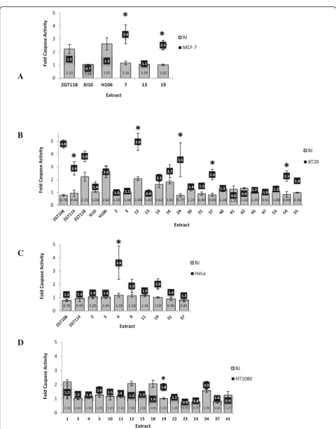
A Caspase-3/7 activity assay was employed to gain insights into the type of apoptosis occurring, namely caspase-dependent or caspase-independent (Fig. 2a-d). The caspase-3/7 results showed a clear trend that a caspase-independent mechanism was mostly responsible for BJ cell death. Extract number 7 induced caspase-dependent apoptosis in MCF-7 cells but had no adverse effect on BJ cell death despite it inducing slight growth inhibition in BJ cells making it a promising drug candi-date for future work. Extract number 19 induced caspase-dependent apoptosis in MCF-7, HeLa, and HT1080 cells but not in BJ cells, even though BJ cells stained positive for phosphatidylserine exposure by APOPercentage assay (Table 3). Caspase-3/7 activity increased in BT20 cells compared to BJ cells in response to extracts ZGT108, ZGT114, 12, 24, 37, 54 and 55. HeLa cells and HT1080 cells displayed significantly in-creased caspase-3/7 activation in response to extracts 4 and 19, respectively (Fig. 2). Interestingly, extract num-ber 55 showed selectivity towards BT20 cells by inhibit-ing its growth and inducinhibit-ing apoptosis via caspase-3/7, neither of which was observed for BJ cells.
Discussion
As a follow-up from our previous studies [6, 7], we screened extracts from 60 marine bacteria isolated from brine pools of the Red Sea. We would like to emphasize
that in our previous two investigations, we could only find extracts active against MCF-7 (ER+) cells, but the current study reports anticancer activities of the extracts isolated from the Red Sea brine-pool harboring bacteria against fibrosarcoma, cervical cancer and particularly BT20 (triple negative) cancer cells. Primary cytotoxicity screening of all extracts against seven cell lines representing five different cancers enabled us to broadly identify potential anticancer extracts from deep-sea microbes. All extracts were also screened against one normal fibroblast cell line (BJ) to further identify those who exhibited selectivity for cancer, but not normal cells.
We investigated whether the brine-pool location had any correlation with anticancer activity induced by the bacterial isolates. Of all isolates from the respective brine-pools, we found that about 50% of all isolates collected from Erba Deep (located at a depth of 2395 m), showed greater than 30% growth inhibition (Fig.1). The most closely related validly described species to these strains are (Table 1): Chromohalobacter israelensis, Salinivibrio siamensis, Idiomarina seosinensis, Pseudoal-teromona scarrageenovora, Pontibacillus halophilus, and Alteromonas macleodii. Our previous work [7] has reported anticancer activity of Chromohalobacter israe-lensis in HeLa cells. A PubMed search did not reveal even a single article that describes the anticancer activity of any of the other five bacterial species. This highlights the fact that the microbes found in the deep-sea brine pools of the Red Sea (especially Erba Deep) may have unique anticancer compounds that can be explored in the future to develop new drugs.
Apoptosis assays (APOPercentage and caspase-3/7 ac-tivity) confirmed that several of these selected (showing > 30% cell growth inhibition) extracts induced apoptotic cell death in cancer cell lines. This secondary screening process identified the extracts that selectively targeted a specific type of cancer via apoptotic cell death. We further investigated if extracts from a particular bacterial species have anticancer activity towards a specific cell line. Intriguingly, all three extracts (10, 15 and 22) that specifically inhibited the growth of fibrosarcoma cells (HT-1080) belonged to strains that were closely related to Salinivibrio costicola, Salinivibrio sharmensis, and Salinicola salaries, respectively, showing an enrichment of genusSalinivibrio.It would be interesting in future to investigate Salinivibrio extracts against other sarcomas as well. In MCF-7 cells, two out of four most active extracts (XI10 and 13) belonged to Pseudoalteromonas mariniglutinosa. This bacterial species had not been tested so far for anticancer activities (PubMed search). Secondary metabolites isolated from Psuedoalteromonas sp. off the coast of Brazil were reported to have potent anticancer activity against a leukemic and melanoma cell line, and the active compound, prodigiosin, was shown
[image:6.595.56.291.85.309.2]A
B
C
D
[image:8.595.57.538.79.692.2]to be selective for cancers overexpressing ErbB-2 [19]. In BT20 cells, extracts (16 and 55) showed selective cell death, and these extracts belong to species Halomonas hamiltoniiand Alcanivorax dieselolei, respectively, none of which have ever been shown to have anticancer activ-ity before. Our observation that Halomonas sp. and Alteromonas sp. contributed to the majority of growth inhibition may be attributed to their ability to produce molecules including exopolysaccharides (EPSs) and Dithiolopyrrolone (DTP), respectively. Exopolysacchar-ides (heterogeneous polymers) isolated fromHalomonas stenophila and Halomonas smyrnensis induced pro-apoptotic effects against human T-leukemia cells [20] and breast cancer MCF-7 cells [21].Alteromonassp. are also well known for producing dithiolopyrrolone (DTP) molecules, which are known potent natural antibiotics; DTP obtained FDA approval as topical antibiotic Bactro-ban® (GlaxoSmithKline) [22]. Not surprising, DTP also exhibits potent anticancer activity. These classes of anti-biotic or polysaccharide type molecules most probably explain the anticancer activity we observed in this study.
In conclusion, our study has identified several micro-bial species that have the potential to kill selectively cancer cells, and interestingly many of these species have never been previously tested for their anticancer activities. Here, we provide seminal baseline data pin-pointing which bacterial species and brine-pools should be targeted for future investigations to isolate anticancer compounds. This work is of particular importance for triple negative breast cancer therapeutic development as no drug exists till date that can effectively cure this aggressive form of breast cancer. Testing of these marine extracts against penta-negative breast cancer cells should be of great interest to future studies.
Abbreviations
DMEM:Dulbecco’s Modified Eagle’s Medium; DTP: Dithiolopyrrolone; ER: Estrogen receptor; FCS: Foetal calf serum; MTT: 3-(4, 5-Dimethylthiazol-2-yl)-2, 5-diphenyltetrazolium bromide; OD: Optical density;
PS: Phosphatidylserine
Acknowledgements
Authors want to thank Dr. Andre Antunes and Dr. Tyas Hikmawan for collecting samples.
Authors’contributions
LE and MK performed biological testing experiments and wrote the manuscript. LE analysed data and compiled tables/figures. SS prepared the extracts for biological evaluations and planned the study with MK. GZ grew the strains in large batches, isolated the strains and provided the taxonomic classification. US planned the exhibition and the cultivation experiments along with the writing of the manuscript. VBB provided general coordination of the study. All authors read and approved the final manuscript.
Funding
Authors thank SEDCO for funding this research.
Availability of data and materials
All 16S rRNA gene sequences of newly isolated strains from this study have been deposited in Genbank. Please refer to Table1for accession numbers.
Ethics approval and consent to participate
Not applicable.
Consent for publication
Not applicable.
Competing interests
The authors declare that they have no competing interests.
Author details
1King Abdullah University of Science and Technology (KAUST), Computational Bioscience Research Center (CBRC), Thuwal, Jeddah 23955-6900, Saudi Arabia.2Key Laboratory of Microbial Resources Collection and Preservation, Ministry of Agriculture, Institute of Agricultural Resources and Regional Planning, Chinese Academy of Agricultural Sciences, Beijing 100081, People’s Republic of China.3King Abdullah University of Science and Technology, Red Sea Research Center, Thuwal, Jeddah 23955-6900, Saudi Arabia.4Department of Microbiology and Cell Science, Institute of Food and Agricultural Sciences, University of Florida, UF/IFAS Fort Lauderdale Research and Education Center, Davie, FL 33314, USA.5School of Molecular and Cell Biology, University of the Witwatersrand, Johannesburg 2050, South Africa.
Received: 24 November 2017 Accepted: 9 June 2019
References
1. Hanahan D, Weinberg RA. Hallmarks of cancer: the next generation. Cell. 2011;144(5):646–74.
2. Ferlay J, Soerjomataram I, Dikshit R, Eser S, Mathers C, Rebelo M, Parkin DM, Forman D, Bray F. Cancer incidence and mortality worldwide: sources, methods and major patterns in GLOBOCAN 2012. Int J Cancer. 2015;136(5):E359–86. 3. Chouchane L, Boussen H, Sastry KS. Breast cancer in Arab populations: molecular characteristics and disease management implications. Lancet Oncol. 2013;14(10):e417–24.
4. Stead LA, Lash TL, Sobieraj JE, Chi DD, Westrup JL, Charlot M, Blanchard RA, Lee JC, King TC, Rosenberg CL. Triple-negative breast cancers are increased in black women regardless of age or body mass index. Breast Cancer Res. 2009;11(2):R18.
5. Al Tamimi DM, Shawarby MA, Ahmed A, Hassan AK, AlOdaini AA. Protein expression profile and prevalence pattern of the molecular classes of breast cancer- a Saudi population based study. BMC Cancer. 2010;10:223. 6. Sagar S, Esau L, Hikmawan T, Antunes A, Holtermann K, Stingl U, Bajic VB,
Kaur M. Cytotoxic and apoptotic evaluations of marine bacteria isolated from brine-seawater interface of the Red Sea. BMC Complement Altern Med. 2013;13:29.
7. Sagar S, Esau L, Holtermann K, Hikmawan T, Zhang G, Stingl U, Bajic VB, Kaur M. Induction of apoptosis in cancer cell lines by the Red Sea brine pool bacterial extracts. BMC Complement Altern Med. 2013;13:344. 8. Dyshlovoy SA, Honecker F. Marine compounds and cancer: where do we
stand? Mar Drugs. 2015;13(9):5657–65.
9. Agrawal S, Acharya D, Adholeya A, Barrow CJ, Deshmukh SK. Nonribosomal peptides from marine microbes and their antimicrobial and anticancer potential. Front Pharmacol. 2017;8:828.
10. Neelam DK, Agrawal A, Tomer AK, Bandyopadhayaya S, Sharma A, Jagannadham MV, Mandal CC, Dadheech PK. A Piscibacillus sp. isolated from a soda Lake exhibits anticancer activity against breast cancer MDA-MB-231 cells. Microorganisms. 2019;7(2).https://doi.org/10.3390/ microorganisms7020034.
11. Antunes A, Ngugi DK, Stingl U. Microbiology of the Red Sea (and other) deep-sea anoxic brine lakes. Environ Microbiol Rep. 2011;3(4):416–33. 12. Craig H. Isotopic composition and origin of the red sea and salton sea
geothermal brines. Science. 1966;154(3756):1544–8.
13. Hartmann M, Scholten JC, Stoffers P, Wehner F. Hydrographic structure of brine-filled deeps in the Red Sea—new results from the Shaban, Kebrit, Atlantis II, and Discovery Deep. Mar Geol. 1998;144(4):311–30.
15. Zhang JH, Chung TD, Oldenburg KR. A simple statistical parameter for use in evaluation and validation of high throughput screening assays. J Biomol Screen. 1999;4(2):67–73.
16. Zhang G, Haroon MF, Zhang R, Dong X, Wang D, Liu Y, Xun W, Stingl U. Ruegeria profundi sp. nov. and Ruegeria marisrubri sp. nov., isolated from the brine-seawater interface at Erba Deep in the Red Sea. Int J Syst Evol Microbiol. 2017;67(11):4624–31.
17. Zhang G, Gu J, Zhang R, Rashid M, Haroon MF, Xun W, Ruan Z, Dong X, Stingl U. Haloprofundus marisrubri gen. nov., sp. nov., an extremely halophilic archaeon isolated from a brine-seawater interface. Int J Syst Evol Microbiol. 2017;67(1):9–16.
18. Kaur M, Esau L. Two-step protocol for preparing adherent cells for high-throughput flow cytometry. Biotechniques. 2015;59(3):119–26. 19. Arthaud ID, Rodrigues FA, Jimenez PC, Montenegro RC, Angelim AL, Maciel
VM, Silveira ER, Freitas HP, Sousa TS, Pessoa OD, et al. Studies on the secondary metabolites of a Pseudoalteromonas sp. isolated from sediments collected at the northeastern coast of Brazil. Chem Biodivers. 2012;9(2):418–27. 20. Ruiz-Ruiz C, Srivastava GK, Carranza D, Mata JA, Llamas I, Santamaria M,
Quesada E, Molina IJ. An exopolysaccharide produced by the novel halophilic bacterium Halomonas stenophila strain B100 selectively induces apoptosis in human T leukaemia cells. Appl Microbiol Biotechnol. 2011;89(2): 345–55.
21. Queiroz E, Fortes ZB, da Cunha MAA, Sarilmiser HK, Barbosa Dekker AM, Oner ET, Dekker RFH, Khaper N. Levan promotes antiproliferative and pro-apoptotic effects in MCF-7 breast cancer cells mediated by oxidative stress. Int J Biol Macromol. 2017;102:565–70.
22. Li B, Wever WJ, Walsh CT, Bowers AA. Dithiolopyrrolones: biosynthesis, synthesis, and activity of a unique class of disulfide-containing antibiotics. Nat Prod Rep. 2014;31(7):905–23.
Publisher’s Note