Normal Cranial Nerves in the Cavernous
Sinuses: Contrast-Enhanced Three-Dimensional
Constructive Interference in the Steady State
MR Imaging
Akiko Yagi, Noriko Sato, Ayako Taketomi, Takahito Nakajima, Hideo Morita, Yoshinori Koyama, Jun Aoki, and Keigo Endo
BACKGROUND AND PURPOSE:Three-dimensional (3D) constructive interference in steady state (CISS) MR imaging is useful for demonstrating cranial nerves (CNs) in the cistern. The purpose of this study was to evaluate normal CNs III, IV, V1, V2, and VI in the cavernous sinuses by using contrast-enhanced, three-dimensional (3D), Fourier transformation CISS MR imaging.
METHODS:In 76 normal cavernous sinuses from 38 patients, detectability of CNs III–VI in the bilateral cavernous sinuses was evaluated by using contrast-enhanced 3D CISS MR imaging. In 40 cavernous sinuses from 20 patients, enhanced 3D CISS and contrast-enhanced T1-weighted MR imaging were compared for the detectability of these CNs.
RESULTS:Each CN was separately demonstrated, and in 11 patients (29%), all CNs in the cavernous sinuses were identified on contrast-enhanced 3D CISS MR imaging. The images depicted the intracavernous segments of CNs III, IV, V1, V2, and VI in 76 (100%), 46 (61%), 70 (92%), 67 (88%), and 73 (96%) of the 76 sinuses, respectively. In comparison of imaging techniques, contrast-enhanced 3D CISS MR imaging had a detection rate significantly higher than that of enhanced T1-weignting imaging (P< .05) in all CNs except for CN III, which was detected in 100% of cases with both techniques.
CONCLUSION:Contrast-enhanced 3D CISS MR imaging provides clear images of each CN in the cavernous segment. This useful method may contribute to the diagnosis of diseases involving the cavernous sinuses, such as Tolosa-Hunt syndrome.
The cavernous sinus contains the venous plexus, in-ternal carotid artery (ICA), periarterial sympathetic nerve fibers, fibrous tissue and cranial nerves (CNs) III (oculomotor nerve), IV (trochlear nerve), V1 (ophthalmic nerve), V2(maxillary nerve), and VI (ab-ducens nerve). Given the inclusion of these major vessels and nerves, the cavernous sinus is a clinically important structure. Visualization of CNs III, IV, V1, V2, and VI in the cavernous sinus has been reported with both MR imaging and CT (1– 8). However, pre-cise evaluation of these CNs in the cavernous sinus is difficult.
Three-dimensional (3D) constructive interference in steady state (CISS) is a high spatial–resolution, refocused, gradient-echo sequence that is flow com-pensated. The 3D CISS sequence depicts small struc-tures surrounded by CSF with high contrast and high spatial resolution; therefore, it is suitable for depict-ing CNs in the cistern. 3D CISS gives the appearance of a heavily T2-weighted sequence. Because the con-trast of CISS sequence is proportional to T2 relax-ation time/T1 relaxrelax-ation time, CSF signal intensity on CISS imaging is higher than that of brain paren-chyma. Therefore, CISS images have an appearance similar to that of T2-weighted images. However, 3D CISS sequences show increased contrast as concen-tration of gadolinium-based contrast agent increases (9). We found that CNs III, IV, V1, V2, and VI in the cavernous sinus were well demonstrated in vivo on 3D CISS imaging after the injection of gadodiamide hy-drate, with the well-enhanced venous plexus of the cavernous sinus playing a similar role to CSF. Using this new method, we studied the normal anatomy in Received May 19, 2004; accepted after revision August 6.
From the Department of Diagnostic Radiology and Nuclear Medicine (A.Y., N.S., A.T., T.N., H.M., Y.K., J.A., K.E.), Gunma University School of Medicine, Gunma, Japan.
Address reprints request to Noriko Sato, M.D., Ph.D., Depart-ment of Diagnostic Radiology and Nuclear Medicine, Gunma Uni-versity School of Medicine, 3–39-15 Showa-machi, Maebashi Gunma, 371-8511, Japan.
©American Society of Neuroradiology
the cavernous sinus and evaluated the detectability of each CN on both contrast-enhanced 3D CISS MR imaging and contrast-enhanced conventional T1-weighted MR imaging.
Methods
Patients
Detectability of intracavernous segments of CNs III, IV, V1,
V2, and VI was evaluated on MR imaging in 76 normal
cav-ernous sinuses. We examined 38 patients (13 men, 25 women; mean age, 51.2 years; range, 16 –78 years) between June 2000 and April 2004. Their presentations and indications included acoustic schwannoma (n⫽11); cholesteatoma (n⫽4); screen-ing for brain metastases in patients with thoracic or abdominal malignancies without other known metastasis (n⫽5); benign nasal tumor (fibroma and schwannoma,n⫽2); neurovascular compression of a facial nerve (n⫽3); Rathke cleft cyst (n⫽2); temporal bone cyst (n ⫽ 1); middle ear adenoma (n ⫽ 1); pineal cyst (n⫽1); pituitary screening (n⫽2); and screening for auditory disturbance, tinnitus, or dizziness (n ⫽ 6). All patients were free of neurologic symptoms for CNs III–VI and had not received chemotherapy or radiation therapy. No pa-tients had paranasal sinusitis. Eleven papa-tients with acoustic schwannoma had undergone surgical resection 3–24 months earlier, without recurrence. Tumor size before surgery was⬍10 mm in all 11 patients and did not involve CNs III–VI. Two patients who underwent pituitary screening had no abnormal MR findings. One was referred for an evaluation of an elevated serum thyroid stimulating hormone level due to antithyroid medication, and the other patient was referred for an evalua-tion of obesity. All patients being screened for auditory distur-bance or tinnitus or dizziness had normal MR imaging results. All participants provided written informed consent before en-tering the study.
MR Imaging
All MR imaging examinations were performed by using a 1.5-T unit (Magnetom Symphony; Siemens, Erlangen, Ger-many) with a regular head coil. All patients underwent con-trast-enhanced 3D CISS MR imaging for the evaluation of bilateral cavernous sinuses. Gadodiamide hydrate (Omniscan; Daiichi Pharmaceutical Co., Tokyo, Japan) was administered at a dose of 0.1 mmol/kg body weight as the intravenous contrast agent. In addition, 20 patients underwent contrast-enhanced coronal T1-weighted MR imaging. Two pulse se-quences were used: 1) 3D CISS (TR/TE/NEX⫽11.84/5.92/1, 70° flip angle, 180 ⫻180-mm (read⫻phase encode) FOV, 39.2-mm slab thickness, 256 ⫻ 224 matrix, 56 three-dimen-sional partitions, one slab, 0.7⫻0.8-mm pixel, 0.7-mm effective section thickness, and imaging time of 4 minutes 28 seconds, and 2) conventional coronal T1-weighted MR imaging (TR/ TE⫽450/15, 200⫻180-mm FOV, 320⫻202 matrix, 3.0-mm section thickness, 0-mm intersection gap).
Image Analysis
Data obtained by using 3D CISS MR imaging were recon-structed in coronal planes by using a section thickness of 0.7 mm. Images were analyzed with a multiplanar reconstruction program (Siemens). We compared the position of a given point in one plane with the same position in two other perpendicular planes. Two neuroradiologists (A.Y., N.S.) analyzed the images collaboratively. Each neuroradiologist made initial evaluations independently, and any disagreements regarding final conclu-sions were resolved by consensus. Normal CNs III–VI were identified in the cavernous sinus when a dark spot was observed in the appropriate location on more than three consecutive sections from contrast-enhanced CISS MR imaging and on
more than one section from contrast-enhanced T1-weighted MR imaging. In addition, we confirmed the continuity of each nerve on reconstructed planes by following the course of each nerve to exclude false-positive results. When CNs were not identified, they were defined as not identified.
In 76 normal cavernous sinuses from 38 patients, detectabil-ity of intracavernous segments of CNs III–VI was examined by using contrast-enhanced 3D CISS MR imaging. To evaluate the influence of age, comparisons were made between two groups: those younger than 55 years and those older than 55 years. In 40 cavernous sinuses from 20 patients, the detectabil-ity of CNs III–VI was compared between contrast-enhanced CISS MR imaging and contrast-enhanced T1-weighted MR imaging.
Statistical Analysis
Statistical comparison of the detectability of CNs with both imaging techniques was performed by using the Fischer exact probability test. Statistical comparison of the detectability of CNs III–VI on contrast-enhanced 3D CISS MR imaging be-tween the two age groups was also performed by using the Fischer exact probability test.P⬍.05 was considered to indi-cate a statistically significant correlation.
Results
Intracavernous segments of CNs III, IV, V1, V2, and VI were identified on contrast-enhanced 3D CISS MR imaging in 76 (100%), 46 (61%), 70 (92%), 67 (88%), and 73 (96%) of the 76 cavernous sinuses, respectively. In 11(29%) of 38 patients, all CNs in the cavernous sinus were identified (Fig 1). No significant differences in each nerve were observed between the younger group (six men, 13 women; age ⱕ 55; mean age, 37.4 years) and the older group (seven men, 12 women; age⬎55 years; mean age, 64.9 years) (Table 1). Our comparison of the detectability of the 40 cav-ernous sinuses showed that intracavcav-ernous segments of CNs III, IV, V1, V2, and VI were identified in 40 (100%), 26 (65%), 37 (93%), 35 (88%), and 38 (95%) cases on contrast-enhanced 3D CISS MR imaging, and in 40 (100%), 12 (30%), 18 (45%), 21 (53%), and 26 (65%) cases on contrast-enhanced T1-weighted MR imaging, respectively (Table 2). CN III was iden-tified in all cases on both contrast-enhanced 3D CISS and contrast-enhanced T1-weighted MR imaging. However, significant differences were noted in all other CNs between the two imaging techniques. En-hanced 3D CISS MR imaging was superior to en-hanced T1-weighted MR imaging in depicting the CNs in the cavernous portions (Fig 2).
Discussion
The anatomy of the cavernous sinus has been stud-ied on dissections and is well explored (10 –17). CN III lies superolateral to the ICA, and CN IV is infe-rior to CN III. CN V1 courses inferolateral to the upper portion of the ICA siphon, and CN V2runs in the inferolateral aspect of the cavernous sinus. CN VI is the only CN to run inside the cavernous sinus, lateral to the ICA.
sinuses. Korogi et al (4) studied dynamic contrast-enhanced MR imaging of the cavernous sinus. In their study, CNs III and IV were defined as a single anatomic structure, as were V1and VI. On dynamic MR imaging, CNs III and IV, V1and VI, and V2all had a detectability rate of 75%. On conventional contrast-enhanced MR imaging, the detectability rates were 62%, 30%, and 28%, respectively. We believe that CN V1and VI should be distinguished,
even on contrast-enhanced T1-weighted MR imaging, because their anatomic courses in the cavernous sinus differ, as we have shown. To our knowledge, no im-aging study has been undertaken to precisely evaluate the CNs in the cavernous sinus in vivo, and we may be the first to demonstrate them clearly and separately. Casselman et al (19) introduced the 3D CISS se-quence to visualize CNs in the internal acoustic canal and at the cerebellopontine angle. The 3D CISS se-quence has been used to demonstrate various other CNs (20 –27). Yousry et al (22) used the 3D CISS sequence to show that 11 of the 12 CNs in their cisternal course were always (100%) identified with a high degree of certainty, with only CN IV varying in cisternal course and being identified in just 47.5% of cases.
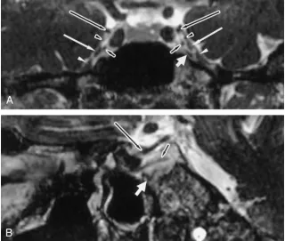
To our knowledge, no studies have demonstrated CNs III–VI in the cavernous sinus on CISS MR im-aging. Shigematsu et al (9) reported that, although 3D CISS gives the appearance of a heavily T2-weighted sequence, 3D CISS sequences show in-creased contrast as concentration of gadolinium-based contrast agent increases. Our study showed that CNs III, IV, V1, V2, and VI in the cavernous sinus were well visualized on contrast-enhanced 3D CISS MR imaging, with the enhanced venous plexus play-ing a role similar to CSF. Venous spaces in the cav-ernous sinus were strongly enhancing, and CNs were shown as nonenhanced spots of hypointensity. In a previous study, it was not possible to detect each CN separately. On the contrarily, we visualized each nerve clearly for the first time. Detectability rates for CNs III, IV, V1, V2, and VI in the cavernous sinus were 100%, 61%, 92%, 88%, and 96%, respectively, on contrast-enhanced CISS MR imaging. These de-tectability rates are higher than those of the previous FIG 1. Contrast-enhanced CISS MR
im-ages (TR/TE⫽11.8/5.9) in a 44-year-old woman whose acoustic schwannoma had been resected without recurrence.
A, Coronal view clearly shows bilateral CNs III (long black arrow), IV (black arrow-head), V1(long white arrow), V2(white
ar-rowhead), and VI (short black arrow) are clearly demonstrated in the normal cav-ernous sinuses. Additional dark spot is shown inferior to left CNs V1and VI (short
white arrow).
[image:3.585.214.534.58.329.2]B, Oblique sagittal view also depicts the left CNs III (long black arrow) and VI (short black arrow) well. Additional dark region shown inAis also identified as a serpigi-nous, dark line without continuity (short white arrow). It is considered to be fibrous tissue.
TABLE 1: Detectability of cranial nerves in patients with normal cavernous sinuses on enhanced 3D CISS MR imaging
Patients
CN
III IV V1 V2 VI
16–55 y (n⫽38)* 38 (100) 24 (63) 36 (95) 32 (84) 38 (100) 56–78 y (n⫽38)†
38 (100) 22 (58) 34 (89) 35 (92) 35 (92)
Pvalue .815 .674 .480 .240
Note.—Data are the number of nerves identified. Data in paren-theses are percentages.
* Mean age, 37.4 y. †Mean age, 64.9 y.
TABLE 2: Detectability of cavernous segments of cranial nerves in 40 normal cavernous sinuses on enhanced imaging
Contrast-Enhanced Imaging
CN
III IV V1 V2 VI
CISS (n⫽40) 40 (100) 26 (65) 37 (93) 35 (88) 38 (95) T1 weighted
(n⫽40)
40 (100) 12 (30) 18 (45) 21 (53) 26 (65)
Pvalue .003 ⬍.001 .001 .001
[image:3.585.54.282.367.438.2] [image:3.585.53.281.514.599.2]study (4). We compared detectability between con-trast-enhanced CISS and concon-trast-enhanced T1-weighted MR imaging and found that it was predom-inantly and significantly higher with the former than with the latter.
CN III is large and was identified on both contrast-enhanced CISS and contrast-contrast-enhanced T1-weighted MR imaging at a rate of 100%. In a previous report, CN IV in the cavernous sinus could not be individu-ally discriminated because of its small size and prox-imity to CN III (18). However, contrast-enhanced CISS MR imaging depicted CN IV in 61% of cases. We found that CN IV did not always run near CN III and sometimes ran near CN V1. van Overbeeke et al (16) reported that, in 75% of their dissections, CN IV was near to CN III; in the other 25%, the nerve was closer to CN V1. Detectability of CNs V1and V2was high in our study. V2 is reportedly easy to identify during routine head MR imaging, given its exit from the skull through the foramen rotundum (18).
In-deed, in our study, V2was easily identified. However, when it was flattened and tightly attached to the dura matter, its differentiation from dura was sometimes difficult. CN VI was second only to CN III in terms of detectabilty, because of its characteristic course inside the cavernous sinus.
On contrast-enhanced CISS MR imaging, some nonenhanced areas were observed in addition to CNs. These were considered to correspond to periarterial sympathetic fibers, fibrous tissues, or branches of the ICA. From the intracavernous segment of the ICA, two main branches arise, the meningohypophyseal trunk and the inferior lateral trunk. The three main branches of the meningohypophyseal trunk are the tentorial artery, the dorsal meningeal artery, and the inferior hypophyseal artery (10). Branches of CN VI were also considered responsible for some nonen-hanced structures. Harris and Rhoton (10) found that the abducens nerve does not always represent a single trunk in the cavernous sinus, and it frequently splits FIG 2. Normal cavernous sinuses in a 46-year-old woman after surgical resec-tion of left acoustic schwannoma without recurrence.
A, Enhanced coronal CISS MR image (TR/TE⫽11.8/5.9) shows bilateral CNs III (long black arrows), IV (black arrowheads), V1(long white arrows), left V2(white
into multiple rootlets numbering anywhere from two to five. Ozveren et al (28) stated that, in autopsy materials, four of 50 cases had a duplicated bilateral abducens nerves, and seven of 50 cases had dupli-cated unilateral abducens nerves. We distinguished the CNs from other structure on the oblique view that was reconstructed during 3D CISS MR imaging (eg, Fig 1B).
Conclusion
Contrast-enhanced 3D CISS MR imaging offers clear images of normal CNs in the cavernous sinus, which has not been shown in vivo. In the comparative study between enhanced CISS and contrast-enhanced T1-weighted MR imaging, the former of-fered significantly higher detectability of CNs. Con-trast-enhanced 3D CISS MR imaging is a useful method, one that will contribute to the diagnosis of diseases involving cavernous sinuses, such as Tolosa-Hunt syndrome.
Acknowledgments
We thank Kazuo Takeuchi, MD, MPH, PhD, from Takasaki University of Health and Welfare, for his support in the statis-tical analysis in the present study.
References
1. Daniels DL, Pech P, Mark L, Pojunas K, Williams AL, Haughton VM. Magnetic resonance imaging of the cavernous sinus. AJR Am J Roentgenol1985;144:1009 –1014
2. Hirsch WL Jr, Hryshko FG, Sekhar LN, et al.Comparison of MR imaging, CT, and angiography in the evaluation of the enlarged cavernous sinus.AJNR Am J Neuroradiol1988;9:907–915 3. Daniels DL, Czervionke LF, Bonneville JF, et al.MR imaging of
the cavernous sinus: value of spin echo and gradient recalled echo images.AJNR Am J Neuroradiol1988;9:947–952
4. Korogi Y, Takahashi M, Sakamoto Y, Shinzato J.Cavernous sinus: correlation between anatomic and dynamic gadolinium-enhanced MR imaging findings.Radiology1991;180:235–237
5. Caillet H, Delvalle A, Doyon D, et al.Visibility of cranial nerves at MRI.J Neuroradiol1990;17:289 –301
6. Laine FJ.Cranial nerves III, IV, and VI.Top Magn Reson Imaging
1996;8:111–130
7. Casselman JW, Dehaene I.Imaging of the IIIrd, IVth, and VIth cranial nerves.Neuroophthalmology1998;19:63– 68
8. Eisenkraft B, Ortiz AO.Imaging evaluation of cranial nerves 3, 4, and 6.Semin Ultrasound CT MRI2001;22:488 –501
9. Shigematsu Y, Korogi Y, Hirai T, et al.Contrast-enhanced CISS
MRI of vestibular schwannomas: phantom and clinical studies.
J Comput Assist Tomogr1999;23:224 –231
10. Harris FS, Rhoton AL Jr. Anatomy of the cavernous sinus: a microsurgical study.J Neurosurg1976;45:169 –180
11. Umansky F, Nathan H.The lateral wall of the cavernous sinus.
J Neurosurg1982;56:228 –234
12. Taptas JN.The so-called cavernous sinus: a review of the contro-versy and its implications for neurosurgeons. Neurosurgery
1982;11:712–717
13. Ono M, Ono M, Rhoton AL Jr, Barry M.Microsurgical anatomy of the region of the tentorial incisura.J Neurosurg1984;60:365–399 14. Kehrli P, Ali M, Reis M Jr, et al.Anatomy and embryology of the
lateral sellar compartment (cavernous sinus) medial wall.Neurol Res1998;20:585–592
15. Dietemann JL, Kehrli P, Maillot C, et al.Is there a dural wall between the cavernous sinus and the pituitary fossa? Anatomical and MRI findings.Neuroradiology1998;40:627– 630
16. van Overbeeke JJ, Jansen JJ, Tulleken CA.The cavernous sinus syndrome: an anatomical and clinical study.Clin Neurol Neurosurg
1988;90:311319
17. Tuccar E, Uz A, Tekdemir I, Elhan A, Ersoy M, Deda H.Anatomical study of the lateral wall of the cavernous sinus, emphasizing dural construction and neural relations.Neurosurg Rev2000;23:45– 48 18. Castillo M.Imaging of the upper cranial nerves I, III-VIII, and the
cavernous sinuses.Magn Reson Imaging Clin N Am2002;10:415– 431 19. Casselman JW, Kuhweide R, Deimling M, Ampe W, Dehaene I, Meeus L.Constructive interference in steady state-3DFT MR im-aging of the inner ear and cerebellopontine angle. AJNR Am J Neuroradiol1993;14:47–57
20. Held P, Fellner C, Fellner F, Seitz J, Strutz J.MRI of inner ear anatomy using 3D MP-RAGE and 3D CISS sequences.Br J Radiol
1997;70:465– 472
21. Lemmerling M, De Praeter G, Mortele´ K, et al.Imaging of the normal pontine cisternal segment of the abducens nerve, using three-dimensional constructive interference in the steady state MRI.Neuroradiology1999;41:384 –386
22. Yousry I, Camelio S, Schmid UD, et al.Visualization of cranial nerves I-XII: value of 3D CISS and T2-weighted FSE sequences.
Eur Radiol2000;10:1061–1067
23. Held P, Nitz W, Seitz J, et al.Comparison of 2D and 3D MRI of the optic and oculomotor nerve anatomy.Clin Imaging2000;24:337–343 24. Held P, Fru¨nd R, Seitz J, Nitz W, Haffke T, Hees H.Comparison of 2-D turbo spin echo and 3-D gradient echo sequences for the detection of the trigeminal nerve and branches anatomy. Eur J Radiol2001;37:18 –25
25. Seitz J, Held P, Strotzer M, et al.MR imaging of cranial nerve lesions using six different high-resolution T1- and T2(*)-weighted 3D and 2D sequences.Acta Radiol2002;43:349 –353
26. Yousry I, Moriggl B, Dieterich M, Naidich TP, Schmid UD, Yousry TA.MR anatomy of the proximal cisternal segment of the troch-lear nerve: neurovascular relationships and landmarks.Radiology
2002;223:31–38
27. Yousry I, Camelio S, Wiesmann M, et al.Detailed magnetic reso-nance imaging anatomy of the cisternal segment of the abducent nerve: Dorello’s canal and neurovascular relationships and land-marks.J Neurosurg1999;91:276 –283