Original Article
Gelsolin-like actin-capping protein is
highly expressed in prostate cancer and
affects cell apoptosis, proliferation and migration
Haiqi Mu1,2, Bo Qiu3, Qipeng Xie4, Yijun Wang2, Cunjin Nan2, Wei Han5, Tong Zhao3, Shan-Chao Zhao1
1Department of Urology, Nanfang Hospital, Southern Medical University, Guangzhou, China; 2Department of Urol-ogy, The Second Affiliated Hospital & Yuying Children’s Hospital of Wenzhou Medical University, Wenzhou, China; 3Department of Pathology, Nanfang Hospital, Southern Medical University, Guangzhou, China; 4Department of Laboratory Medicine, The Second Affiliated Hospital & Yuying Children’s Hospital of Wenzhou Medical University, Wenzhou, China; 5Cancer Research Institute, Southern Medical University, Guangzhou, China
Received February 19, 2016; Accepted July 12, 2016; Epub January 1, 2017; Published January 15, 2017
Abstract: Objective: To investigate the expression of Gelsolin-like actin-capping protein (CapG) and its biological functions in prostate cancer. Methods: CapG expression was examined in 90 cases of prostate cancer (PCa) and 30 cases of adjacent normal prostate (NP) by immunohistochemistry (IHC). Then, the correlation between CapG expres-sion and clinicalpathological characteristics was assessed in PCa. In addition, CapG expresexpres-sion was examined in PC3, DU145 and Lncap cells by qRT-PCR and Western blot. When CapG expression was knocked down by specific siRNA, cell apoptosis, proliferation and migration were evaluated, respectively. Results: The positive rate of CapG expression in prostate cancer and adjacent normal prostate tissues were 54.4% and 16.7%, and the difference was statistically significant (P<0.001). Meanwhile, CapG expression was significantly correlated with PSA level, Gleason score, T stage and lymph node metastasis in prostate cancer (P<0.001). Furthermore, higher expression of CapG was observed in PC3 cells compared with that in DU145 and Lncap cells by qRT-PCR and Western blot. CapG down-regulation significantly inhibited the proliferation and migration, but promoted the apoptosis in PC3 and Lncap cells. Conclusions: CapG is highly expressed in prostate cancer and affects cell apoptosis, proliferation and migration.
Keywords: Prostate cancer, CapG, apoptosis, proliferation, migration
Introduction
Prostate cancer (PCa) is one of most common malignancy, which has increasing incidence and mortality rates in men all over the word [1, 2]. Meanwhile, PCa is a major cause of cancer related deaths in China [3, 4]. Although the outcome of PCa patients with localized or re- gional disease has been markedly improved,
the 5-year survival rate remains unsatisfied [5,
6]. Furthermore, the mechanism of carcino- genesis and progression of PCa have not yet been fully understood. Thus, molecular
mark-ers associated with PCa should be identified,
which may be helpful for the diagnosis and therapy of PCa.
Gelsolin-like actin-capping protein (CapG), a member of the gelsolin protein family, is an
actin binding protein, which is crucial for mo- dulating the motility of cells [7, 8]. Studies de- monstrated that CapG was involved in various types of carcinomas [9-11]. Higher expression of CapG was observed in breast cancer [12], non-small cell lung cancer [11] and ovarian car-cinoma [13]. However, loss of CapG protein expression was found in stomach cancer cell lines, lung cancer cell lines and melanoma cell lines [14]. In addition, CapG plays key roles in regulating cell division [15], motility [16], migra-tion and invasiveness [13]. However, it is not
fully identified about the relationship between
CapG expression and prostate cancer.
CapG affects cell apoptosis, proliferation and migration in prostate cancer
Materials and methods
Patient and tissue samples
A total of 90 cases of PCa tissues and 30 cases of adjacent normal prostate tissues were collected from Nanfang hospital and the
Second Affiliated Hospital of Wenzhou
Medi-cal University between January 2013 and De- cember 2015. All samples were obtained by surgery resection and none of patients recei- ved chemotherapy, radiotherapy or immuno-therapy before surgery. The clinicopathological characters of patients, including age, smoke history, PSA level, Gleason score, T stage and lymph node metastasis were obtained by hos-pital records. This study was supported by the Medical Ethics Committee of Nanfang hospi- tal of southern medical university and the Se-
cond Affiliated Hospital of Wenzhou
Medi-cal University, also supported with patients’ approval.
Immunohistochemistry (IHC)
All samples were formalin-fixed,
paraffin-em-bedded and cut with two-micrometer sections. Sections were dipped into xylene and rehydrat-ed with gradrehydrat-ed alcohol under standard pro- cedures. Subsequently, antigen retrieval was heated with 0.01 mol/L citrate buffer (pH 6.0)
for five minutes. After incubation with 3%
hy-drogen peroxide and 5% goat serum indivi- dually, sections were incubated with polyclo- nal antibody CapG antibody (dilution, 1:150) (Proteintech Co, USA) for 2 h at room tem- perature. After washing with PBS (Phosphate Buffered Saline), sections were incubated with biotinylated secondary antibody for 30 minutes. Then, sections were developed by 3,3’-diaminobenzidine (DAB) and stained with hematoxylin. Goat serum substituted primary antibody as negative control.
According to the staining intensity and the percentage of positive-staining cells, CapG ex-
pression was classified into negative and
po-sitive expression. Staining intensity was record-ed as absent, weak, moderate and strong. The percentage of positive-staining cells divided into less than and more than 25%. Only these sections with moderate or strong staining and more than 25% of positive-staining cells were regarded as CapG positive expression. Others
were definited as CapG negative expression.
Cell culture and transfection
Human prostate cancer cell lines PC3, DU145 and Lncap were purchased from American Type Culture Collection and cultured with RP- MI-1640 (Gibco, USA) supplemented with 12% fetal bovine serum (HyClone, USA) at 37°C in a humid atmosphere with 5% CO2. Using Lipo- fectamine2000 Transfection Reagent (Invitro- gen, USA), cells were transfected with
CaPG-specific siRNA and those in normal controls
(NC) were transfected with scramble sequence. RNA preparation and quantitative reverse transcription-polymerase chain
After transfection for 48 h, cells were collect- ed to extract total RNA using the Isogen (Nip- pon Gene, Tokyo, Japan). Then the concentra-tion of total RNA was quantitatively measured and reversely transcribed into cDNA using the reverse transcription kit (Takara Biotech- nology, Japanese). The gene expression was
quantified by using SYBR Green PCR kit (Toyobo,
Osaka, Japan). The PCR reaction was conduct-ed as follow: 95°C for 30 s, 95°C for 5 s and 60°C for 30 s. Each sample was repeatedly measured three times and the mRNA levels of genes were calculated using the 2-ΔΔCt. The primer sequences of GAPDH were 5’-GCATC- CTGCACCACCAACTG-3’ and 5’-GCCTGCTTCAC- CACCTTCTT-3’. The primer sequence of CapG were 5’-CGAACACTCAGGTGGAGATT3’ (F) and 5’-CGAACACTCAGGTGGAGATT-3’ (R).
Western blot
After transfection for 48 h, cells were collected, homogenized with RIPA lysis buffer (Thermo
Flow cytometry analysis of apoptosis
After transfection for 48 h, cells were
harvest-ed for the apoptosis analysis by flow cytometry.
Simply, 1×10⁵ cells was collected and wash-
ed with PBS. Then, cells were resuspended in
400 μl binding buffer and stained with 10 μl propidium iodide and 5 μl Annexin V-FITC
(BestBio, Shanghai, China) for 15 min at room temperature. Stained cells were measured by
flow cytometry. Each test was repeated for
three times.
CCK-8 cell proliferation assay
Cell proliferation was determined by CCK-8
assay as previously described [17]. In briefly,
2×10³ cells were cultured in a 96-multiwell
plates, and transfected with CaPG-specific
siRNA and scramble sequence, respectively. After 24 h transfection, cells in each well were
incubated with 10 μl CCK-8 (5 mg/ml) solution
for 4 h at 37°C. Then, the optical density (OD) in each well was determined by using a Ther- momax microplate reader (Molecular Devices, Sunnyvale, CA, USA) at 450 nm. Each test was repeated for three times.
Cell migration
3×10⁴ cells were cultured with RPMI-1640
(Gibco, USA) supplemented with 5% fetal bovine serum (HyClone, USA) in 24-well inserts and transfected with control RNA or CapG siRNA.
The inserts filled in RPMI-1640 medium with
25% fetal bovine serum was considered as an
attractant. After 48 h, cells were fixed in 95%
ethanol solution and stained with Giemsa’s staining solution. Then, migrated cells were analyzed under optical microscopy. Each exper-iment was repeated for three times.
Statistical analysis
All data were analyzed using SPSS 17.0 soft-ware (SPSS Inc., Chicago, IL, USA). Pearson’s
chi-square test (χ2) was used to evaluate the
CapG expression in prostate cancer and nor-mal prostate and to assess the correlation between CapG expression and clinicalpatho-logical characters. Independent-sample t-test was used to compare the difference in two
groups. The statistically significant difference
[image:3.612.95.524.72.323.2]was P<0.05.
CapG affects cell apoptosis, proliferation and migration in prostate cancer
Results
CapG is highly expressed in prostate cancer and correlated with clinicalpathological char-acters
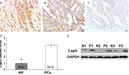
IHC results shown that positive CapG expres-sion in prostate cancer tissues was located in the cytoplasm of cells and stained as yellow brown (Figure 1A and 1B). While positive CapG expression was hard to be detected in adjacent normal prostate tissues (Figure 1C). The posi-tive rates of CapG expression in prostate can-cer and adjacent normal prostate were 54.4% and 16.7%, and the difference was statistically
significant (Table 1, P<0.001). These results indicated that CapG was highly expressed in prostate cancer compared with those in adja-cent normal prostate. To further validate the observation, we detected the expression of CapG in 3 cases of fresh prostate cancer
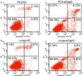
tis-qRT-PCR and Western blot results indicated CapG expression was successfully down-regu-lated in PC3 and Lncap cells (Figure 2B). After CapG down-regulation, the apoptotic rate was
significantly increased in PC3 and Lncap cells
(Figure 3, P<0.05). Meanwhile, CapG depletion
significantly inhibited cell proliferation com -pared with those in normal controls (Figure 4A, P<0.001). Furthermore, the migrated number of cells was markedly reduced when CapG expression was down-regulated in PC3 and Lncap cells (Figure 4B, P<0.001).
Discussion
In this study, we are the first to evaluate the
expression of CapG in PCa and normal prostate tissues. IHC results indicated that CapG was highly expressed in PCa compared with normal prostate, indicating that high expression of Table 1. Expression of CapG in prostate cancer and
normal prostate tissues
Tissue types N CapG (%) P value
Negative Positive
Prostate cancer 90 41 (45.6) 49 (54.4) <0.001 Normal prostate 30 25 (83.3) 5 (16.7)
Table 2. Correlation between CapG expression and clinical pathological characters in prostate cancer
Clinicalpathological
characteristics N
CapG
P value Negative Positive
Age (years)
≤58 59 29 30 0.380
>58 31 12 19
Smoke history 1.000
Negative 33 15 18
Positive 57 26 31
PSA (ng/ml) <0.001
>10 42 29 13
≤10 48 12 36
Gleason score
≤7 45 31 14 <0.001
>7 45 10 35
T stage
T1-T2 37 30 7 <0.001
T3-T4 53 11 42
Lymph node metastasis
Negative 62 37 25 <0.001
Positive 28 4 24
sues and matched normal prostate tis-sues by qRT-PCR and Western blot. As expected, qRT-PCR and Western blot re- sults also indicated that higher express- ion of CapG was found in prostate cancer in comparison with those in normal pros-tate (Figure 1D and 1E), which was con- sistent with IHC results.
Then, we evaluated the correlation be- tween CapG expression and clinicalpatho-logical characteristics in prostate cancer. As shown in Table 2, significant
correla-tion between CapG expression with PSA level, Gleason score, T stage and lymph node metastasis was recorded in pro- state cancer (P<0.05). However, CapG ex-
pression wasn’t significantly associated
with age and smoke history (P>0.05). CapG down-regulation affects cell apopto-sis, proliferation and migration
Figure 2. CapG expression was measured in human prostate cancer cell lines. A: CapG expression was measured in PC3, Du145 and Lncap cell lines by qRT-PCR and Western blot. B: CapG down-regulation in PC3 and Lncap cells was confirmed by qRT-PCR and Western blot. *: P<0.001.
Figure 3. Effect of CapG down-regulation on cell apoptosis was evaluated by flow cytometry. A: The apoptotic rates in PC3-NC and PC3-siCapG cells were 3.14%±0.27 and 14.78%±1.16, respectively, and the difference was statistically significant (P<0.001). B: The apoptotic rates in Lncap-NC and Lncap-siCapG cells were 3.90%±0.33 and 11.10%±1.72, respectively, and the difference was statistically significant (P<0.001).
CapG might be correlated with the formation of PCa. Meanwhile, the same eviden- ce that CapG highly express-
ed in PCa was confirmed by
qRT-PCR and Western blot. These observations indicated that abnormal high-expres-sion of CapG was contribu- ted to the formation of PCa, which might be helpful for the diagnosis of PCa. Our da- ta was also consistent with the observations in non-small cell lung cancer [11], breast cancer [12] and ovarian car- cinoma [13], indicating that CapG was a carcinogenic ge- ne. However, Watari et al. [14] reported that CapG was lost in the tumorigenic cell line, such as lung cancer, stomach cancer and melanoma, and might be a candidate tumor suppressor.
[image:5.612.92.374.378.636.2]clin-CapG affects cell apoptosis, proliferation and migration in prostate cancer
icalpathological characteristics in PCa. Results
exhibited that CapG expression was significant -ly correlated with PSA level, Gleason score, T stage and lymph node metastasis. Higher expression of CapG was found in patients with higher level of PSA (>10 ng/ml). Meanwhile, CapG expression was markedly increased in patients with higher Gleason score (>7) and T
stage (T3-T4), and was also significantly elevat -ed in patents with lymph node metastasis. It is
well known that PSA is beneficial for the diagno -sis and prognostic prediction in patients with PCa [18, 19]. Gleason score, T stage and lymph node metastasis were implicated with the pro-gression of PCa and might be served as prog-nostic factors in patients with PCa [20-22]. Thus, these data further validated that CapG was a carcinogenic gene in PCa and higher expression of CapG was relative to the progres-sion of PCa.
At present, CapG has been reported to be con-nected with cell invasion, division and motility [7, 9, 12, 13, 15, 16]. However, the effect of CapG expression on cell apoptosis, prolifera-tion and migraprolifera-tion was little to know. In present study, we observed that CapG expression was higher in PC3 cell lines compared with that in Du145 and Lncap cell lines, suggesting that CapG was highly expressed in human PCa cell lines. Subsequently, we found that CapG
deple-tion significantly promoted the apoptosis in
PC3 and Lncap cells, while markedly inhibited the proliferation and migration compared with those in normal controls. Meanwhile, Li et al. [23]. also reported that CapG was expressed in PC3, Du145 and Lncap cell lines, and CapG
downregulation significantly inhibited the prolif -eration, invasion and metastasis of DU145
cells. Thus, these data confirmed that high
CapG expression was contributed to cell
[image:6.612.92.517.77.402.2]eration and migration, which might promote the progression of PCa. Meanwhile, these data fur-ther validated the observations in PCa tissues, and supported that CapG might be served as a therapeutic target in Pca. In addition, CapG
downregulation significantly suppressed the
migration and invasion in ovarian carcinoma and breast cancer [13, 24].
In summary, our results indicate that CapG is highly expressed in PCa and affects cell apop-tosis, proliferation and migration. However, the mechanism of CapG in PCa remains unknown. Further investigations are needed.
Acknowledgements
This work was supported by a National Natural Science Foundation of China (No.81328017), a Natural Science Foundation of Guangdong Province (2016A030313583), two Science and Technology Planning Projects of Guangdong Province (No.2013B051000050, No.2014A- 020212538), and two grants from President Foundation of Nanfang Hospital, Southern Me- dical University (No.G2011014, No.2013Z011). Disclosure of conflict of interest
None.
Address correspondence to: Shan-Chao Zhao, De- partment of Urology, Nanfang Hospital, Southern Medical University, Guangzhou 510515, China. E-mail: [email protected]
References
[1] Petrakis D, Pentheroudakis G, Kamina S, Pappa L, Papadiotis E, Malamou-Mitsi V and Pavlidis N. An unusual presentation of a pa-tient with advanced prostate cancer, massive ascites and peritoneal metastasis: Case report and literature review. J Adv Res 2015; 6: 517-521.
[2] Zhao L, Yu N, Guo T, Hou Y, Zeng Z, Yang X, Hu P, Tang X, Wang J and Liu M. Tissue biomarkers for prognosis of prostate cancer: a systematic review and meta-analysis. Cancer Epidemiol Biomarkers Prev 2014; 23: 1047-1054. [3] Lei F, Zhang L, Li X, Lin X, Wu S, Li F and Liu J.
Overexpression of prostate tumor overexpre- ssed 1 correlates with tumor progression and predicts poor prognosis in breast cancer. BMC Cancer 2014; 14: 457.
[4] Jiang X, Li X, Huang H, Jiang F, Lin Z, He H, Chen Y, Yue F, Zou J, He Y, You P, Wang W, Yang
W, Zhao H, Lai Y, Wang F, Zhong W and Liu L. Elevated levels of mitochondrion-associated autophagy inhibitor LRPPRC are associated with poor prognosis in patients with prostate cancer. Cancer 2014; 120: 1228-1236. [5] Dimakakos A, Armakolas A and Koutsilieris M.
Novel tools for prostate cancer prognosis, diag-nosis, and follow-up. Biomed Res Int 2014; 2014: 890697.
[6] Rachner TD, Thiele S, Gobel A, Browne A, Fuessel S, Erdmann K, Wirth MP, Frohner M, Todenhofer T, Muders MH, Kieslinger M, Rauner M and Hofbauer LC. High serum levels of Dickkopf-1 are associated with a poor prog-nosis in prostate cancer patients. BMC Cancer 2014; 14: 649.
[7] De Corte V, Van Impe K, Bruyneel E, Boucherie C, Mareel M, Vandekerckhove J and Gettemans J. Increased importin-beta-dependent nuclear import of the actin modulating protein CapG promotes cell invasion. J Cell Sci 2004; 117: 5283-5292.
[8] Van Impe K, De Corte V, Eichinger L, Bruyneel E, Mareel M, Vandekerckhove J and Gettemans J. The Nucleo-cytoplasmic actin-binding pro-tein CapG lacks a nuclear export sequence present in structurally related proteins. J Biol Chem 2003; 278: 17945-17952.
[9] Renz M and Langowski J. Dynamics of the CapG actin-binding protein in the cell nucleus studied by FRAP and FCS. Chromosome Res 2008; 16: 427-437.
[10] Tonack S, Patel S, Jalali M, Nedjadi T, Jenkins RE, Goldring C, Neoptolemos J and Costello E. Tetracycline-inducible protein expression in pancreatic cancer cells: effects of CapG over-expression. World J Gastroenterol 2011; 17: 1947-1960.
[11] Zhu WY, Hunag YY, Liu XG, He JY, Chen DD, Zeng F, Zhou JH and Zhang YK. Prognostic evaluation of CapG, gelsolin, P-gp, GSTP1, and Topo-II proteins in non-small cell lung cancer. Anat Rec (Hoboken) 2012; 295: 208-214. [12] Renz M, Betz B, Niederacher D, Bender HG
and Langowski J. Invasive breast cancer cells exhibit increased mobility of the actin-binding protein CapG. Int J Cancer 2008; 122: 1476-1482.
[13] Glaser J, Neumann MH, Mei Q, Betz B, Seier N, Beyer I, Fehm T, Neubauer H, Niederacher D and Fleisch MC. Macrophage capping protein CapG is a putative oncogene involved in migra-tion and invasiveness in ovarian carcinoma. Biomed Res Int 2014; 2014: 379847.
hu-CapG affects cell apoptosis, proliferation and migration in prostate cancer
man cancer cells by new candidate tumor sup-pressor gene CapG. Oncogene 200; 625: 7373-7380.
[15] Hubert T, Van Impe K, VandekerckhoveJ and Gettemans J. The actin-capping protein CapG localizes to microtubule-dependent organelles during the cell cycle. Biochem Biophys Res Commun 2009; 380: 166-170.
[16] Tonack S, Patel S, Jalali M, Nedjadi T, Jenkins RE, Goldring C, Neoptolemos J and Costello E. Tetracycline-inducible protein expression in pancreatic cancer cells: effects of CapG over-expression. World J Gastroenterol 2011; 17: 1947-1960.
[17] Bao JM, He MY, Liu YW, Lu YJ, Hong YQ, Luo HH, Ren ZL, Zhao SC and Jiang Y. AGE/RAGE/ Akt pathway contributes to prostate cancer cell proliferation by promoting Rb phosphorylation and degradation. Am J Cancer Res 2015; 5: 1741-1750.
[18] Giannarini G, Scott CA, Moro U, Pertoldi B, Beltrami CA and Selli C. Are PSA density and PSA density of the transition zone more accu-rate than PSA in predicting the pathological stage of clinically localized prostate cancer? Urol Oncol 2008; 26: 353-360.
[19] Eminaga O, Hinkelammert R, Titze U, Abbas M, Eltze E, Bettendorf O and Semjonow A. The presence of positive surgical margins in pa-tients with organ-confined prostate cancer re -sults in biochemical recurrence at a similar rate to that in patients with extracapsular ex-tension and PSA </= 10 ng/ml. Urol Oncol 2014; 32: 32 e17-25.
[20] Amin A, Partin A and Epstein JI. Gleason score 7 prostate cancer on needle biopsy: relation of primary pattern 3 or 4 to pathological stage and progression after radical prostatectomy. J Urol 2011; 186: 1286-1290.
[21] Deng FM, Mendrinos SE, Das K and Melamed J. Periprostatic lymph node metastasis in pros-tate cancer and its clinical significance. Histo-pathology 2012; 60: 1004-1008.
[22] Carlsson SV, Tafe LJ, Chade DC, Sjoberg DD, Passoni N, Shariat SF, Eastham J, Scardino PT, Fine SW and Touijer KA. Pathological features of lymph node metastasis for predicting bio-chemical recurrence after radical prostatecto-my for prostate cancer. J Urol 2013; 189: 1314-1318.
[23] Li B, Guo KK, Li CY, Li HL, Zhao PP, Chen K and Liu CX. Influence of suppression of CapG gene expression by siRNA on the growth and metas-tasis of human prostate cancer cells. Genet Mol Res 2015; 14: 15769-15778.