lesion. This study aimed to investigate the relationship between HDL function and HSP65 in AS by observing differ-ent doses of HSP65 impact on macrophage cholesterol efflux rate in apolipoprotein E gene deleted mice and HDL anti-inflammatory, antioxidant function. Healthy male ApoE-/- mice at 8 weeks were fed with high-fat diet. On the 16th
week, blood lipid, serum IFN-γ, IL-10, MPO, PON1 levels were tested. HSP65 antibody titer and peritoneal macro -phages 3H-TC switching-out rate were determined. Liver tissue ABCA1, ABCG1, SR-BI, and LXR-α protein expression
were detected by Western blot. TG, TC, and LDL-C level elevated, while HDL-C decreased significantly in group B, C, D, and E compared with group A (P < 0.05). Group E showed lower HDL-C level than group B (P < 0.05). HSP65 antibody titer, IFN-γ, and IL-10 level in group B were similar to group A (P < 0.05). HSP65 antibody titer and IFN-γ level increased, whereas IL-10 reduced in group C, D, and E compared with group B with dose-dependent (P < 0.05). Macrophages 3H-TC switching-out rate, PON1 activity, and liver tissue ABCA1, ABCG1, SR-BI, and LXR-α protein level
declined obviously, while MPO activity increased significantly in group B, C, D, E compared with group A (P < 0.05). HSP65 induced inflammatory immunoreaction, damaged HDL function, and declined macrophage cholesterol efflux rate through downregulating liver tissue ABCA1, ABCG1, SR-BI, and LXR-α protein with dose-dependent.
Keywords: Atherosclerosis, HDL, HSP, LXR-α, macrophage cholesterol efflux rate
Introduction
Atherosclerosis (AS) is an important reason to cause multiple cardiac-cerebral vascular dis-eases. It is featured as dyslipidemia, vascular endothelial cells injury, and vascular chronic
inflammatory immune response [1, 2]. Present
study showed that AS was promoted by
oxida-tive modification of monocyte-derived-macro -phages, lipoprotein, T lymphocytes, and normal
cell components in the arterial wall [3, 4].
Hi-gh-density lipoprotein (HDL) has various
func-tions including anti-oxidation, anti-inflamma -tion, promoting reverse cholesterol transport (RCT), antithrombus, and improving endothelial function. AS was negatively correlated with
HDL-C, though HDL-C cannot accurately reflect HDL function [5, 6]. Under acute and chronic inflammation reaction condition, HDL composi -tion and func-tion changes lead to its lack of normal function. Thus, exploring HDL function
changes has important significance for AS and
CHD risk assessment [7, 8]. The main physio -logical function of heat shock protein (HSP) is featured as protein structure catalyst that has critical role in cell proliferation, cell cycle regula-tion, and apoptosis. HSP can activate autoim-mune reaction to promote cytokines secretion, because of HSP cross reaction between differ-ent species and highly conserved sequence. The pathogenesis of B and T cell response
indi-cated that HSP was related to various inflam -matory response and autoimmune reaction. HSP65 has strong immunogenicity that overex-pressed in infection. It can promote AS
patho-logical process through the inflammatory reac -tion. Animal experiments showed that AS pla-
que area increased significantly in mice with HSP65 immunity, and proinflammatory factor
expression within the plaque elevated obviously
[9, 10]. Inflammatory stimulation can promote
is study intended to discuss the relationship between HDL function changes and HSP65 in AS through injecting different doses of HSP65 to ApoE-/- mice to observe its impact on
macro-phage efflux rate and HDL function.
Materials and methods
Experimental animal and grouping
Ten healthy male C57BL/6J mice at 8 weeks old and weighted 18~22 g were fed normally as the control group (A). Forty healthy male ApoE -/-mice provided by the Chinese academy of
med-ical sciences, animal experiment center (license SYXK-2013-0025). The mice were maintained in SPF grade laboratory animal center. ApoE -/-mice were randomly divided into phosphate buffer solution (PBS) group (B), 6.25, 12.5, and
25 μg HSP65 group (C, D, E). The mice in group
A were fed with common diet for 16 weeks, while the mice in group B, C, D, and E received high-fat diet for 16 weeks. They received PBS or HSP65 back subcutaneous injection at the 3rd or 16th week at 20 ml/kg.
[image:2.629.99.530.78.515.2]Mice were used for all experiments, and all pro -cedures were approved by the Animal Ethics
Committee of Central Hospital of Chongqing Three Gorges.
Test drugs and reagents
HSP65 was from StressMarq. RAW264.7 mac -rophages were provided by ATCC. 3H-TC was
purchased from Perkin Elmer. IFN-γ and IL-10 ELISA kits, MPO, PON1, and total cholesterol
kits were bought from Nanjing Jiancheng Bio- engineering Institute. ABCA1 primary antibody
was from Novus. ABCG1, SR-BI, and LXR-α pri -mary antibodies were from Abcam.
Specimen collection
On the 16th week, after fasted for 12 h, the blood was extracted from heart to isolate serum. Peritoneal macrophage and liver tissue were stored in liquid nitrogen. Aorta was
sepa-rated under the microscope and fixed with 10%
formaldehyde.
Serum lipid, IFN-γ, IL-10, MPO, PON1 level de -tection
Blood lipid was tested by automatic biochemis-try analyzer: total cholesterol (TC), triglyceride (TG), low density lipoprotein (LDL-C), HDL-C.
IFN-γ and IL-10 were detected by ELISA. MPO
and PON1 levels were tested according to the manual.
Serum HSP65 antibody titer determination
According to previous report [11], antibody (pro
-tein content 1 μg/ml) was diluted by PBS and
DMEM containing 0.2% BSA, 1 μCi/ml 3H-TC
and 30 μg/ml oxidized LDL [12, 13]. After 24 h
incubation and washed by PBS for 2 times, the
macrophages were added with DMEM contain
-ing 2.8% mice serum DMEM for 6 h. After treat -ed by NaOH at room temperature for half an hour, the cell lysis was collected and detected
using liquid flash counter. 3H-TC switching-out rate = 3H-TC radiation quantity in cytokine/(3 H-TC radiation quantity in cytokine + intracellu- lar).
Western blot
Mice liver tissue was cracked by lysis and pro -tein content was determined by BCA kit. The protein was separated by SDS-PAGE and trans-ferred to PVDF membrane. After blocked for 1 h, the membrane was incubated in primary antibody (ABCG1 1:1000, ABCA1 1:500, SR-BI
1:500, LXR-α 1:500) at 4°C overnight. And
then the membrane was incubated in second-ary antibody (1:15000) for 1 h and developed by luminescence reagent. The image was ana-lyzed by Quantity One software.
Aortic plaques determination
Aorta was extracted and fixed. After dehydra -tion, hyaliniza-tion, embedding, sectioning, and HE staining, the slice was observed under the microscope and analyzed by Image-plus 6.0 software. The area ratio of plaque and blood vessel was calculated.
Statistical analysis
SPSS 19.0 was applied to perform normality
test. Measurement data in normal distribution
[image:3.629.100.294.80.239.2]was presented as mean ± standard deviation (_X ± S). One-way ANOVA and LSD test was used
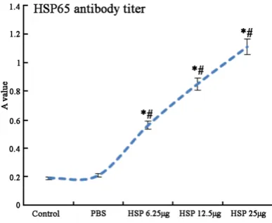
Figure 2. Serum HSP65 antibody titer comparison. *P < 0.05, compared with control; #P < 0.05,
for mean value comparison. P < 0.05 was
con-Results
Blood lipid level comparison
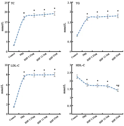
Serum TG, TC, LDL-C levels in group B, C, D, E
increased, while HDL-C decreased significantly
compared with group A (P < 0.05). No obvious difference was observed in serum TG, TC, LDL-C among group B, C, D, and E (P > 0.05). Group E showed markedly lower HDL-C level than that in group B (P < 0.05) (Figure 1).
Serum HSP65 antibody titer comparison
Group B showed no statistical difference of HSP65 antibody titer with group A (P > 0.05).
HSP65 antibody titer increased significantly in
[image:4.629.102.524.80.254.2]group C, D, and E compared with group B with
[image:4.629.102.528.295.465.2]Figure 3. Serum IFN-γ and IL-10 comparison. *P < 0.05, compared with control; #P < 0.05, compared with PBS.
Figure 4. Serum MPO and PON1 comparison. *P < 0.05, compared with control; #P < 0.05, compared with PBS; &P
< 0.05, compared with HSP 6.25 μg.
[image:4.629.102.294.521.670.2]Serum IFN-γ and IL-10 comparison
AS progress is associated with Th1/Th2
imbal-ance. As an inflammation suppressor, IL-10 is
mainly secreted by Th2. It plays an anti-AS effect through downregulating cell adhesion
[image:5.629.102.527.81.666.2]IFN-γ is a proinflammatory factor mainly secret -ed by Th1. It can promote AS by collaborative-
ly inducing inflammatory factor expression. Compared with group A, IFN-γ and IL-10 showed no significant difference in group B (P > 0.05), while IL-10 declined and IFN-γ elevated in group
C, D, and E group with dose dependent (Figure 3).
Serum MPO and PON1 comparison
PON1 level reduced while MPO level rise in
group B compared with group A (P < 0.05). Following dose increasing, PON1 declined and
MPO elevated in group C, D, and E with
dose-dependent (Figure 4).
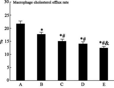
Macrophage cholesterol efflux rate compari -son
Macrophage cholesterol efflux rate decreased
obviously in group B compared with group A (P
< 0.05). Group C, D, and E presented significant macrophage cholesterol efflux rate reduction
with dose-dependent (Figure 5).
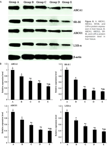
ABCA1, ABCG1, SR-BI, and LXR-α protein ex
-pression in liver tissue
ABCA1, ABCG1, SR-BI, and LXR-α protein
ex-pression level decreased markedly in group B compared with group A (P < 0.05), whereas they reduced obviously in group C, D, and E (Figure 6).
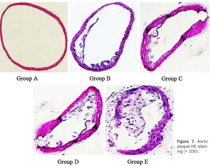
Aortic plaque HE staining
HE staining revealed that HE staining showed smooth intima and integrate endothelial cells in group A. Intima thickening and plaque area increasing could be observed in group B, C, D, and E (Figure 7).
Discussion
The risk of cardiovascular disease reduces 2%
following HDL-C level elevates 1 mg/dL. Several
CHD patients show HDL-C reduction [14, 15].
Clinical trial showed that the incidence of car-diovascular events cannot be reduced by in-creasing HDL-C level. Cholesterol transport
[image:6.629.103.528.80.416.2]matory antioxidation play an important role in
AS process. HDL can play its anti-inflammation
role by inhibiting monocyte adhesion and de- creasing endothelial cell adhesion molecule expression. HDL exerts antioxidant effect th- rough inhibiting LDL oxidation of phospholipids and inactivated generated phospholipids, while
PON1 and MPO are the most important
en-zymes in the process. PON1 has various func-tions including preventing lipid peroxide gen- eration, inhibiting LDL oxidation, hydrolysis of cholesterol ester hydroperoxide type
phospho-lipid, and eliminating LPO in LDL. MPO may
cause HDL losing anti-AS effect by oxidizing apolipoprotein A-I in tissue and blood
circula-tion [19, 20]. Detecting MPO and PON1 activity can reflect the antioxidant function of HDL. Our
study revealed that HSP65 subcutaneous immunization on ApoE-/- mice can cause serum
IL-10 decline, IFN-γ elevation, decrease PON1 activity, and elevate MPO activity with
dose-dependent, suggesting that HDL function ch-
ange is associated with inflammation. HSP65
subcutaneous immunization on ApoE-/- mice
may induce inflammation and promote HDL
function changes, further leading to AS prog-ress. In this study, HSP65 subcutaneous
injec-tion reduced macrophage efflux rate with
dose-dependent, indicating that HSP65 impaired reverse cholesterol transport mediated by HDL
and promoted AS except inducing
inflamma-tion.
HSP can be divided into different subgroups in accordance with amino acid sequence homol-ogy and relative molecular weight, such as
HSP70, HSP60, and HSP90. Multiple studies
have shown that oxidative stress and oxidized low density lipoprotein can induce HSP overex-pression in macrophages and endothelial cells
cess. The main effect of ABCG1 is to mediate cholesterol transporting to the mature HDL. ABCA1 can promote intracellular pre-HDL for-mation by combining phospholipids to free cho-lesterol and apolipoprotein A I, which is the rate-limiting step in the process. The real HDL receptor is SRB I, while the later mediates in-
tracellular cholesterol efflux and liver tissue
uptake HDL-C, promoting bidirectional
choles-terol efflux between HDL and cells [22-24]. LXR-α is one of the major nuclear receptors for
regulating cholesterol metabolism that can
promote intracellular free cholesterol efflux
through regulating ABCG1 and ABCA1
expres-sion. Inflammatory immune response can
re-duce cholesterol transport protein expression
level, and inflammatory factors IL-1, IFN-γ, and TNF-α can reduce ABCG1 expression level [23, 24]. This study revealed that HSP65 subcuta -neous injection on ApoE-/- mice gradually de-
clined macrophage efflux rate with dose-depen -dent, decreased ABCA1, ABCG1, SR-BI, and
LXR-α protein expression in liver tissue, sup
-pressed serum IL-10 level, and elevated IFN-γ
level, suggesting that HSP65 can cause
macro-phage efflux rate reduction and ABCA1, ABCG1, SR-BI, and LXR-α protein downregulation in
liver tissue. In conclusion, HSP65 can reduce
macrophage cholesterol efflux rate with dose
dependent by down-regulating ABCA1, ABCG1,
SR-BI, and LXR-α protein expression, inducing inflammation, and damaging HDL function.
Disclosure of conflict of interest
None.
Chongqing 404000, China. Tel: +86-23-58122622; Fax: +86-23-58122622; E-mail: luoyiihello@sina. com
References
[1] Yao ST, Zhao L, Miao C, Tian H, Yang NN, Guo SD, Zhai L, Chen J, Wang YW, Qin SC. [Endoplasmic reticulum stress mediates oxi -dized low density lipoprotein-induced scaven-ger receptor A1 upregulation in macrophages]. Sheng Li Xue Bao 2014; 66: 612-618. [2] Sun H, Shen J, Liu T, Tan Y, Tian D, Luo T, Lai W,
Dai M, Guo Z. Heat shock protein 65 promotes atherosclerosis through impairing the proper-ties of high density lipoprotein. Atherosclerosis 2014; 237: 853-861.
[3] Li H, Huang S, Wang S, Zhao J, Su L, Zhao B, Zhang Y, Zhang S, Miao J. Targeting annexin A7 by a small molecule suppressed the activity of phosphatidylcholine-specific phospholipase C in vascular endothelial cells and inhibited ath-erosclerosis in apolipoprotein E(-)/(-)mice. Cell Death Dis 2013; 4: e806.
[4] Raizman JE, Chen YX, Seibert T, Hibbert B, Cuerrier CM, Salari S, Zhao X, Hu T, Shi C, Ma X, Simard T, Caravaggio J, Rayner K, Bowdish D, Moore K, O’Brien ER. Heat shock protein-27 attenuates foam cell formation and atherogen-esis by down-regulating scavenger receptor-A expression via NF-kappaB signaling. Biochim Biophys Acta 2013; 1831: 1721-1728. [5] Cuerrier CM, Chen YX, Tremblay D, Rayner K,
McNulty M, Zhao X, Kennedy CR, de BelleRoche J, Pelling AE, O’Brien ER. Chronic over-expres -sion of heat shock protein 27 attenuates ath-erogenesis and enhances plaque remodeling: a combined histological and mechanical as-sessment of aortic lesions. PLoS One 2013; 8: e55867.
[6] Jang EJ, Jung KY, Hwang E, Jang YJ. Ch- aracterization of human anti-heat shock pro-tein 60 monoclonal autoantibody Fab frag-ments in atherosclerosis: genetic and func-tional analysis. Mol Immunol 2013; 54: 338-346.
[7] Yao S, Zong C, Zhang Y, Sang H, Yang M, Jiao P, Fang Y, Yang N, Song G, Qin S. Activating tran-scription factor 6 mediates oxidized LDL-in-duced cholesterol accumulation and apoptosis in macrophages by up-regulating CHOP expres-sion. J Atheroscler Thromb 2013; 20: 94-107. [8] Almanzar G, Ollinger R, Leuenberger J,
Onestingel E, Rantner B, Zehm S, Cardini B, van der Zee R, Grundtman C, Wick G. Auto- reactive HSP60 epitope-specific T-cells in early human atherosclerotic lesions. J Autoimmun 2012; 39: 441-450.
[9] Huang CY, Shih CM, Tsao NW, Chen YH, Li CY, Chang YJ, Chang NC, Ou KL, Lin CY, Lin YW, Nien CH, Lin FY. GroEL1, from Chlamydia pneu-moniae, induces vascular adhesion molecule 1 expression by p37(AUF1) in endothelial cells and hypercholesterolemic rabbit. PLoS One 2012; 7: e42808.
[10] Salari S, Seibert T, Chen YX, Hu T, Shi C, Zhao X, Cuerrier CM, Raizman JE, O’Brien ER. Ex-tracellular HSP27 acts as a signaling molecule to activate NF-kappaB in macrophages. Cell Stress Chaperones 2013; 18: 53-63.
[11] Long J, Lin J, Yang X, Yuan D, Wu J, Li T, Cao R, Liu J. Nasal immunization with different forms of heat shock protein-65 reduced high-choles-terol-diet-driven rabbit atherosclerosis. Int Im- munopharmacol 2012; 13: 82-87.
[12] Zhao SP, Yang J, Li J, Dong SZ, Wu ZH. Effect of niacin on LXRalpha and PPARgamma expres-sion and HDL-induced cholesterol efflux in adi -pocytes of hypercholesterolemic rabbits. Int J Cardiol 2008; 124: 172-178.
[13] Khera AV, Cuchel M, de la Llera-Moya M, Rodrigues A, Burke MF, Jafri K, French BC, Phillips JA, Mucksavage ML, Wilensky RL, Mohler ER, Rothblat GH, Rader DJ. Cholesterol efflux capacity, high-density lipoprotein func -tion, and atherosclerosis. N Engl J Med 2011; 364: 127-135.
[14] Truman JP, Al Gadban MM, Smith KJ, Jenkins RW, Mayroo N, Virella G, Lopes-Virella MF, Bielawska A, Hannun YA, Hammad SM. Diff-erential regulation of acid sphingomyelinase in macrophages stimulated with oxidized low-density lipoprotein (LDL) and oxidized LDL im-mune complexes: role in phagocytosis and cy-tokine release. Immunology 2012; 136: 30-45.
[15] Isa SA, Ruffino JS, Ahluwalia M, Thomas AW, Morris K, Webb R. M2 macrophages exhibit higher sensitivity to oxLDL-induced lipotoxicity than other monocyte/macrophage subtypes. Lipids Health Dis 2011; 10: 229.
[16] Leng X, Zhan R, Wang Y, Liu X, Gong J, Gao X, Wu L, Wang L, Zhao Y, Wang X, Zhang Z, Pang W, Qian L. Anti-heat shock protein 70 autoanti-body epitope changes and BD091 promotes atherosclerosis in rats. Cell Stress Chaperones 2010; 15: 947-958.
[17] Breder I, Coope A, Arruda AP, Razolli D, Milanski M, Dorighello Gde G, de Oliveira HC, Velloso LA. Reduction of endoplasmic reticulum stre- ss--a novel mechanism of action of statins in the protection against atherosclerosis. Athero-sclerosis 2010; 212: 30-31.
-regulated protein 78 prompts scavenger re-ceptor A-mediated secretion of tumor necrosis factor-alpha by RAW 264.7 cells. Clin Exp Pharmacol Physiol 2009; 36: 940-944.