Prophylactic Bactericidal Orthopedic Implants – Animal
Testing Study
Richard A. Wysk1, Wayne J. Sebastianelli1, Rohan A. Shirwaiker2, Gregory M. Bailey1, Charumani Charumani2, Mary Kennett2, Amy Kaucher2, Rachel Abrahams2, Thomas A. Fuller1*, Patricia Royer, Robert C. Voigt1 and Paul H. Cohen1
1ArgentumCidalElectrics (ACE), Inc., Lewistown, USA; 2The Pennsylvania State University, University Park, USA. Email: *[email protected]; *[email protected]
Received 3 January 2010; revised 18 February 2010; accepted 22 April 2010.
ABSTRACT
This paper summarizes preliminary rat studies aim- ed at identifying the effectiveness of using electrically stimulated silver as a bactericidal agent for indwell-ing residual hardware devices (RHD). A variety of bactericidal indwelling devices were designed, fabric- ated and surgically inserted into the medullary cavity of live rats. The rats were inoculated with Staphyloco- ccus aureus to try and induce osteomyelitis. A total of 37 surgeries were performed by implanting the rats with both control and potentially bactericidal devices. As surgical procedures and devices were improved, it appeared that the implants produced antibiotic effe- cts in the animals. All of the control animals and all of the animals where the device failed tested positive for Staphylococcus aureus growth. Of the rats with operational bactericidal devices (those that survived the surgery and incubation period), half tested nega-tive for Staphylococcus aureus. The device designs are discussed in this paper along with the test procedures, operating practices and results. A statistical analysis of the results, which shows a very high confidence le- vel in the effectiveness of electrically stimulated silver as a bactericidal agent/antibiotic, is also presented.
Keywords:Antibacterial; Antimicrobial; Bactericidal; Si- lver; Ionic Silver; Residual Hardware Devices (RHD); Animal Testing
1. INTRODUCTION
Replacing arthritic joints has improved the quality of life for millions of Americans. Over the past decade, there has been an increaseinthenumber of totalhipandknee replacement surgeriesperformed inthe U.S. In 2005, a total of 808,000 total hip and knee replacements were performed in the U.S [1]. By 2030, the total number of
replacements is projected to be more than 4 million [2]. Of these surgeries, 0.3 – 2% result in deep bone infect- ions (osteomyelitis), according to current data [3,4]. While this rate is fairly low, the cost associated with mitigating deep bone infections far exceeds the cost of the initial replacement surgery.
Patients who develop osteomyelitis must undergo diff- icult and expensive treatment. Total mitigation of osteo- myelitic infection is typically achieved after: 1) implant device removal, 2) local debridement of infected area, 3) insertion of a spacer prosthetic, 4) an aggressive six we- ek course of limb protection and aggressive antimicrob- ial therapy, and 5) a second joint arthroplasty. The prim- ary objective of this research is to improve the quality of life by reducing pathogenic bacteria in in vivo joint rep-
lacements using the antimicrobial properties of silver stimulated by an electric current. The long-term goal is to augment current implants with antimicrobial surface technology, thus allowing treatment of RHD associated osteomyelitic infections without the removal of prosth- etic implant or hardware device.
Significant research has been performed recently on antimicrobial colloids composed of silver nano-particles stabilized by polymers or other agents [5-7]. Many stud-ies have shown these materials to display antimicrobial efficacy to a wide spectrum of microorganisms [8-10]. Our in vitro laboratory studies have shown these new materials to exhibit acceptable early antibacterial prop-erties; however in water-rich environs, these materials may quickly lose their effectiveness (typically minutes to hours). After years of investigating the bactericidal effects of silver, the proper device configuration required for silver to be an effective bactericide has been identi-fied. The key is to continually produce a controlled rele- ase of silver ions (Ag+) and expose bacteria to these ions for extended time periods.
to fight infections, and that drug resistant infections kill more Americans every year than AIDS and breast cancer combined. Our hypothesis is that ionized antimicrobial silver and silver forms can be used to kill a wide variety of bacteria, i.e., both standard strains of bacteria as well
as methicillin-resistant bacteria. We base this hypothesis
on the long history of bactericidal metals, particularly silver, gold and copper, that have been used in medicine for years as antimicrobial agents in the form of wound dressings and debriding agents [8,9,12-14]. The histori-cal problem has been that these bactericidal metals seem to work in certain situations while showing little effect in others. Based on three plus years of in vitro testing, we have identified the key science and engineering ele-ments that allow bactericidal metals, particularly silver, to work effectively. The use of antimicrobial silver is particularly promising because in spite of being used for more than 100 years, there has been no evidence of a definitive pattern for silver resistant infections [15].
The principal focus of this research is to describe a new system designed to make replacement prostheses antiba- cterial. In the following section, we will describe the pr- oposed system that is identified as effective against most bacteria which cause osteomyelitic infections. A few device design iterations along with a final device design that was used in our animal testing are also described. Finally, the protocol used for animal testing and the re-sults that were obtained in the animal tests are presented.
2. THE ANTIMICROBIAL SYSTEM
A system was developed to provide prophylactic and an- tibiotic protection for both soft tissues as well as bone against common bacteria, e.g., strains of Escherichia co- li, Staphylococcus aureus, Pseudomonas aeruginosa, Enterococcus faecalis, and methicillin-resistant Staphy-lococcus aureus (MRSA) and fungi e.g. Candida albi-cans. Our in vitro and early animal testing has shown
that a properly configured device that is stimulated by a very small amount of electrical current is both an effecti- ve bactericide as well as a fungicide. The central hypot- hesis behind the proposed research is that electrically ionized antimicrobial silver and its alloys can be used as a safe and effective means of eliminating microorgan-isms associated with biofilms on RHDs. The key to this technology is using the bacteria-rich environment to carry silver ions and complete an electrical circuit in the device configuration shown in Figure 1. This configura-tion creates a regional antibiotic environment around the RHD (inhibition zone diameter ≈ 20mm in a petri dish), and appears to work for different concentrations of bac-terial colonies.
3. IN VITRO STUDIES
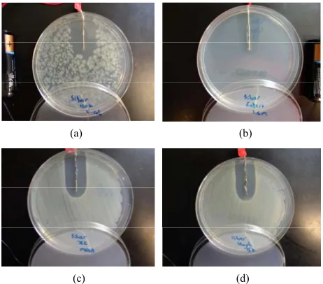
For more than three years, we have experimented with si
[image:2.595.310.541.349.446.2]lver stimulated by small currents to produce silver ions. We have demonstrated that our configuration is effective against all common harmful bacteria and fungi that were tested in the laboratory. Our system shows reproducible results for a variety of bacteria and fungi, as shown in Figure 2. In another test, we inserted our device in the medulary cavity of a rat tibia and then embedded the tibia in Mueller Hinton agar (MHA) inoculated with Ps- eudomonas aeruginosa to determine if our system wor-
ked in this simulated in vivo environment. The results of
this test are shown in Figure 3. These results are sign- ificant in that the bactericidal kill region that is created from within the rat tibia extends well into the agar. This demonstrates that our Ag+ system can penetrate the bone – a problem for most antibiotics.
4. IN VIVO STUDIES
—ANIMAL TESTING
[image:2.595.309.540.479.682.2]The properties of silver have been known for quite some time. Chronic exposure to silver, termed argyria, is man- ifested by an irreversible gray or blue-gray discoloration of the skin and mucous membranes. When the body is
Figure 1. Proposed antimicrobial system concept.
(a) (b)
(c) (d)
Kill Zone
Figure 3. Simulated in vivo testing results of the proposed
system on Pseudomonas Aeruginosa.
exposed to silver challenge the liver acts as a filter mec- han ism, collecting over 90% of the absorbed silver and eliminating it in the feces via liver and biliary tract excr- etion [16,17]. As such the liver cells will experience the greatest levels of toxic elements borne by the blood. The cellular toxic levels of hepatocytes were determined to be both time and concentration dependant but could be appreciated at 30 M [18]. These results were confirmed by Rungby in 1990 when he proposed a range of 30-70 M silver concentration as that needed to show appreci- able cytotoxic effects within the hepatocytes [19]. With- in all of our designs we will keep local ionic concentrate- ons at or below toxic concentrations by controlling the electrical circuitry associated with the implant device.
Two separate sets of animal testing studies were con-ducted to investigate the effectiveness of an in vivo
elec-trically stimulated silver device that was inserted into the tibia’s medullary cavity as a bactericidal agent. The pro-tocol for generating osteomyelitis in the animals paral-leled that described by Lucke et al. [20]. As described in this reference, rat tibias were inoculated with Staphylo-coccus aureus to try to create osteomyelitis. Surgical
procedures were developed to install and retain the pro-posed bactericidal devices within the rat tibia.
The protocol to grow the inocula was adopted from Lucke et al. [21]. Staphylococcus aureus (ATCC number
29213), used as the pathogen, was grown overnight in 9 ml tryptic soy broth (TSB) (caseinpepton–soybean flour– peptone–solution; Oxoid Ltd., Basingstoke, Hampshire, UK). From this culture, 100 µl aliquots were transferred to sterile tubes containing 3 ml of TSB. These tubes were then incubated for 3 h at 37°C to obtain log-phase growth. After incubation, the tubes were centrifuged for 10 min at 3000 rpm, the supernatant was decanted, and the remaining pellet was washed twice with phosphate-
buffered saline (PBS). Under spectrophotometric control the bacterial sediment was added to PBS until a McFar- land standard of six was obtained. Colony- forming units (CFU) per ml were confirmed by several plate counts with the use of a spiralplater (Spiral System Inc., Cincin- nati, OH). This procedure was repeated 20 times. Suspe- nsions were split into portions and deep frozen at –80°C until the day of surgery. To quantify a possible loss of viable bacteria following the freeze–thaw cycle the CFU/ml were reconfirmed after each cycle of defrosting. In addition, each time a rat was inoculated, a 10 µl sam-ple of inoculant was analyzed to determine its bacterial concentration. The concentration was adjusted to fit into the desired range (102 - 104 CFU/ml) using McFarland Standards and was confirmed by plate counts.
Surgeries in both sets of studies were performed at the Penn State Central Biology Labs. All through the testing studies, several different bactericidal devices designed were inserted into the tibial canal of 4-month-old Spra-gue Dawley rats through a proximal incision site and tibial cannulation. A similar surgical protocol was empl- oyed for all the hardware insertions with only slight de-vice-specific modifications being made to the procedures as necessary. For each of the surgeries, one leg was sha- ved and scrubbed with betadine and alcohol prep. Each animal was placed on sterile drapes and their bodies were covered with sterile sheets; the prepped leg was sepa-rately draped in a sterile manner.A small incision (~ 5 mm) of skin and fascia at the proximal tibial metaphysis was created to provide access to the tibial periosteum. The medullary cavity of the proximal metaphysis was ex- posed using a 1 mm surgical drill, leaving the surround-ing periosteum intact. A steel Kirschner wire (Ø 0.8 mm) was inserted into the medullary cavity and pushed forw- ard distally for smooth dilatation of the cavity for a leng- th of approximately 8-10 mm distally, and removed. Sta- phylococcus aureus wasinjected into the medullary
cav-ity to initiate osteomyelitis. Animals were closely monit- ored post-surgery and analgesics were administered ini-tially on a regular schedule. No antibiotics were given to any of the animals since they would have masked the re- sults of the test.
Length of device med.
Initial bench top testing revealed that early prototype device designs performed well; however, biocompatibil-ity within lab animals remained a question. Iterative tes- ting of three device designs was performed to determine the one that was most compatible with the lab rats. Three separate, but similar, device-specific surgical procedures were used during this phase of the study.
4.1. Overview of Surgical Procedure for Animal Testing Studies #1
The first set of animal testing studies was performed in 2007 in which three iterative designs of bactericidal dev- ices and control devices were fabricated and inserted into the tibia medullary cavities of 21 living rats. Table 1 identifies the specifics of the iterative design changes. Sixteen of these animals survived the surgery and the two week incubation period following the surgery. Of the 16 rats, 5 were implanted with control devices (titan- ium wire). The inoculation process proved very succe- ssful as all 5 of the control animals without a bactericidal device developed Staphylococcus aureus based bone in-
fections. Of the remaining 11 rats, only 9 animals had devices which remained in the tibia during the testing period. 3 of those 9 devices failed during the indwelling period leaving only 6 animals as prime targets for meas-uring efficacy of the device. Of these 6 possible cases, 3 of the rats with working devices were found to be staph
free. All of the other animals with non-silver control devi- ces or failed silver devices that were inoculated and euthanized cultured positive for Staphylococcus aureus.
This preliminary study showed that the devices worked in at least half of the cases where they remained in the tibia and continued to be operational. The quantity of
staphylococcus aureus present in the tibia was not
quan-tified so it is possible that some of the devices judged ineffective may have significantly reduced the amount of bacteria present at the end of testing.
During this entire animal testing sequence, the surgi-
cal procedures were continually refined making small iterative improvements for two purposes: 1) to keep for-eign-borne microbes from impacting the testing and 2) to improve the device for animal comfort and performance. For surgeries performed on the first 6 rats, the staphylo-coccus (~10 - 20 µl containing 103 CFUs) was injected
via syringe into the medullary cavity prior to inserting the device. For the remainder of the surgeries, the wire tip of the device was dipped into the solution of staphy-lococcus aureus prior to inserting it into the tibia. This
refinement was necessary because the medullary canal could not hold all of the fluid injected and the overflow ran into the soft tissue surrounding the incision site. The overflow of the injection fluid caused, in addition to the desired osteomyelitic infection, a soft tissue infection surrounding the incision site. Details of each of the pro-cedures, blood reports and tibia X-ray reports were re-corded. Ketmine/Xylazine (100 mg/kg) and IP Isoflurane (10 mg/kg) were used as anesthetics.
All three device designs used in this set of study and their surgical results are discussed below. One failure in this set of testing study was that the epoxy capsule used to enclose the device battery within the animal became infected in the majority of the animals.
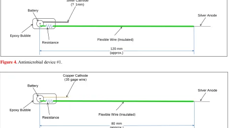
4.1.1. Antimicrobial Device #1—Design and Surgical Summary
The first set of surgeries was performed on 6 rats. The first implant design had a stiff silver wire cathode (Ø 0.8 mm, ~ 30 mm length) and a stiff silver wire anode (Ø 0.8 mm, ~ 40 mm length and then cut to fit the tibia) that was inserted into the tibial canal. The anodic wire was connected to the 1.5 V battery via a 35 gage insulated copper wire. The conductive path for the two wires was the highly resistive soft rat tissue between the anode and cathode. The distance between the two wires (anode and cathode) was ~ 70 - 90 mm when implanted, and the total length of the flexible wire was as much as 120 mm. A small epoxy cap was used to prevent the battery and wires from eroding through the external skin layers. The
Table 1. Device design summary for 2007 animal testing.
Section of Text
Device number
Figure
number Anode configuration Cathode configuration
Battery encasement
4.11 1 4 Stiff silver wire - connected to battery by flexible 35 gauge copper wire Stiff silver wire 100-120 mm epoxy
4.12 2 5 Stiff silver wire - connected to battery by flexible 35 gauge copper wire Flexible copper wire 80-100 mm epoxy
4.13 3 6 Stiff silver wire - connected to battery by flexible 35 gauge copper wire
Silver painted wire, wrapped around insulated
portion of anode wire
device design for these surgeries is shown in Figure 4. The anodic tip of the device was inserted into the tibial canal; the conductive wire and battery pack was stored under the dermatological layer of the rat’s flank. This de- sign was tested at two different levels of current – high amperage (without resistor) and low amperage (with resistor), and with a similar control device in which the battery was removed and the silver wire was replaced with a titanium wire (2 rats).
Thus, six rats were fit with implants during the first set of surgical trials; two devices with no resistor, two de- vices with 1 M-Ω resistor and two devices with titanium wire (control). All 6 rats survived both the surgery and the two week incubation period post-surgery. After the two week period, all 6 rats were euthanized and studied to determine if the devices had eliminated the bacteria (especially the staphylococcus aureus). All of the
de-vices were checked for continuity and voltage after re-moval. Three of the four devices were deemed to be functional. 2 of these 3 rats with functional devices did not show any staph growth.
4.1.2. Antimicrobial Device #2—Design and Surgical Summary
The second set of surgeries was performed on 9 rats. In this set of surgeries, the cathode of the implanted device was modified by replacing the stiff sliver wire with a flexible copper wire. The additional flexibility was
desired for rat comfort as the original design (stiff wire) dug into the rat’s muscle tissue, causing a point of irrita-tion. The total length of the implant was reduced to ~80-100 mm in order to facilitate ease of insertion. The inoculation during the second surgery set was performed by dipping the wire into the staphylococcus aureus
solu-tion to induce a staph infection within the medullary
cavity. The device used for the second set of surgeries is shown in Figure 5.
Nine rats were surgically implanted with the follow-ing: three devices with no resistor (high amperage), three with a 1 M-Ω resistor (low amperage) and three with a titanium control. In this group, 2 of 9 rats died. The rem- aining 7 rats survived the observation period and were euthanized after two weeks. A few of the devices had their cathode tipping out of the rat skin, thus breaking the continuity while in vivo. Devices were checked for
continuity after the animals were euthanized and only three appeared functional. Unfortunately, the design changes created a device that could “float” within the animals, allowing the devices to migrate out of the me-dullary cavity during the post-surgical phase of the study. Due to migration, none of the devices remained in the tibial canal when the postmortems were performed. Drawbacks of this design included breakage of the cop-per wire and poking of the cathode through the rat skin. Resistive soft tissue between the anode-cathode separa-tion was also a problem.
+ Battery
Silver Cathode (? 1mm)
Silver Anode
Flexible Wire (Insulated) Resistance
Epoxy Bubble
[image:5.595.68.529.433.554.2] [image:5.595.66.532.439.699.2]120 mm (approx.)
Figure 4. Antimicrobial device #1.
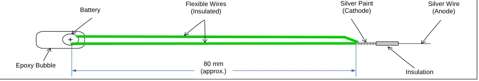
[image:5.595.64.532.585.705.2]4.1.3. Antimicrobial Device #3—Design and Surgical Summary
In order to alleviate the migration of the device within the test animal, a third device design was created. The an- ode and cathode were integrated on the same wire and were separated by a small insulation strip. In this config- uration, electrons are drawn from the silver paint (cath-ode) to the anode over the insulation through the cond- ucting media. This design was accompanied by a change in electrode polarities resulting in two types of devices – anodic and cathodic. The length of the flexible wire was limited to ~80-90 mm. Similar to the previous sets of su- rgeries, bacterial inoculation was created by dipping de- vices into the staphylococcus aureus solution. The third
device design is shown in Figure 6.
Within this phase of the study, 6 rats were surgically implanted using two implants of each kind: three with high amperage anodic devices, two with low amperage anodic devices and one with a high amperage cathodic device. In this group, 3 of the 6 rats died post surgery. The remaining 3 rats survived for the full observation period and were euthanized after one week. All the de-vices were checked for continuity and voltage after re-moval and all were functional.
Several modifications to the implant device and surg- ical procedures were made. The device changes appe- ared to have corrected most of the early drawbacks. The surgical procedure was modified to include a stitch to hold the device in place. The result was that none of the devices floated within the rat’s body. Only 1 of the 3 rats which survived was staph free. This could have been due
to the limited separation between the electrodes. This device appeared to be working as evident by the lack of
puss on gross examination during the device removal; however a quantitative CFU count was not performed.
A few potential device design concerns remained for this device. It was possible that the amount of fluids sur- rounding the implant was insufficient to facilitate proper movement of the antimicrobial Ag+ ions. Another possi-bility was that the flow of antimicrobial Ag+ ions was localized since ions take the path of least resistance and in this case the path of least resistance translated into the path of least distance over the insulation (about 5 mm).
4.2. Overview of Surgical Procedure for Animal Testing Studies #2
The second set of animal testing surgeries was perform- ed in 2008. A total of 16 surgeries were performed with 4 rats used as controls (titanium wire/silver wire without current) and 12 rats implanted with three iterations of antimicrobial designs. Table 2 identifies the specifics of the iterative design changes. Thirteen of these animals survived the surgery and an incubation period and could be harvested for pathology. Three of these thirteen ani-mals (2 with antimicrobial devices and 1 with control) were harvested within the one week incubation period following the surgery due to declining health. Thus, 3 an- imals with control and 10 animals with the antimicrobial devices were left as prime targets for measuring the ef-ficacy of the devices. All control rats tested positive for
Staphylococcus aureus growth. Where the devices failed,
all of the animals also tested positive for Staphylococcus aureus. Of the 10 rats that had antimicrobial devices
implanted and survived the surgery and incubation pe-riod, 4 rats were clear of Staphylococcus aureus. The Staph growth was not quantified in this study.
Battery
Epoxy Bubble 80 mm
(approx.) Flexible Wires (Insulated)
Insulation Silver Paint
(Cathode)
[image:6.595.58.540.495.577.2]Silver Wire (Anode)
Figure 6. Antimicrobial device #3.
Table 2. Device design summary for 2008 animal testing.
Section of Text
Device number
Figure
number Anode configuration Cathode configuration
Length of device
Battery encasement
4.21 4 7 Stripped end of Teflon insulated silver wire Stripped end of Teflon insulated silver wire, wrapped around insulated portion of anode wire 60 mm epoxy
4.22 5 8 Stiff silver wire connected directly to battery Stripped end of copper wire, wrapped around section of anode, insulated via heat shrink wrap 60 mm Silicone
4.23 6 9 Stiff silver wire connected directly to battery
Stripped end of Teflon insulated silver wire, wrapped around section of anode, insulated via heat shrink wrap
Refinements in this set of surgeries were focused on keeping foreign-borne microbes from impacting the tes- ting, and improving the device for animal comfort and performance. A surgical protocol similar to the one used in the first set of surgeries was followed. Two primary modifications in the surgeries were made: 1) the wire device was inserted in the primal portion of the tibia so that a longer portion of the device could be inserted, and 2) the animals were placed in a plastic surgical sack so that airborne bacteria could be contained. In this set of surgeries, the medullary cavity of the primal metaphysis was exposed using a 16 gage needle to produce a hole. 5 µl of Staphylococcus aureus (104 CFU/ml) was injected
via micro-syringe into the medullary cavity prior to inse- rting the device. The micro-syringe eliminated any over-flow of the bacteria. Similar to the first set of surgeries, all animals were closely monitored post-surgery and an- algesics were administered initially on a regular sched-ule. No antibiotics were given to any of the animals to eliminate masking of test results. The epoxy capsule us- ed to enclose the device battery within the animal in our early testing was replaced by a silicone shell to minimize discomfort to the animals.
4.2.1. Antimicrobial Device #4—Design and Surgical Summary
The fourth implant design used during the first set of surgeries in the second study set was a modification of antimicrobial device #3 used during the first set of study. In this design, the anode and cathode were integrated on the same wire. Unlike device #3 where the anode was a silver wire soldered to 35 gage copper wire, the anode in this design was the stripped end of Teflon coated silver wire directly connected to the positive terminal of a 1.5 V battery. Similarly, the silver painted cathode of device #3 was replaced by the stripped end of another Teflon co- ated silver wire directly connected to the negative batt- ery terminal. The electrodes were separated by Teflon insulation on the anodic wire. The electrons were drawn from the silver anode to the silver cathode over the insu-
[image:7.595.310.539.450.540.2]lation by the conducting media. A small silicone cap was used to encase the battery and its soldered connections. Length of the device was limited to 60 mm. The design was used with two combinations of currents – high am-perage (no resistor) and low amam-perage (100 KΩ resistor). Figure 7 shows schematic drawing of the high amperage device design.
Four rats were implanted with this device and all surv- ived the surgery and incubation period. One of the 4 rats implanted with this device tested staph free. This result could be attributed to the quality of the fabricated device as all devices seemed to separate at the coiled portion. Although this design was more robust in construction than the previous ones, a more effective release of ions was desired. This could be achieved by replacing one of the coated electrode wires with a bare one. Commercial silicone used to encase the battery was not bio-compati-ble and caused irritation to the rats.
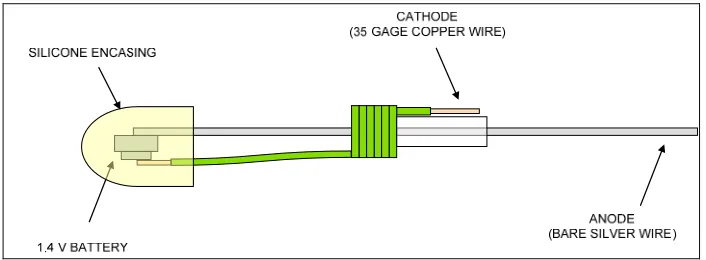
4.2.2. Antimicrobial Device #5—Design and Surgical Summary
Since anodic device designs were being used, the Teflon coated silver anode was replaced by a bare silver wire. Thirty-five gage copper wire was used as the cathode. Insulated shrink tubing was fitted onto the bare silver an- ode to separate the electrodes. Commercial silicone was replaced with medical grade sterile silicone for the bat-tery encasing. Length of the device was limited to 60 mm. These devices were only used with high amperage (no resistor). The device design is shown in Figure 8.
[image:7.595.122.475.576.706.2]Figure 7. Antimicrobial device #4.
Of the 4 rats implanted with antimicrobial devices, 1 died before pathology was performed. One of the rem- aining 3 with working antimicrobial devices tested Staph
free. In analyzing the device used in these surgeries, we concluded that the 35 gage copper wire should be re-placed with Teflon coated silver wire as the cathode as the copper wire seemed to unravel and oxidize. The sili-cone encasing should be reduced in size by about 20%. The length of the device should be smaller for ease of insertion.
4.2.3. Antimicrobial Device #6—Design and Surgical Summary
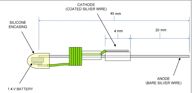
This device was slightly modified from the previous ver- sion. Bare silver wire continued to be used as the anode. Teflon coated silver wire replaced 35 gage copper wire as the cathode. In addition to the shrink tubing used as insulation between the anode and cathode, another piece of the tubing was introduced to hold the anodic and ca-thodic wires together. The size of medical grade silicone encapsulation was reduced. Length of the device was reduced to 45 mm. These devices were also used with high amperage (no resistor). The schematic design of the device is shown in Figure 9.
Four rats were implanted with this device out of which 3 survived through the post surgery incubation period.
Two of the remaining 3 rats were found to be Staph free.
Of the 10 rats that had antimicrobial implant devices and survived through the incubation period post surgery in animal testing studies #2, the rats implanted with these devices looked the healthiest. This design eliminated all drawbacks of the previous versions and seemed very robust.
5. STATISTICAL ANALYSIS
[image:8.595.111.490.405.588.2]Statistical testing was conducted in order to determine any significant statistical difference between the devices used in surgeries. Because several of the animals died pr- ior to harvest and 3 of the devices did not function after they were implanted, unequal test samples resulted for all categories. This made performing a paired test impos- sible so an F-Test was conducted to determine significa- nce. The first test conducted compared all effective con-trol devices to all effective antimicrobial devices. The term effective here means that the devices remained in the tibia post-surgery and were extracted from the live rats after an incubation period. These results are summa-rized in Table 3. Using a Fischer’s F-Test at 85% confi-dence level, we can conclude that there is a statistically signify- cant difference between antimicrobial and con-trol devices.
[image:8.595.112.485.640.722.2]Figure 9. Antimicrobial device #6.
Table 3. Summary of all devices for all surgeries (total)
Staph aureus free Staph aureus Total
Antimicrobial device 7 12 19
Control Device 0 7 7
During animal testing studies #1, antimicrobial de-vices were found to be electrically disconnected proxi-mal to the tibial cavity insertion point of 2 rats, render-ing the devices electrically ineffective. If these 2 devices are considered as control instead of antimicrobial devic- es, the F-test concludes that there is a statistically sign- ificant difference between antimicrobial devices and con- trol devices at 95% confidence level. In addition, each device design was compared with others to determine any statistical significance. Strictly statistically speaking, only antimicrobial device #1 and antimicrobial device #6 showed significant difference from the controls among all six design iterations. Although 2 out of 3 animals implanted with working antimicrobial device #1 tested
staph free, the device tip often penetrated the soft tissue
and caused irritation to the animals. On the other hand, antimicrobial device #6 also resulted in 2 out of 3 ani-mals being staph free but eliminated the problem
associ-ated with damaging the soft tissue.
6. DISCUSSION AND CONCLUSIONS
An early conjecture in this research was that a bacteri-cidal environment would be created if we could get bac-teria to conduct Ag+. The hope was to create this envi-ronment within a bone because getting traditional antibi-otics to penetrate the bone while the antibiantibi-otics are still viable can be very difficult. A basic design concept was developed and had to go through several iterations gov-erned by rat comfort after implant as well as bactericidal performance. Interestingly, the first and the last device design iterations showed the greatest efficacy.
Based on the surgeries, pathology results and statisti-cal analysis, the rat osteomyelitic model described in lit- erature is validated, since all animals without an antimic- robial device were infected. More importantly, the resu- lts show that properly configured electrically stimulated silver is an effective bactericidal agent for indwelling devices. Of all the surgeries performed using the bacteri-cidal devices, there is a statistically significant difference between using no device and an antimicrobial device. As the antimicrobial devices and surgical procedures were refined throughout the study, the effectiveness of the devices was found to be improved. In the last set of sur-geries, 67% of the harvested animals were free of Sta- phylococcus aureus even after they were inoculated with
the bacteria and given no antibiotics. The bactericidal device as configured has a definite ability to reduce/ eliminate bacterial infection. Using such a bactericidal device in conjunction with a standard treatment of anti-biotics should have a profound effect on the number of residual hardware associated bacterial infections.
7. ACKNOWLEDGEMENTS
The technology and designs tested within this study are protected under U. S. Patent as owned by ArgentumCidalElectrics, Inc.. It is only with their support that device modifications and manufacturing could be properly completed and controlled.
REFERENCES
[1] Iorio, R., Robb, W.J., Healy, W.L., Berry, D.J., Hozack, W.J., Kyle, R.F., Lewallen, D.G., Trousdale, R.T., Jira-nek, W.A., Stamos, V.P. and Parsley, B.S. (2008) Or-thopaedic surgeon workforce and volume assessment for total hip and knee replacement in the united states: Pre-paring for an epidemic. The Journal of Bone and Joint Surgery, 90(7), 1598-1605.
[2] Kurtz, S., Ong, K., Lau, E., Mowat F. and Halpern, M. (2007) Projections of primary and revision hip and knee arthroplasty in the United States from 2005 to 2030. The Journal of Bone and Joint Surgery, 89(4), 780-785. [3] Cumming, D. and Parker, M.J. (2007) Urinary
catheteri-sation and deep wound infection after hip fracture sur-gery. International Orthopaedics, 31(4), 483-485.
[4] Fitzgerald Jr.R.H. (1992) Total hip arthroplasty sepsis: Prevention and diagnosis. Orthopedic Clinics of North America, 23(2), 259-264.
[5] Furst, A. and Schlauder, M.C. (1978) Inactivity of two noble metals as carcinogens. The Journal of Environ-mental Pathology, Toxicology and Oncology, 1(1), 51- 57.
[6] Solberg, B.D., Gutow, A.P. and Baumgaertner, M.R. (1999) Efficacy of gentamycin impregnated resorbable hydroxyapatite cement in treating osteomyelitis in a rat model. Journal of Orthopaedic and Trauma, 13(2), 102-
106.
[7] Furno, F., Morley, K.S., Wong, B., Sharp, B.L., Arnold, P.L., Howdle, S.M., Bayston, R., Brown, P.D., Winship P.D. and Reid, H.J. (2004) Silver nanoparticles and polymeric medical devices: A new approach to preven-tion of infecpreven-tion? The Journal of Antimicrobial Chemo-therapy, 54(6), 1019-1024.
[8] Klasen, H.J. (2000) Historical review of the use of silver in the treatment of burns Part 1: Early uses. Burns, 26(2), 117-130.
[9] Klasen, H.J. (2000) Historical review of the use of silver in the treatment of burns Part 2: Renewed interest for sil-ver. Burns, 26(2), 131-138.
[10] Samuel, U. and Guggenbichler, J.P. (2004) Prevention of catheter-related infections: The potential of a new nano- silver impregnated catheter. The International Journal of Antimicrobial Agents,23(Suppl 1), 75-78.
[11] Langreth, R. and Herper, M. (2006) Germ Warfare. Forbes. http://www.forbes.com/forbes/2006/0619/060.html. [12] Melaiye, A. and Youngs, W.J. (2005) Silver and its
ap-plication as an antimicrobial agent. Expert Opinion on TherapeuticPatens, 15(2), 125-130.
[13] Price, W.R. and Wood, M. (1996) Silver nitrate burn dressing: Treatment of seventy burned persons. American Journal of Surger , 112(5), 674-680.
microbicides in preventing infections in healthcare. In:
Block SS. Disinfection, sterilization, and preservation, Li-
ppincott Williams & Wilkins, Philadelphia, PA, 415-430. [15] Slawson, R.M., Van Dyke, M.L., Lee, H. and Trevors, T. J. (1992) Germanium and silver resistance, accumulation and toxicity in microorganisms. Plasmid, 27(1), 72-79.
[16] Alexander, J. and Aaseth, J. (1981) Hepatobillary trans-port and organ distribution of silver in the rat as influ-enced by selenite. Toxicology, 21(3), 179-186
[17] Fowler, B.A. and Nordberg, G.F. (2007) Silver. In: Friberg, G.F. Nordberg and V. Vouk (Eds.), Handbook on the toxicology of Metals, 2nd Edition, Elsevier,
Am-sterdam, 521-531.
[18] Baldi, C., Minoia, C., Di Nucci, A., Capodagio, E and Manso, L. (1988) Effects of silver in isolated rat
hepato-cytes. Toxicology Letters, 41(3), 261-268
[19] Rungby, J. (1990) An experimental study of silver on the nervous system and on aspects of its general cellular toxicity. Danish Medical Bulletin, 37(5), 442-449
[20] Lucke, M., Wildemann, B., Sadoni, S., Surke, C., Sch- iller, R., Stemberger, A., Raschke, M., Haas, N.P. and Schmidmaier, G. (2005) Systematic versus local applica-tion of gentamicin in prophylaxis of implant-related os-teomyelitis in a rat model. Bone, 36(5), 770-778.
[21] Lucke, M., Schmidmaier, G., Sadoni, S., Wildemann, B., Schiller, R., Stemberger, A., Haas, N.P. and Raschke, M. (2003) A new model of implant related osteomyelitis in rats. Journal of Biomedical Materials Research, 67B(1),