Sequence and phylogenetic analysis of the gene
for surface layer protein,
slpA
, from 14 PCR
ribotypes of
Clostridium difficile
De´irdre Nı´ Eidhin,
1Anthony W. Ryan,
1Rachael M. Doyle,
1,23
J. Bernard Walsh
1,2and Dermot Kelleher
1Correspondence De´irdre Nı´ Eidhin [email protected]
Department of Clinical Medicine and Dublin Molecular Medicine Centre, Trinity College Dublin, Trinity Centre for Health Sciences1and Mercer’s Institute for Research in Ageing2,
St James’s Hospital, James’s St., Dublin 8, Ireland
Received 17 June 2005 Accepted 23 September 2005
Clostridium difficileis the commonest cause of antibiotic-associated diarrhoea, with the hospitalized elderly being at particular risk. The organism makes a crystalline surface protein layer (S-layer), encoded by theslpAgene, the product of which is cleaved to give two mature peptides which associate to form the layer. The larger peptide (high molecular weight; HMW), derived from the C-terminal portion of the precursor, is relatively conserved, whereas the smaller peptide (low molecular weight; LMW), derived from the N-terminal portion of the precursor, is a dominant antigen which substantially forms the basis for serotyping of isolates. PCR ribotyping is a more discriminatory typing method, based on the intergenic rRNA. We obtained the sequence forslpAand some flanking DNA from a collection ofC. difficilestrains of 14 ribotypes isolated from elderly patients. Sequences from different ribotypes were compared with one another and with published sequences. Sequences fromC. difficileribotypes 046 and 092 were identical. Sequences from ribotype pairs 005 and 054, 012 and 046/092, 014 and 066 and 031 and 094 differed by 1–3 nt in theslpAgene. There were ultimately nine ribotypes or groups of ribotypes with very differentslpAsequences, particularly in the region encoding the LMW peptide. The sequence from ribotype 002 was very different from previously published sequences. The DNA segment sequenced included the 59315 bp of asecAhomologue, encoding a putative transport protein required for peptide secretion across the plasma membrane. The amino acid sequences of the predicted HMW peptides were aligned and a neighbour-joining tree was produced using 10 000 bootstrap replicates. The predicted SecA N-terminal region was similarly analysed. For both SlpA and SecA, a strong association was found between ribotypes 012, 046/092, 017, 031 and 094. Ribotypes 001 and 078 formed part of this clade for SlpA but not SecA, indicating independent evolution forslpAandsecA, presumably because they come under different selection pressures.
INTRODUCTION
Clostridium difficileis now the leading cause of nosocomial diarrhoea among hospitalized patients undergoing anti-biotic treatment and is associated with substantial morbidity and mortality. The spectrum of disease ranges from mild diarrhoea to pseudomembranous colitis, which can be fatal (Kelly & LaMont, 1998). The infection routinely requires
isolation of affected patients, additional antibiotic therapy and a prolongation of hospital stay, which has implications for patient turnover and health economics (Kyneet al., 2002).
C. difficilemakes a crystalline protein surface layer (S-layer), a structural feature of many bacteria. S-layers have been ascribed various roles including nutrient uptake, exclusion of noxious substances, antiphagocytosis and colonization (Sa´ra & Sleytr, 2000). InC. difficile, the S-layer is the pre-dominant surface antigen, and a strong serum IgG response has been found among convalescent patients (Doyle, 2004; Pantostiet al., 1989). There are a number of variant types which are serologically distinct, substantially forming the basis for serotyping ofC. difficilestrains (Delme´eet al., 1986, 1990).
3Present address:St Columcille’s Hospital, Loughlinstown, Co. Dublin, Ireland.
The GenBank/EMBL/DDBJ accession number for the slpA and flanking sequences ofC. difficileisolates are DQ060625–DQ060643. Compositional data on SlpA from different PCR ribotypes are available as supplementary material in JMM Online.
46204G2006 SGM Printed in Great Britain 69
The S-layer is encoded by the slpA gene, the product of which contains a cleavable signal sequence and is further cleaved to give two mature peptides which then associate to form the S-layer (Calabiet al., 2001; Karjalainenet al., 2001). The peptide derived from the N-terminal region of the precursor is smaller and more highly variable, whereas the peptide derived from the C-terminal region is relatively conserved. For convenience, the two products are respec-tively known as the low molecular weight (LMW) and high molecular weight (HMW) peptides. The LMW peptide appears to be the main serotyping antigen (Poxtonet al., 1999). The HMW peptide has sequence similarity to the N-acetylmuramoyl-L-alanine amidase fromBacillus subtilis, and has been shown to possess amidase activity (Calabi et al., 2001). A number of other genes encoding putative amidases, known asslpA paralogues, occur in the vicinity ofslpA(Calabi & Fairweather, 2002).
A recent study (Doyle, 2004) described the isolation and typing ofC. difficilefrom a large number of patients who developed diarrhoea while attending the care of the elderly unit at St James’s Hospital, Dublin. A total of 14 types were identified within this population by PCR ribotyping, which is based on polymorphism of the intergenic DNA between the regions encoding 16S and 23S rRNA (O’Neill et al., 1996). Since 116 PCR ribotypes of C. difficile have been identified to date (Stubbset al., 1999), this typing method is considered more discriminatory than serotyping, which only distinguishes 21 types (Delme´eet al., 1990). Our pri-mary objective was to sequence theslpAgene from the PCR ribotypes identified and to compare the sequences with published data.
Calabiet al. (2001) reported a large ORF immediately down-stream ofslpAwith strong sequence similarity to the secA gene of other bacterial species. ThesecAproduct is an essen-tial component of the general secretory pathway in the Bacteria. It is a large protein which interacts with nascent proteins and with other components of the export pathway and provides energy for translocation by ATP hydrolysis (Schmidt & Kiser, 1999). This multifunctionality is reflected in a high degree of sequence conservation forsecAbetween species. The sequence we obtained from each isolate included 315 bp from the 59end of this gene. The consistent presence of a short segment of a conserved housekeeping gene in proximity toslpAprovided reassurance that we had sequenced the genuineslpAallele in each case and not one of its many paralogues. We also constructed phylogenetic trees for segments of the translated slpA and secA genes and compared their variability between ribotypes.
METHODS
C. difficileisolates and culture. C. difficileisolates were selected from a collection of all patient isolates obtained from July 1998 to December 1999 at the care of the elderly unit at St James’s Hospital (Doyle, 2004). Isolates were typed by Jon Brazier at the Anaerobe Reference Unit, Public Health Laboratory Service (since renamed the Health Protection Agency), University Hospital of Wales, Cardiff,
and identified by toxin production and PCR ribotype according to the scheme of O’Neillet al. (1996), based on the variable 16S–23S intergenic spacer region. The strains, identified by their Anaerobe Reference Unit designation, are listed in Table 1. Where possible, more than one isolate was selected, separated by date of isolation or ward or both. Three such well-separated isolates were selected for ribotype 001 (which accounted for approximately half of all isolates) and two each for ribotypes 002, 005 and 012. Single isolates were tested for the remaining ribotypes, either because only one isolate was available or because isolates occurred close together. Cultures were grown in anaerobic jars on Columbia blood agar (Lab M) with 7 % defibrinated horse blood, fastidious anaerobe broth (Lab M) or pre-reduced brain heart infusion broth containing 0?5 % (w/v) thioglycolate (BHI-TG). Stocks were maintained in cooked meat medium (Oxoid), made up in fastidious anaerobe broth.
Preparation of SlpA fromC. difficile. SlpA was prepared from early stationary phase cultures grown in BHI-TG by extraction with 8 M urea as described by Cerquetti et al. (2000). Extraction was done in the presence of Complete protease inhibitor cocktail (Roche Diagnostics). Extracts were dialysed against 50 mM Tris/HCl pH 7?4 and the protein content was measured by Bradford assay. Samples (5mg total protein) were visualized by SDS-PAGE on 12 % total monomer gels stained with Coomassie brilliant blue R. Relative molecular mass standards (Sigma wide molecular weight range) were included on each gel.
DNA isolation, amplification and sequencing. DNA was isolated from overnight BHI-TG-grownC. difficilecultures using the Gentra Puregene DNA isolation kit for yeast and Gram-positive bacteria with an additional proteinase K step (400mg ml21added to the lytic buffer, incubation for 1 h at 55uC followed by 10 min at 80uC) and omission of RNase treatment. DNA was amplified by PCR using HotStarTaqpolymerase (Qiagen), with an initial DNA denaturation step of 15 min at 95uC followed by 30 cycles of denaturation for 1 min at 94uC, annealing at 37uC for 30 s and extension at 68uC for 2 min 45 s. A final extension was done at 72uC for 10 min. The primers used were (forward) 2107F (59-ATGGATTATTATAGA-GATGTGAG-39) or 227F (59-AATATAATGTTGGGAGG-39) and (reverse)+562R (59-ACCTTCACCAGTTTTCAT-39). Primer227F was used to amplify DNA from ribotypes 002, 010, 014 and 066. Product purity, size and yield were checked on 0?8 % agarose gels using lambda DNA cut with EcoRI and BamHI or with EcoRI andHindIII as standard. DNA products were cloned in the pBAD ThioTOPO vector and transformed into competentEscherichia coli Top10, supplied as One Shot competent cells[genotype F2 mcrA D(mrr–hsdRMS–mcrBC)w80lacZDM15DlacX74recA1 deoR araD139 D(ara–leu)7697galU galK rpsL(StrR)endA1 nupG], as recommended by the manufacturer (Invitrogen). Recombinants were selected on LB agar supplemented with ampicillin and checked by restriction digestion and plasmid DNA was isolated for sequencing. Sequencing was also carried out on PCR products directly. DNA was sequenced commercially at the Biochemistry Department, University of Cambridge. We designed the custom primers required to complete sequencing from both strands.
0?2. Secondary structure prediction was based on the consensus from theSOPM(Geourjon & Dele´age, 1994),HNN(Guermeur, 1997),DPM (Dele´age & Roux, 1987), DSC (King & Sternberg, 1996), GOR IV (Garnier et al., 1996), PHD (Rost & Sander, 1994), PREDATOR (Frishman & Argos, 1996) andSIMPA96 (Levin, 1997) tools, using the NPS interface (Combet et al., 2000). Internal peptide repeats were detected using theRADARtool (Heger & Holm, 2000). Molecular mass and pI of predicted mature peptides were calculated by the Compute pI/Mw tool of the ExPASy proteomics server of the Swiss Institute of Bioinformatics (http://us.expasy.org/tools/pi_tool.html; Gasteigeret al., 2003). Codon usage was calculated by the CODONFREQUENCYtool from the Wisconsin Package (Accelrys Inc.) and values for relative synonymous codon usage were calculated. Relative synonymous codon usage is defined as the observed occurrence of a given codon divided by the expected occurrence. Values close to 1 are indicative of a lack of bias. Rho-independent terminators were detected by the TERMINATORtool from the Wisconsin package (Brendel & Trifonov, 1984). To study evolutionary relationships between ribotypes, amino acid sequences were aligned using CLUSTAL W as before. Pairwise Poisson correction distances, which correct for multiple substitu-tions at the same site, were calculated from the resulting alignment and unrooted neighbour-joining trees were drawn from the resulting distance matrix using MEGA2 software (Kumar et al., 2001). Bootstrap analyses of the phylogeny were performed using 10 000 bootstrap replications.
RESULTS
Amplification and sequencing ofslpA gene and flanking DNA
TheslpAgene and flanking DNA was sequenced from strains of all 14 ribotypes isolated from patients at St James’s
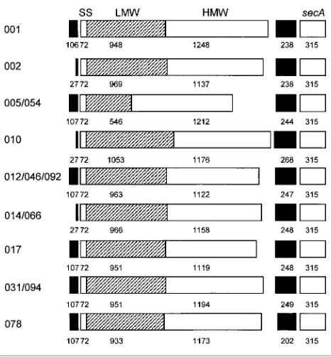
Hospital over a 16 month period (Table 1). Forward pri-mer2107F, based on non-coding sequence starting at posi-tion 2107 from the slpA gene from strain 630 (ribotype 012), was not successful in amplifying sequences from all ribotypes. Forward primer227F, used to amplify DNA from ribotypes 002, 010, 014 and 066, was based on a 17 nt stretch beginning 26–27 nt upstream of theslpAgene which was conserved among the remaining ribotypes. A single reverse primer, +562R, based on nt 298–315 of the secA homologue from strain 630 (Calabiet al., 2001), was used throughout. A single product was obtained from each isolate under the PCR conditions chosen. Products varied from 2496 to 2927 bp depending on ribotype (Fig. 1). Each fragment contained a complete ORF of 1830 to 2301 bp, presumed to beslpA, and terminated in a fragment con-taining 315 bp from the 59end of another ORF, predicted to be asecAgene based on similarity to the allele in the geno-me sequence. The intergenic DNA between the two ORFs varied from 202 to 268 bp. The upstream DNA segment was 106–107 bp for products generated from primer2107F and 26–27 bp for products generated from primer227F.
Comparison of sequences of slpA and flanking DNA from Dublin isolates
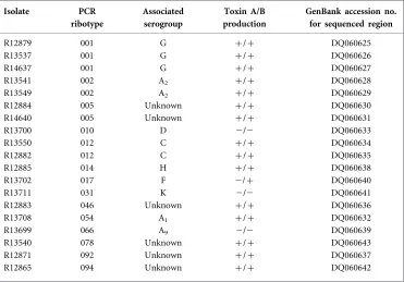
[image:3.595.54.425.113.372.2]Sequences obtained from different isolates of the same ribo-type were all identical. The fragments from riboribo-types 046 and 092 were also identical, and their sequences were treated as one in subsequent analyses. The sequences from some ribotypes were almost identical, with 1–3 nt differences Table 1.C. difficilestrains used in this study
Isolates were obtained from St James’s Hospital, Dublin (1998–2000) (Doyle, 2004). Serogroups are as assigned by Stubbset al. (1999) and Brazier (2001).
Isolate PCR
ribotype
Associated serogroup
Toxin A/B production
GenBank accession no. for sequenced region
R12879 001 G +/+ DQ060625 R13537 001 G +/+ DQ060626 R14637 001 G +/+ DQ060627 R13541 002 A2 +/+ DQ060628 R13549 002 A2 +/+ DQ060629 R12884 005 Unknown +/+ DQ060630 R14640 005 Unknown +/+ DQ060631
R13700 010 D 2/2 DQ060633
R13550 012 C +/+ DQ060634 R12882 012 C +/+ DQ060635 R12885 014 H +/+ DQ060638 R13702 017 F 2/+ DQ060640
R13711 031 K 2/2 DQ060641
R12883 046 Unknown +/+ DQ060636 R13708 054 A1 +/+ DQ060632 R13699 066 A9 2/2 DQ060639 R13540 078 Unknown +/+ DQ060643 R12871 092 Unknown +/+ DQ060637 R12865 094 Unknown +/+ DQ060642
http://jmm.sgmjournals.org 71
which always occurred inslpA(Table 2). These differences always translated to amino acid differences, often radical. Fig. 2(a) shows aCLUSTAL Walignment of the translatedslpA ORFs from the different ribotypes, with residue differences between pairs of nearly identical peptides underlined. Thus ribotype pairs 005 and 054 differed by 3 nt and 2 amino acid residues and pairs 012 and 046/092, 014 and 066 and 031 and 094 each differed by 1 nt and 1 amino acid residue. From the 14 ribotypes, we thus identified nine classes which varied substantially in sequence.
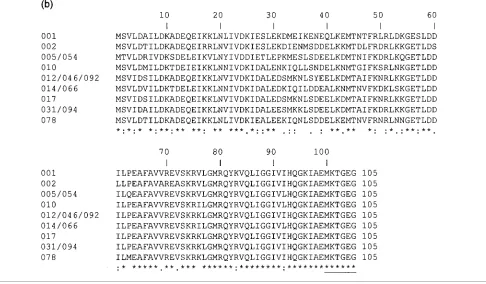
As noted by others, the sequence of the predicted HMW peptide was far more strongly conserved than that of the LMW peptide, with the alignment showing substantial blocks of similar or identical sequence interspersed with stretches which varied in length and in sequence between ribotypes. The pattern was quite different for the available 105 residues of the SecA homologue sequence (Fig. 2b), which showed complete alignment and strong conservation (over 62 % identical residues).
Comparison with other published slpA sequences
Sequence differences betweenslpAgenes from the ribotype groups identified among the Dublin isolates are summa-rized in Table 2 and comparisons are made with published sequences. Many of the strains from which the slpA gene
has been sequenced are known by serogroup only and, since there are more ribotypes than serogroups, a serogroup designation alone is often ambiguous. Moreover, not all ribotypes have been assigned to serogroups. Nonetheless, all publishedslpAsequences from ribotype 012 (or serogroup C) and 017 (or serogroup F) strains are identical to the cor-responding sequences from our collection. Indeed, strong similarity was generally found between sequences from strains of a given serogroup. Serogroup A was an exception. This grouping is based primarily on a shared flagellar anti-gen, and subgroups are based on cross-reactivity of bac-teria from which flagella have been removed mechanically (Delme´eet al., 1990). It is therefore to be expected thatslpA sequences from strains of different serogroup A subgroups may vary. Surprisingly, the sequence from a serogroup A10 strain showed strong similarity to the sequence from serogroup A1 strains. The strain in question, the only A10 strain for which anyslpA sequence is available, is peculiar among serogroup A strains in that it has no flagella and does not react with anti-flagellin serum (Delme´e et al., 1990). Also surprisingly, we found only 1 nt difference between slpA from ribotype 014 (serogroup H) and ribotype 066 (serogroup A9). We infer that limitations of the serogroup-ing method may result in discrepancies such as these.
We noted a close similarity between sequences from ribo-types 005, 016 and 054. Ribotype 005 has not been assigned to a serogroup, but itsslpAsequence closely resembles that of a serogroup A1 strain (Karjalainenet al., 2001) and that of ribotype 054, which has been assigned to serogroup A1. No sequence strongly similar to theslpAfrom ribotype 002 was found among publishedslpAsequences.
Prediction of post-translational cleavage sites
The predicted N-terminal amino acid sequence was well conserved for SlpA and showed the hallmarks of a Gram-positive cleavable sequence (Fig. 2a; van Welyet al., 2001). For most ribotypes, both the neural network and hidden Markov models predicted the cleavage site just C-terminal of A24. This prediction concurs with experimental pre-diction based on N-terminal sequencing of the native SlpA LMW peptide for ribotypes 001, 012 and 017 and possibly 010, 014 and 066 (Calabiet al., 2001; Cerquettiet al., 2000). For ribotypes 002, 005 and 054, there were slight discre-pancies between the predictors, with only the neural network predicting the cleavage site after position A24 for ribotype 002 and only the hidden Markov model making a similar prediction for ribotypes 005 and 054. This am-biguity may have been caused by the three consecutive alanine residues at positions 24–26 in these ribotypes (J. D. Bendtsen, personal communication), since alanine is strongly favoured in the position preceding the cleavage site. Position 24 occurs at the end of a highly conserved stre-tch of 7 amino acid residues with the consensus sequence SAAPVFA, which seems unlikely to be coincidental.
[image:4.595.44.277.62.313.2]The position of the secondary cleavage site, to cleave the precursor SlpA into the LMW and HMW peptides, was
Table 2. Comparison of similarslpAsequences from Dublin (SJH) isolates and comparison with publishedslpAsequences
All Dublin isolates of a given ribotype had identical sequences; numbers of strains are indicated in parentheses. Serogroups and ribotypes are listed according to known correlations between ribotype and serogroup from Stubbs et al. (1999) and Brazier (2001) unless indicated. Since there are more serogroups than ribotypes and not all ribotypes have been assigned to a serogroup, it is more tenuous to use knowledge of serogroup to predict ribotype.
SJH ribotype (n)
Associated serogroup
Compared strain
Origin Ribotype Serogroup Sequence differences Reference*
nt aa
001 (3) G R8366 UK 1D G 0 0 AJ300676 (Calabiet al., 2001)
ATCC 43599 Belgium 001, 115 GD 8 1 AF448128(Karjalainenet al., 2002)
96-392 France 001, 115 GD 8 1 AF448129(Karjalainenet al., 2002)
005 (2) Unknown ATCC 43594 Belgium 021, 054, 075 A1D 4 (includes
3 extra nt)
4 AF458877(Karjalainenet al., 2002)
R13708 Ireland 054D A1 3 2 This study
054 (1) A1 167 USA 016d Unknown 4 3 AF478570 (Calabi & Fairweather, 2002)
TO005 Canada 039, 067 A10D 12 (includes gaps and
extra nt)
19 (due to frame-shift) AF458878(Karjalainenet al., 2002)
010 (1) D ATCC 43597 Belgium 010 DD 0 0 AF458880(Karjalainenet al., 2002)
90-111 France 010 DD 0 0 AF458881(Karjalainenet al., 2002)
93-136 France 010 DD 0 0 AF458882(Karjalainenet al., 2002)
Y UK 010d D 4 3 AF478571 (Calabi & Fairweather, 2002)
012 (2) C 630 Switzerland 012D C 0 0 Sanger sequencing project
C253 Italy 012 CD 0 0 AJ291709 (Karjalainenet al., 2001)
ATCC 43596 Belgium 012 CD 0 0 AF448123(Karjalainenet al., 2002)
R12883 Ireland 046D Unknown 1 1 This study
R12871 Ireland 092D Unknown 1 1 This study
046 (1) Unknown R12871 Ireland 092D Unknown 0 0 This study
066 (1) A9 R12885 Ireland 014D H 1 1 This study
ATCC 43600 Belgium 014, 020 HD 2 1 AF448365(Karjalainenet al., 2002)
89-638 France 014, 020 HD 2 1 AF448366(Karjalainenet al., 2002)
90-204 France 014, 020 HD 2 1 AF448367(Karjalainenet al., 2002)
017 (1) F R7404 UK 017D F 0 0 AJ300677 (Calabiet al., 2001)
ATCC 43598 Belgium 017 FD 0 0 AF448125(Karjalainenet al., 2002)
GAI 95600 Japan 017 FD 0 0 AF448126(Karjalainenet al., 2002)
GAI 95601 Japan 017 FD 0 0 AF448127(Karjalainenet al., 2002)
094 (1) Unknown R13711 Ireland 031D K 1 1 This study
ATCC 43602 Belgium 031, 053, 057 KD 3 (includes 1 extra nt,
1 nt gap)
18 (due to frame-shift) AF448368(Karjalainenet al., 2002)
94-416 France 031, 053, 057 KD As above As above AF448369(Karjalainenet al., 2002)
48-515 Belgium 031, 053, 057 KD As above As above AF448370(Karjalainenet al., 2002)
078 (1) Unknown 9354 France Unknown AD (unknown subgroup) 3 2 AF448120(Karjalainenet al., 2002)
*Accession numbers in italics represent partial sequences (1017–1185 nt from 59 region ofslpA).
DFrom listed reference.
dPersonal communication from Neil Fairweather.
http://jmm.sgmjournals.org
73
Variants
of
slpA
gene
from
C.
predicted from comparison with published N-terminal amino acid sequence of the mature HMW peptide (Calabi et al., 2001; Cerquetti et al., 2000). For previously un-published sequences, prediction was based on comparison with published sequences (Fig. 2a). Cleavage is generally
predicted to occur N-terminal to an alanine or serine residue and C-terminal to a consensus motif TKS or TYX. Cleavage might actually occur some way upstream of this site, with some residues lost from the N terminus of the peptide during maturation. An absolutely conserved GKR motif
http://jmm.sgmjournals.org 75
occurs close to the predicted C termini of all the LMW peptides, which are otherwise generally dissimilar (Fig. 2a). The GKR motif is generally followed by a leucine residue, or occasionally valine, and is preceded by a conserved tyrosine at position28, an aromatic residue at23 and hydrophilic and hydrophobic residues at positions27 and24, respec-tively. The pattern appears in all other published sequences, although, presumably due to the heterogeneity of the LMW peptide, computer-generated alignments do not always recognize it (Calabi & Fairweather, 2002; Karjalainenet al., 2002).
Internal repeats in the HMW peptide
The RADARprogram identified an 11 residue motif in the HMW peptide, beginning 40–50 residues from the predicted N terminus and repeated twice at approximately 100–120 residue intervals (Fig. 2a). Four positions were absolutely conserved in all three repeats and a fifth position contained strongly similar residues. Another motif, repeated once, was
found N-terminal of the second and third repeats. Both types of repeat occurred in regions of the sequence which were strongly conserved between ribotypes, so that a given repeat showed more similarity between ribotypes than to other repeats within a ribotype. For example, the unvarying DR of the principal motif was followed by the dissimilar residues isoleucine, tyrosine and glutamine in repeats 1, 2 and 3, respectively.
Molecular mass and isoelectric point prediction for the mature peptides
[image:8.595.53.539.67.351.2]Fig. 3 shows an SDS-PAGE gel of crude SlpA preparations from all ribotypes. SlpA is seen as two dominant bands for all but ribotypes 005 and 054, where a single strong band was observed at approximately 45 000. Calabi & Fairweather (2002) report a similar absence of the second band for strain 167, which is ribotype 016 (Neil Fairweather, personal com-munication) and has an almost identicalslpAsequence to that of ribotype 054. As expected, migration profiles of SlpA
peptides were identical for ribotypes with identical or nearly identicalslpAsequences. Relative molecular mass (exclusive of glycosylation) and pI were calculated using ExPASy soft-ware (Supplementary Table S1 available in JMM Online). The SlpA peptides tended to migrate more slowly than predicted, probably due to the high content of acidic amino acid residues or to post-translational modification. The pI range for the LMW peptides was within the typical range (4–6) for bacterial surface layer proteins (Sleytret al., 1999) and was consistently more acidic for the HMW peptide (4?46–4?69) than for the LMW peptide (4?83–5?09).
Amino acid composition
There was broad similarity when amino acid composition of the mature SlpA peptides was compared between ribo-types. Supplementary Fig. S1(a) (available in JMM Online) shows a comparison of the average composition of the pre-dicted mature peptides, the translated slpA ORF and the global average of a compilation of 148C. difficile coding sequences (http://www.kazusa.or.jp/codon/). Typical fea-tures of bacterial surface layer proteins were present (Pum et al., 2000; Sa´ra & Sleytr, 2000; Sleytret al., 1999), i.e. high content of acidic amino acids, no cysteine and very little methionine, low arginine content and little or no histidine. Lysine, alanine, aspartic acid and valine were the most abundant amino acids, and the last three were present at strikingly high levels compared with the global average. The aromatic amino acid content was generally low. Among functionally similar groups of amino acids, there was a general preference for the same residue in both peptides, e.g. both peptides showed a strong preference for aspartate over glutamate and for tyrosine over phenylalanine, in contrast to the global average. A striking exception was threonine, which was much more abundant in the LMW peptide. Supplementary Fig. S1(b) shows a comparison for groups of functionally similar amino acids. The HMW peptide had a higher content of acidic and hydrophobic amino acids and a lower content of aromatic amino acids. The LMW peptide
from ribotypes 005 and 054, although much smaller than that from the other ribotypes (180 residues compared to ¢311 residues), contained numbers of asparagines close to the mean for all ribotypes, possibly indicating that a mini-mum number of these residues is essential to maintain the structural integrity of the protein.
Codon usage
Codon usage was analysed for the slpA genes of all ribo-types and for theslpAsegments encoding predicted mature peptides. Codon usage patterns were compared with those of the same compilation of 148 sequences used for compa-rison of amino acid composition (Supplementary Fig. S2a). The species bias in favour of codons containing minimal G and C was generally stronger for the slpA gene, which is predicted to be highly expressed. Where codons permitted a choice of A or T in the wobble position, there was an occa-sional variation in preference. However, the combined fre-quencies of synonymous codons with a higher AT content generally well exceeded those of codons with a higher GC content.
A comparison of codon usage between the 59and 39regions of slpA, encoding the LMW and HMW peptides, respec-tively (Supplementary Fig. S2b), showed some differences in usage patterns for several amino acids, while maintaining the general bias in favour of A or T in the wobble position. Thus GCA was strongly favoured over GCT for alanine in the LMW peptide, with the reverse being true of the HMW peptide; and GTA strongly favoured over GTT for valine in the LMW peptide, with the reverse being true for the HMW peptide. Analogous differences occurred for serine and leucine, which have six codons each, while maintaining the AT-rich bias. Phenylalanine was an exception, with TTC preferred to TTT for phenylalanine in the HMW peptide, though not the LMW peptide.
Flanking DNA
[image:9.595.53.289.67.194.2]In strain 630,slpA is flanked upstream by 213 nt of non-coding DNA preceded by an slpA paralogue and down-stream by 247 nt of non-coding DNA followed by thesecA homologue (Calabiet al., 2001). In our sequences, 106–107 nt of upstream sequence is available for most ribotypes and was found to be identical for ribotypes 012, 046, 092, 017 and 078, which differed by a single nucleotide from ribotypes 031 and 094 and showed greater divergence from ribotypes 001 and 005/054 (Fig. 4a). These ribotypes shared a 22 nt stretch a short distance upstream of theslpAstart codon, from which it was possible to design a primer to amplify the relevant sequence from the remaining ribotypes. This shared sequence included a polypurine stretch at positions29 to216 (28 to 215 for ribotypes 005 and 054), containing the motif GGGAGG, strongly suggestive of a ribosome-binding site (Shine–Dalgarno box) in composition and location. No rho-independent terminators were identified in any of the upstream sequences, either the 107 nt available from the fragments sequenced from 10 ribotypes or the 213 nt of
Fig. 3. SDS-PAGE profiles of crude SlpA preparations from different ribotypes of C. difficile. Preparations from ribotypes with similar or identical slpA sequences are in adjacent lanes (underlined). Relative molecular mass markers and their posi-tions (6103) are shown on the left.
http://jmm.sgmjournals.org 77
intergenic DNA from strain 630 (ribotype 012) known from the Sanger sequencing project. It is tentatively inferred that transcription of the upstreamslpAparalogue terminates by a rho-mediated process. Rho-dependent terminators are not readily identifiable in sequences (Henkin, 1996).
The distance between theslpAandsecAgenes varied from 202 nt (for ribotype 078) to 268 nt (for ribotype 010). Fig. 4(b) shows an alignment of the sequences. TheslpA gene terminated in TAA for all ribotypes except 078, where it ended in TAG. The first half of the sequence was relatively unconserved, especially for ribotypes 010 and 078, and was considerably shorter in the latter. Sequences encoding potential mRNA stem and loop structures, reminiscent of rho-independent terminators, were found in this region. Rho-independent terminators show similarity between widely distributed bacterial genera (Vermat et al., 2002). Potential regions of dyad symmetry were 10–13 bp, with an unpaired loop of 3–4 nt, and typically contained three or four GC pairs, which would stabilize the structure of the transcript. Two such structures were found in all ribo-types except 010 and 078, which had one each. In rho-independent terminators, the region of dyad symmetry commonly gives way to a 39run of non-pairing Ts, which is predicted to facilitate the release of the transcript, and the consensus sequence TCTG (Brendel & Trifonov, 1984). The terminators predicted for slpA were generally found to contain either the T-trail, the consensus sequence TATG/ TGTG or occasionally both. A region of quite strong con-servation was identified (59 % identity) from approximately nt2110 to nt28 to211 upstream ofsecA, ending in a very likely Shine–Dalgarno box forsecA(AGGAGG).
Phylogenetic analysis
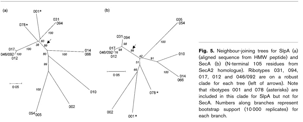
Evolutionary relationships between ribotypes based on the slpAgenes were examined by aligning the HMW peptides usingCLUSTAL Wand constructing neighbour-joining trees (Fig. 5a). Alignment gaps were omitted from the phylo-genetic analysis. The LMW peptide was not included in the analysis, as there is very little overall sequence conservation between ribotypes in this region and two of the ribotypes contain a much shorter peptide than the others. The same analysis was carried out with the available N-terminal 105 residues of sequence from the SecA homologue (Fig. 5b), so that variation in SlpA, presumed to be an antigen under evolutionary pressure to diversify, could be compared with variation in SecA, an essential protein which is not surface exposed.
[image:11.595.50.561.532.738.2]The trees suggest different evolutionary histories for slpA and secA, consistent with either recombination and/or positive selection. Recombination would produce new antigenic variants that could enable a strain to evade host defences, which could undergo positive selection. In parti-cular, both trees show a robust clade (indicated by an arrow) containing ribotypes 012, 046/092, 017, 031 and 094. How-ever, ribotypes 001 and 078 (indicated by asterisks) are also contained within this clade for the SlpA tree. This is con-sistent with recombination between lineages on the slpA gene. There are other differences between the trees, but these are less well supported statistically. Furthermore, the secA clade containing ribotypes 012, 046/092, 017, 031 and 094 exhibits considerably shorter branch lengths than the cor-responding slpA clade, consistent with the housekeeping role of SecA.
Fig. 5. Neighbour-joining trees for SlpA (a) (aligned sequence from HMW peptide) and SecA (b) (N-terminal 105 residues from SecA2 homologue). Ribotypes 031, 094, 017, 012 and 046/092 are on a robust clade for each tree (left of arrows). Note that ribotypes 001 and 078 (asterisks) are included in this clade for SlpA but not for SecA. Numbers along branches represent bootstrap support (10 000 replicates) for each branch.
Fig. 4.(a) Sequence immediately upstream fromslpA. The left-hand primers used to amplify the DNA are shown by arrows, to indicate where the sequence information may not be completely accurate, and the predicted Shine–Dalgarno box forslpA is underlined. (b) Intergenic region betweenslpAandsecAgenes. The positions of possible rho-independent terminators for theslpA gene are underlined, along with the predicted Shine–Dalgarno box for thesecAgene.
http://jmm.sgmjournals.org 79
DISCUSSION
We have sequenced theslpAgene and flanking DNA from C. difficile ribotypes isolated from patients in St James’s Hospital, Dublin over a 16-month period. The most fre-quently occurring ribotypes were 001, 012 and 017, andslpA had already been sequenced from these. However, for the first time, we report complete DNA sequences forslpAfrom strains formally assigned to ribotypes 002, 005, 010, 014, 031, 046, 054, 066, 078, 092 and 094. DNA sequence obtained from two or three isolates of each of the more common ribotypes was always identical, indicative of clonal spread. We also provide information on flanking DNA, including the 59315 nt from a putativesecAgene. The availability of sequence from an adjacent conserved gene provides reassur-ance that a single amplicon has been sequenced in all strains. Given the existence of numerous paralogues ofslpAin the genome, this is not a trivial consideration.
We show a strong relatedness between theslpA sequences from ribotypes 005, 016 and 054, between ribotypes 012, 046 and 092, between ribotypes 014 and 066 and between ribotypes 031 and 094. It is useful to have this sequence information onslpA, which is strongly related to serogroup designation and complements ribotyping, which is based on non-coding DNA that is subject to different evolutionary pressure. Since there are more ribotypes than serogroups, it is not surprising that different ribotypes should have similar serogrouping antigens. However, the sequence data we have acquired indicate how very alike someslpAsequences from different ribotypes are, at both the nucleotide and amino acid sequence levels. This near-identity occurs alongside important strain differences, such as toxin production. Thus ribotype 031, which is deficient in both A and B toxins, has an slpA with one nucleotide difference compared with ribotype 094, which produces both toxins. Similarly,slpAs from ribotypes 066 (non-toxigenic) and 014 (toxin A+and B+) differ by a single nucleotide. The sequence from ribotype 002 strains (equivalent serogroup A2 according to Stubbset al., 1999) does not appear to resemble closely that published for any other strain.
An international effort is being made to coordinate typing methods in order to correlate the dominant types in out-breaks ofC. difficiledisease around the world. Serogrouping, which is largely based on differences between SlpA variants, is complicated by the existence of flagellated strains, since flagellar antigens may cross-react and production of flagella can vary with culture age and conditions. Serogroup A strains share serologically cross-reactive flagellins and sub-groups were originally distinguished by SDS-PAGE profiles of major peptides, probably SlpA, in whole-cell preparations (Delme´e et al., 1986). The role of flagellar antigens was appreciated later (Delme´eet al., 1990) and it became pos-sible to distinguish the A subgroups by slide-agglutination on removal of the flagella by sonicating the bacteria. How-ever, it was also noted that strains of some other serogroups were flagellated and that cross-reactive flagellins occurred widely. Although it was claimed that non-serogroup-A
strains had fewer flagella, and did not cross-react with group A strains in slide-agglutination, it is conceivable that mis-takes could occur with heavily flagellated non-group-A strains.
Although some of the present work highlights the diver-sity of SlpA in different ribotypes and serogroups, some features appear consistently, notably the conserved stretch of sequence in the leader peptide, the GKRL/V motif and adjacent conserved residues in the vicinity of the secondary cleavage site and the five blocks of conserved sequence in the HMW peptide, interspersed with stretches of variable length and sequence. We also detected the DR-containing repeat motif, originally identified by Calabi & Fairweather (2002) in a comparison of SlpA molecules from six strains, in all of our sequences.
The GKRL/V motif is intriguing, given that GKR represents the longest stretch of absolutely conserved sequence in the LMW peptide and occurs near the precursor cleavage site. A considerable number of proteins from eukaryotes and viruses are cleaved post-translationally C-terminal to a pair of basic residues. These include mammalian growth factors and receptor proteins and glycoproteins from HIV and varicella zoster virus (reviewed by Seidah & Chre´tien, 1999). Some bacterial toxins are activated by cleavage at pairs of basic residues by host proteases, but cleavage of bacterial proteins by bacterial proteases at such sites has not been widely reported. It is possible that the conservation of the site we have identified has more to do with binding of the relevant protease or transient association with the plasma membrane to facilitate cleavage at the distal sites already predicted experimentally.
There are several reports (Calabiet al., 2001; Cerquettiet al., 2000) that SlpA is glycosylated, probably more extensively on the HMW peptide, but the nature and extent of gly-cosylation are unknown. The widespread occurrence of glycosylation among bacterial proteins, including S-layers, has only recently been accepted and its potential role in virulence appreciated (reviewed by Scha¨ffer & Messner, 2001; Schmidtet al., 2003; Spiro, 2002). Clostridial S-layers were among the first bacterial glycoproteins to be described (Sleytr & Thorne, 1976). In bacteria, glycopeptide linkages may beN-glycosyl on asparagine residues or, more often,O -glycosyl on serine, threonine and tyrosine residues. The sequence and structural requirements for glycosylation sites seem less well defined in prokaryotes than in eukaryotes, making their prediction from primary sequence virtually impossible, at least for the moment (Scha¨ffer & Messner, 2001). Although it is known that the toxins mediate their activity by glycosylation of a critical threonine residue on the host Rho family of GTPases (reviewed by Spiro, 2002), at least two toxin-negative strains are reported to have a glycosylated SlpA (Cerquetti et al., 2000), implying the existence of a separate mechanism for SlpA glycosylation.
C. difficileproteins. Some of these, e.g. the unusually high content of acidic amino acids and the absence of cysteine, are features of S-layers in general. There is a certain bias in favour of amino acids with smaller side-groups where these are functionally similar, i.e. aspartate is favoured over glutamate, asparagine over glutamine and alanine and valine over other hydrophobic amino acids, possibly because of the need for economy or possibly because of constraints imposed by the crystalline structure. Conversely, some amino acids, although present at a low level, may be critically important. Thus arginine is rare in SlpA, but where it occurs it is often in a relatively conserved region. Of the seven to ten arginines found in the HMW peptide, four positions are absolutely conserved and three of these are associated with the repeated DR motif. In the LMW peptide, which has two to five arginines, one is always found in the GKR motif.
S-layers composed of two peptides have been described for a few other organisms, but the SlpA fromC. difficileis the only reported incidence of a two-component S-layer derived from a single precursor peptide. The reason for this pheno-menon is not clear. Although it is known that the LMW and HMW peptides have different lattice structures (Cerquetti et al., 2000), detailed tertiary structure comparisons are not possible at this time. Secondary structure analyses (not shown) indicated that both peptides were composed predominantly of random coil (44–49 %) interspersed with alpha helical regions (28–34 %) and extended strand (19–22 %) and were not generally informative. We therefore compared the amino acid composition of the LMW and HMW peptides. This generally reflected the overall com-position of the translated ORF, with the greatest difference noted for threonine, which was quite abundant in the LMW peptide at an average of just over 11 %, compared with 5?8 and 5?7 %, respectively, for the HMW peptide and the global average. A survey of published eubacterial S-layer sequences shows that a high threonine content (>9 %) is quite com-mon, with the highest content (18?2 %) for Caulobacter crescentus, for reasons which have not been explained. The LMW peptide also had a higher content of aromatic amino acids, although SlpA had a rather low content of aromatic amino acids overall. We noted that the LMW peptide had two absolutely conserved tyrosines in close proximity (Fig. 2a), one absolutely conserved phenylalanine and one position which always contained either residue. It is tem-pting to suppose that the aromatic amino acids might be involved in binding of the LMW to a host ligand via a carbohydrate receptor or that they play an essential role in stabilizing the crystalline lattice structure.
Given the high rate of protein synthesis needed to maintain the integrity of the S-layer during growth and division (Sleytr & Messner, 1983), it is not surprising that codon usage inslpAreflects the strong AT-rich bias ofC. difficile. The differences in codon usage between the LMW and HMW peptides (albeit within this bias) may reflect different evolutionary origins or perhaps an influence of DNA secondary structure.
A short stretch of presumably non-coding DNA upstream of slpA was fairly well conserved among 10 ribotypes, but differed sufficiently to prevent the use of the original left-hand primer in ribotypes 002, 010, 014 and 066. The intergenic region fromslpAto secAvaried substantially in sequence, particularly the moiety closest to theslpA gene. The fragment from ribotype 078, which had an upstream sequence similar or identical to those of nine other ribo-types, seems truncated in the region immediately down-stream of slpA, lacking a predicted terminator and even employing a different termination codon. A recombination event in this region might explain why ribotype 078 is found on different clades in the bootstrap analyses of SlpA and SecA (Fig. 5). In ribotype 010, for which we have very little sequence upstream ofslpA, theslpA–secAintergenic region, which is relatively long, also appears to encode a single terminator. This ribotype is found on a fairly strong clade with ribotypes 014 and 066 for SecA in the bootstrap analyses, but is not strongly associated with any other ribotype for SlpA. There is little variation in the DNA sequence either upstream or downstream from slpA in ribotypes 012, 046/092, 017, 031 and 094, and all of these are quite tightly linked on both trees. However, ribotype 001, which shows small to moderate differences in upstream and downstream flanking sequences, is part of this clade for SlpA, but not for SecA. A strain that acquires a novel surface layer protein by recombination, thereby generating a new strain, presumably has greater ability to evade a host’s immune response. Toxin production, resulting in profuse diarrhoea, is an effective means of dispersing spores and spreading the disease. This may be the case with ribotype 001, which appears to have acquired a novelslpAgene by recombination with another clade and is responsible for approximately half of C. difficile infections in the UK (Brazier, 1998) and in St James’s Hospital (Doyle, 2004).
A sequence similarity search of the published genome forC. difficile630 has revealed the presence of a secondsecAallele located approximately 1 Mb from the gene flanking slpA and in the opposite orientation. Although uncommon, two secA alleles have been found in a number of genera, including Mycobacterium and Listeria, where the second SecA (SecA2) appears to have a specific role in the export of virulence-related proteins (Pallenet al., 2003). The product of the gene flanking slpA in C. difficile 630 (genome sequence) more closely resembles the SecA2 of other species, being the smaller of the two proteins and lacking a region predicted to interact with the chaperone SecB (Fekkeset al., 1997). Although it may play a subsidiary role, the predicted SecA2 does show strong similarity to its counterpart in other species, notably Listeria monocytogenes and Streptococcus parasanguinis(approx. 40 % identical residues), indicating a high degree of conservation between genera. SinceC. difficile is not known to grow outside a human or mammalian host in nature, ifsecA2is required for colonization, it is de facto an essential gene. The location of a gene for a major transport protein immediately downstream from slpA is unlikely to be coincidental, and it is known that S-layer
http://jmm.sgmjournals.org 81
expression inAeromonas salmonicidadepends on an ATP-dependent transport protein encoded by a downstream gene (Chu & Trust, 1993). Interestingly, in Listeria monocyto-genes, SecA2 is believed to be responsible for the secretion of two autolysins, NamA and p60, and deletion of eithersecA2 or either of the structural genes leads to accelerated clear-ance of the organism from spleens and livers of infected mice (Lenzet al., 2003). The HMW peptide ofC. difficile SlpA has N-acetylmuramoyl amidase activity, and slpA is located among a cluster of genes encoding similar domains (Calabi et al., 2001). Although C. difficile autolysins are predicted to be involved in wall remodelling during cell growth and division (Calabiet al., 2001), it is possible that they may have additional roles in virulence.
ACKNOWLEDGEMENTS
This work was supported by grants from Enterprise Ireland (ATRP-01/ 165) and from the Higher Education Authority. The authors wish to thank Ms Patricia O’Brien and the staff at the Microbiology Laboratory, St James’s Hospital, for advice on C. difficile culture. Thanks are also due to Dr Denis Shields, Royal College of Surgeons in Ireland, for help with interpreting neighbour-joining trees and to Neil Whitehead, DNA sequencing laboratory, Biochemistry Dept, University of Cambridge, for providing excellent support and service with DNA sequencing. Thanks to Dr Jon Brazier of the Anaerobe Reference Laboratory at the HPA in Cardiff for PCR ribotyping of isolates. We are grateful to Drs Neil Fairweather and Emanuela Calabi, Imperial College, London, for sharing unpublished data on ribotyping. Thanks to Dr Julian Parkhill and colleagues of theC. difficile(Strain 630) Sequencing Group at the Sanger Institute (http://www.sanger. ac.uk/Projects/C_difficile/) for providing open access to the genome sequence and for permission to publish our in silico findings with respect tosecAhomologues.
REFERENCES
Bendtsen, J. D., Nielsen, H., von Heijne, G. & Brunak, S. (2004). Improved prediction of signal peptides: SignalP 3.0.J Mol Biol340, 783–795.
Brazier, J. S. (1998). The epidemiology and typing of Clostridium difficile.J Antimicrob Chemother41 (Suppl. C), 47–57.
Brazier, J. S. (2001).Typing of Clostridium difficile. Clin Microbiol Infect7, 428–431.
Brendel, V. & Trifonov, E. N. (1984).A computer algorithm for testing potential prokaryotic terminators.Nucleic Acids Res12, 4411–4427. Calabi, E. & Fairweather, N. (2002).Patterns of sequence conserva-tion in the S-layer proteins and related sequences in Clostridium difficile.J Bacteriol184, 3886–3897.
Calabi, E., Ward, S., Wren, B., Paxton, T., Panico, M., Morris, H., Dell, A., Dougan, G. & Fairweather, N. (2001). Molecular charac-terization of the surface layer proteins fromClostridium difficile.Mol Microbiol40, 1187–1199.
Cerquetti, M., Molinari, A., Sebastianelli, A., Diociaiuti, M., Petruzzelli, R., Capo, C. & Mastrantonio, P. (2000). Characteriza-tion of surface layer proteins from different Clostridium difficile clinical isolates. Microb Pathog28, 363–372.
Chu, S. & Trust, T. J. (1993).AnAeromonas salmonicidagene which influences A-protein expression inEscherichia coliencodes a protein
containing an ATP-binding cassette and maps beside the surface array protein gene.J Bacteriol175, 3105–3114.
Combet, C., Blanchet, C., Geourjon, C. & Deleage, G. (2000). NPS@: network protein sequence analysis. Trends Biochem Sci 25, 147–150.
Dele´age, G. & Roux, B. (1987).An algorithm for protein secondary structure prediction based on class prediction.Protein Eng1, 289–294. Delme´e, M., Laroche, Y., Avesani, V. & Cornelis, G. (1986). Comparison of serogrouping and polyacrylamide gel electrophoresis for typingClostridium difficile. J Clin Microbiol24, 991–994. Delme´e, M., Avesani, V., Delferriere, N. & Burtonboy, G. (1990). Characterization of flagella ofClostridium difficile and their role in serogrouping reactions.J Clin Microbiol28, 2210–2214.
Doyle, R. M. (2004).Humoral immune response to Clostridium difficile associated disease. MD thesis, Trinity College Dublin, Ireland. Fekkes, P., van der Does, C. & Driessen, A. J. (1997).The molecular chaperone SecB is released from the carboxy-terminus of SecA during initiation of precursor protein translocation.EMBO J16, 6105–6113. Frishman, D. & Argos, P. (1996). Incorporation of non-local interactions in protein secondary structure prediction from the amino acid sequence.Protein Eng9, 133–142.
Garnier, J., Gibrat, J. F. & Robson, B. (1996). GOR method for predicting protein secondary structure from amino acid sequence. Methods Enzymol266, 540–553.
Gasteiger, E., Gattiker, A., Hoogland, C., Ivanyi, I., Appel, R. D. & Bairoch, A. (2003). ExPASy: the proteomics server for in-depth protein knowledge and analysis.Nucleic Acids Res31, 3784–3788. Geourjon, C. & Dele´age, G. (1994).SOPM: a self-optimized method for protein secondary structure prediction.Protein Eng7, 157–164. Guermeur, Y. (1997).Combinaison de classifieurs statistiques, applica-tion a` la pre´dicapplica-tion de structure se´condaire des prote´ines. PhD thesis, Universite´ Paris 6, France (in French).
Heger, A. & Holm, L. (2000).Rapid automatic detection and align-ment of repeats in protein sequences.Proteins41, 224–237. Henkin, T. M. (1996). Control of transcription termination in prokaryotes.Annu Rev Genet30, 35–57.
Karjalainen, T., Waligora-Dupriet, A. J., Cerquetti, M., Spigaglia, P., Maggioni, A., Mauri, P. & Mastrantonio, P. (2001). Molecular and genomic analysis of genes encoding surface-anchored proteins from Clostridium difficile.Infect Immun69, 3442–3446.
Karjalainen, T., Saumier, N., Barc, M. C., Delme´e, M. & Collignon, A. (2002).Clostridium difficilegenotyping based onslpAvariable region in S-layer gene sequence: an alternative to serotyping.J Clin Micro-biol40, 2452–2458.
Kelly, C. P. & LaMont, J. T. (1998).Clostridium difficile infection. Annu Rev Med49, 375–390.
King, R. D. & Sternberg, M. J. E. (1996). Identification and application of the concepts important for accurate and reliable pro-tein secondary structure prediction.Protein Sci5, 2298–2310. Kumar, S., Tamura, K., Jakobsen, I.-B. & Nei, M. (2001).MEGA2: molecular evolutionary genetics analysis software.Bioinformatics17, 1244–1245.
Kyne, L., Hamel, M. B., Polavaram, R. & Kelly, C. P. (2002).Health care costs and mortality associated with nosocomial diarrhea due to Clostridium difficile.Clin Infect Dis34, 346–353.
Lenz, L. L., Mohammadi, S., Geissler, A. & Portnoy, D. A. (2003). SecA2-dependent secretion of autolytic enzymes promotes Listeria monocytogenespathogenesis.Proc Natl Acad Sci U S A100, 12432–12437.
O’Neill, G. L., Ogunsola, F. T., Brazier, J. S. & Duerden, B. I. (1996). Modification of a PCR ribotyping method for application as a routine typing scheme forClostridium difficile.Anaerobe2, 205–209. Pallen, M. J., Chaudhuri, R. R. & Henderson, I. R. (2003).Genomic analysis of secretion systems.Curr Opin Microbiol6, 519–527. Pantosti, A., Cerquetti, M., Viti, F., Ortisi, G. & Mastrantonio, P. (1989).Immunoblot analysis of serum immunoglobulin G response to surface proteins ofClostridium difficilein patients with antibiotic-associated diarrhea.J Clin Microbiol27, 2594–2597.
Poxton, I. R., Higgins, P. G., Currie, C. G. & McCoubrey, J. (1999). Variation in the cell surface proteins ofClostridium difficile.Anaerobe 5, 213–215.
Pum, D., Neubauer, A., Gyorvary, E., Sa´ra, M. & Sleytr, U. B. (2000). S-layer proteins as basic building blocks in a biomolecular construction kit.Nanotechnology 11, 100–107.
Rost, B. & Sander, C. (1994).Combining evolutionary information and neural networks to predict protein secondary structure.Proteins19, 55–72. Sa´ra, M. & Sleytr, U. B. (2000).S-layer proteins.J Bacteriol182, 859–868.
Scha¨ffer, C. & Messner, P. (2001). Glycobiology of surface layer proteins.Biochimie83, 591–599.
Schmidt, M. G. & Kiser, K. B. (1999).SecA: the ubiquitous component of preprotein translocase in prokaryotes.Microbes Infect1, 993–1004. Schmidt, M. A., Riley, L. W. & Benz, I. (2003). Sweet new world: glycoproteins in bacterial pathogens.Trends Microbiol11, 554–561. Seidah, N. G. & Chre´tien, M. (1999).Proprotein and prohormone convertases: a family of subtilases generating diverse bioactive polypeptides.Brain Res848, 45–62.
Sleytr, U. B. & Messner, P. (1983). Crystalline surface layers on bacteria.Annu Rev Microbiol37, 311–339.
Sleytr, U. B. & Thorne, K. J. (1976).Chemical characterization of the regularly arranged surface layers of Clostridium thermosacchar-olyticum and Clostridium thermohydrosulfuricum. J Bacteriol 126, 377–383.
Sleytr, U. B., Messner, P., Pum, D. & Sa´ra, M. (1999).Crystalline bacterial cell surface layers (S layers): From supramolecular cell structure to biomimetics and nanotechnology. Angew Chem Int Ed Engl38, 1035–1054.
Spiro, R. G. (2002).Protein glycosylation: nature, distribution, enzy-matic formation, and disease implications of glycopeptide bonds. Glycobiology12, 43R–56R.
Stubbs, S. L., Brazier, J. S., O’Neill, G. L. & Duerden, B. I. (1999). PCR targeted to the 16S-23S rRNA gene intergenic spacer region of Clostridium difficile and construction of a library consisting of 116 different PCR ribotypes.J Clin Microbiol37, 461–463.
Thompson, J. D., Higgins, D. G. & Gibson, T. J. (1994).CLUSTAL W: improving the sensitivity of progressive multiple sequence alignment through sequence weighting, position-specific gap penalties and weight matrix choice.Nucleic Acids Res22, 4673–4680.
van Wely, K. H., Swaving, J., Freudl, R. & Driessen, A. J. (2001). Translocation of proteins across the cell envelope of Gram-positive bacteria.FEMS Microbiol Rev25, 437–454.
Vermat, T., Vandenbrouck, Y., Viari, A. & d’Aubenton Carafa, Y. (2002).Prediction, distribution and evolution of intrinsic transcrip-tion terminators in bacterial genomes. InJOBIM 2002, pp. 137–142. Edited by J. Nicolas & C. Thermes. Rennes: IMPG.
http://jmm.sgmjournals.org 83