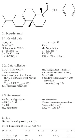
Crystal structure of 7-isopropyl-1,4a,
N
-
trimethyl-1,2,3,4,4a,4b,5,6,7,8,10,10a-
dodecahydrophenanthrene-1-carbox-amide
Li Liu, Xin-Yan Yan and Xiao-Ping Rao*
Institute of Chemical Industry of Forest Products, Chinese Academy of Forestry, Key Laboratory of Biomass Energy and Material, Jiangsu Province, National Engineering Lab. for Biomass Chemical Utilization, Key and Lab. on Forest Chemical Engineering, SFA, Nanjing, 210042, People’s Republic of China. *Correspondence e-mail: [email protected]
Received 17 September 2015; accepted 21 September 2015
Edited by E. R. T. Tiekink, University of Malaya, Malaysia
In the title compound, C26H37NO, a new derivative of dihydroabietic acid, the two cyclohexene rings adopt half chair conformations, whereas the cyclohexane ring has a chair conformation. Each of the methyl groups is in an axial position with respect to the tricyclic hydrophenanthrene residue. In the crystal packing, methylene-C—H (phenyl) interactions lead to supramolecular helical chains along [010]; the amide-H atom does not form a significant intermolecular interaction owing to steric pressure.
Keywords:crystal structure; dihydroabietic acid derivative; C—H
interactions.
CCDC reference:1426243
1. Related literature
For crystal structure of dihydroabietic acid derivatives, see: Rao et al. (2009); Rao (2010). For the biological activity of rosin acid derivatives, see: Fonsecaet al.(2004); Gonzale´zet al.
(2010); Rao et al.(2008); Sepulvedaet al.(2005); Xinget al.
(2013).
2. Experimental
2.1. Crystal data
C26H37NO Mr= 379.57
Orthorhombic,P212121 a= 26.223 (5) A˚ b= 5.9230 (12) A˚ c= 14.493 (3) A˚
V= 2251.0 (8) A˚3 Z= 4
MoKradiation
= 0.07 mm1 T= 293 K
0.200.200.10 mm
2.2. Data collection
Enraf–Nonius CAD-4 diffractometer
Absorption correction: scan (CAD-4 Software; Enraf–Nonius, 1985)
Tmin= 0.987,Tmax= 0.993 4707 measured reflections
4122 independent reflections 2080 reflections withI> 2(I) Rint= 0.099
3 standard reflections every 200 reflections
intensity decay: 1%
2.3. Refinement
R[F2> 2(F2)] = 0.079 wR(F2) = 0.193 S= 1.01 4122 reflections
254 parameters
H-atom parameters constrained max= 0.21 e A˚
3
min=0.30 e A˚
[image:1.610.308.561.78.545.2]3
Table 1
Hydrogen-bond geometry (A˚ ,).
Cg1 is the centroid of the C21–C26 ring.
D—H A D—H H A D A D—H A
C3—H3A Cg1i
0.97 2.82 3.705 (5) 151
Symmetry code: (i)xþ1 2;y
1 2;z.
Data collection:CAD-4 Software(Enraf–Nonius, 1985); cell refine-ment:CAD-4 Software; data reduction:XCAD4(Harms & Wocadlo, 1995); program(s) used to solve structure: SHELXS97 (Sheldrick, 2008); program(s) used to refine structure:SHELXL97(Sheldrick, 2008); molecular graphics:ORTEP-3 for Windows(Farrugia, 2012); software used to prepare material for publication:SHELXL97.
Acknowledgements
This research was supported by a grant from the National Natural Science Foundation of China (31470596).
Supporting information for this paper is available from the IUCr electronic archives (Reference: TK5389).
data reports
Acta Cryst.(2015).E71, o801–o802 doi:10.1107/S2056989015017648 Liuet al.
o801
References
Enraf–Nonius (1985).CAD-4 Software. Enraf-Nonius, Delft, The Netherlands. Farrugia, L. J. (2012).J. Appl. Cryst.45, 849–854.
Fonseca, T., Gigante, B., Marques, M. M., Gilchrist, T. L. & De Clercq, E. (2004).Bioorg. Med. Chem.12, 103–112.
Gonza´lez, M. A., Pe´rez-Guaita, D., Correa-Royero, J., Zapata, B., Agudelo, L., Mesa-Arango, A. & Betancur-Galvis, L. (2010).Eur. J. Med. Chem.45, 811– 816.
Harms, K. & Wocadlo, S. (1995).XCAD4. University of Marburg, Germany. Rao, X. P. (2010).Acta Cryst.E66, o2725.
Rao, X. P., Song, Z. Q. & He, L. (2008).Heteroat. Chem.19, 512–516. Rao, X. P., Song, Z. Q., Shang, S. B. & Wu, Y. (2009).Acta Cryst.E65, o2804. Sepulveda, B., Astudillo, L., Rodriguez, J., Yanez, T., Theoduloz, C. &
Schmedahirschmann, G. (2005).Pharmacol. Res.52, 429–437. Sheldrick, G. M. (2008).Acta Cryst.A64, 112–122.
supporting information
sup-1
Acta Cryst. (2015). E71, o801–o802supporting information
Acta Cryst. (2015). E71, o801–o802 [doi:10.1107/S2056989015017648]
Crystal structure of 7-isopropyl-1,4a,
N
-trimethyl-1,2,3,4,4a,4b,5,6,7,8,10,10a-dodecahydrophenanthrene-1-carboxamide
Li Liu, Xin-Yan Yan and Xiao-Ping Rao
S1. Comment
Rosin acid derivatives exhibit wide range of biological activities, such as antifungal and antitumor (Fonseca et al., 2004; Rao et al., 2008; Gonzaléz et al., 2008; Xing et al., 2013.) activities. Nitrogen derivatives of rosin acid have been studied as cytotoxic reagents and they are found to have high activity in reducing blood serum cholesterol levels in animals
(Sepulveda et al., 2005). In this work, we describe the crystal structure of the title compound.
Dihydroabietic acid is one of the main component of hydrogenated rosin, which can be isolated from hydrogenated
rosin by recrystallization. In this work, we have obtained the single crystal structure of the title compound, the tricyclic
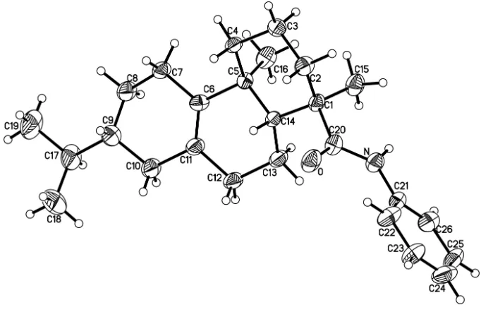
hydrophenanthrene nuclei had the similar crystal structure with dihydroabietic acid derivatives (Rao et al., 2009; Rao, 2010). The two cyclohexenes adopt half chair conformations, whereas the cyclohexane has a chair conformation (Fig. 1).
The two methyl groups are in axial positions with respect to the tricyclic hydrophenanthrene nuclei. The structures of
related dihydroabietic acid derivatives are known (Rao et al. 2009; Rao, 2010)
S2. Experimental
A mixture of dihydroabietic acid (0.1 mol), oxalyl chloride (0.11 mol) and dichloromethane (40 ml) was stirred at 313 K
for 4 h. After distilling off the solvent, the residue was added to aniline (0.2 mol) in toluene (60 ml) solution. The mixture
was reacted for 24 h at room temperature. The solvent was distilled off, and upon recrystallization from acetone, white
crystals of the title compound were obtained (yield 53%, M.p. 422 K). Single crystals were grown from acetone.
S3. Refinement
H atoms were positioned geometrically and refined as riding atoms, with C—H = 0.96 Å and Uiso(H) = 1.5Ueq(C) for
methyl H atoms, and C—H = 0.97–0.98 Å and Uiso(H) = 1.2Ueq(C) for all other H atoms. The absolute structure was not
Figure 1
Molecular structure of the title compound, with H atoms represented by small spheres of arbitrary radius and
displacement ellipsoids at the 30% probability level.
7-Isopropyl-1,4a,N-trimethyl-1,2,3,4,4a,4b,5,6,7,8,10,10a-dodecahydrophenanthrene-1-carboxamide
Crystal data
C26H37NO
Mr = 379.57
Orthorhombic, P212121
a = 26.223 (5) Å
b = 5.9230 (12) Å
c = 14.493 (3) Å
V = 2251.0 (8) Å3
Z = 4
F(000) = 832
Dx = 1.120 Mg m−3
Mo Kα radiation, λ = 0.71073 Å Cell parameters from 25 reflections
θ = 9–13°
µ = 0.07 mm−1
T = 293 K Block, white
0.20 × 0.20 × 0.10 mm
Data collection
Enraf–Nonius CAD-4 diffractometer
Radiation source: fine-focus sealed tube Graphite monochromator
ω/2θ scans
Absorption correction: ψ scan
(CAD-4 Software; Enraf–Nonius, 1985)
Tmin = 0.987, Tmax = 0.993
4707 measured reflections
4122 independent reflections 2080 reflections with I > 2σ(I)
Rint = 0.099
θmax = 25.4°, θmin = 1.6°
h = −31→31
k = 0→7
l = 0→17
3 standard reflections every 200 reflections intensity decay: 1%
Refinement
Refinement on F2
Least-squares matrix: full
R[F2 > 2σ(F2)] = 0.079
wR(F2) = 0.193
S = 1.01
254 parameters 0 restraints
Primary atom site location: structure-invariant direct methods
supporting information
sup-3
Acta Cryst. (2015). E71, o801–o802Hydrogen site location: inferred from neighbouring sites
H-atom parameters constrained
w = 1/[σ2(F
o2) + (0.040P)2 + 2.3P]
where P = (Fo2 + 2Fc2)/3
(Δ/σ)max < 0.001
Δρmax = 0.21 e Å−3
Δρmin = −0.30 e Å−3
Absolute structure: nd
Special details
Geometry. All e.s.d.'s (except the e.s.d. in the dihedral angle between two l.s. planes) are estimated using the full
covariance matrix. The cell e.s.d.'s are taken into account individually in the estimation of e.s.d.'s in distances, angles and torsion angles; correlations between e.s.d.'s in cell parameters are only used when they are defined by crystal symmetry. An approximate (isotropic) treatment of cell e.s.d.'s is used for estimating e.s.d.'s involving l.s. planes.
Refinement. Refinement of F2 against ALL reflections. The weighted R-factor wR and goodness of fit S are based on F2,
conventional R-factors R are based on F, with F set to zero for negative F2. The threshold expression of F2 > σ(F2) is used
only for calculating R-factors(gt) etc. and is not relevant to the choice of reflections for refinement. R-factors based on F2
are statistically about twice as large as those based on F, and R- factors based on ALL data will be even larger.
Fractional atomic coordinates and isotropic or equivalent isotropic displacement parameters (Å2)
x y z Uiso*/Ueq
H13A −0.0443 0.2538 −0.1003 0.067* H13B 0.0038 0.1190 −0.0668 0.067* C14 −0.06178 (17) 0.0224 (9) 0.0016 (3) 0.0416 (13) H14A −0.0541 −0.1391 0.0060 0.050* C15 −0.0514 (2) 0.3815 (10) 0.1043 (4) 0.0629 (16) H15A −0.0868 0.4197 0.1110 0.094* H15B −0.0380 0.4529 0.0500 0.094* H15C −0.0329 0.4328 0.1575 0.094* C16 −0.1422 (2) 0.2680 (10) −0.0338 (4) 0.0597 (16) H16A −0.1367 0.3567 0.0207 0.089* H16B −0.1782 0.2562 −0.0454 0.089* H16C −0.1259 0.3393 −0.0854 0.089* C17 −0.1645 (3) −0.4587 (13) −0.3682 (4) 0.081 (2) H17A −0.1606 −0.3274 −0.4087 0.097* C18 −0.1258 (3) −0.6326 (14) −0.4005 (4) 0.109 (3) H18A −0.1333 −0.6755 −0.4629 0.164* H18B −0.1275 −0.7632 −0.3614 0.164* H18C −0.0922 −0.5688 −0.3977 0.164* C19 −0.2188 (3) −0.5441 (16) −0.3825 (4) 0.114 (3) H19A −0.2230 −0.5928 −0.4452 0.170* H19B −0.2425 −0.4247 −0.3696 0.170* H19C −0.2252 −0.6687 −0.3417 0.170* C20 0.0111 (2) 0.0508 (10) 0.1082 (4) 0.0501 (14) C21 0.09969 (19) 0.2039 (9) 0.1201 (4) 0.0455 (13) C22 0.1229 (2) 0.0189 (13) 0.1586 (4) 0.0669 (18) H22A 0.1035 −0.1027 0.1788 0.080* C23 0.1762 (2) 0.0137 (13) 0.1673 (4) 0.074 (2) H23A 0.1921 −0.1103 0.1940 0.089* C24 0.2042 (2) 0.1900 (14) 0.1367 (4) 0.082 (2) H24A 0.2395 0.1847 0.1415 0.098* C25 0.1812 (2) 0.3788 (12) 0.0981 (4) 0.0692 (18) H25A 0.2005 0.5010 0.0781 0.083* C26 0.1281 (2) 0.3796 (11) 0.0904 (4) 0.0614 (16) H26A 0.1119 0.5038 0.0642 0.074*
Atomic displacement parameters (Å2)
U11 U22 U33 U12 U13 U23
supporting information
sup-5
Acta Cryst. (2015). E71, o801–o802C9 0.070 (4) 0.093 (5) 0.024 (3) −0.015 (4) −0.004 (3) −0.004 (3) C10 0.052 (4) 0.098 (5) 0.041 (3) −0.009 (4) −0.004 (3) −0.012 (3) C11 0.042 (3) 0.069 (4) 0.046 (3) −0.005 (3) −0.002 (3) 0.000 (3) C12 0.041 (3) 0.111 (5) 0.041 (3) −0.017 (4) 0.005 (3) −0.012 (4) C13 0.038 (3) 0.083 (4) 0.047 (3) −0.011 (3) 0.002 (2) −0.005 (3) C14 0.034 (3) 0.049 (3) 0.042 (3) −0.006 (2) 0.001 (2) −0.003 (3) C15 0.055 (4) 0.062 (4) 0.072 (4) −0.005 (3) −0.017 (3) −0.003 (3) C16 0.053 (4) 0.062 (4) 0.064 (4) 0.001 (3) −0.016 (3) 0.010 (3) C17 0.097 (5) 0.108 (6) 0.038 (3) −0.031 (5) −0.006 (3) −0.007 (4) C18 0.131 (7) 0.141 (8) 0.055 (4) −0.017 (6) 0.018 (5) −0.035 (5) C19 0.102 (6) 0.178 (9) 0.061 (4) −0.048 (6) −0.022 (4) −0.022 (6) C20 0.049 (3) 0.049 (3) 0.052 (3) −0.004 (3) −0.009 (3) 0.009 (3) C21 0.039 (3) 0.050 (3) 0.047 (3) −0.004 (3) −0.002 (3) 0.003 (3) C22 0.039 (4) 0.100 (5) 0.062 (4) −0.009 (4) −0.002 (3) 0.018 (4) C23 0.041 (4) 0.093 (5) 0.087 (5) −0.005 (4) −0.009 (3) 0.015 (4) C24 0.041 (4) 0.135 (7) 0.069 (5) 0.002 (4) 0.002 (3) −0.005 (5) C25 0.050 (4) 0.095 (5) 0.063 (4) −0.030 (4) 0.005 (3) 0.008 (4) C26 0.055 (4) 0.082 (5) 0.047 (3) −0.007 (3) −0.005 (3) 0.009 (3)
Geometric parameters (Å, º)
C8—H8B 0.9700 C23—C24 1.351 (9) C9—C10 1.519 (7) C23—H23A 0.9300 C9—C17 1.544 (7) C24—C25 1.388 (9) C9—H9A 0.9800 C24—H24A 0.9300 C10—C11 1.506 (7) C25—C26 1.397 (7) C10—H10A 0.9700 C25—H25A 0.9300 C10—H10B 0.9700 C26—H26A 0.9300
supporting information
sup-7
Acta Cryst. (2015). E71, o801–o802C8—C7—H7B 109.0 C17—C19—H19B 109.5 H7A—C7—H7B 107.8 H19A—C19—H19B 109.5 C9—C8—C7 111.3 (5) C17—C19—H19C 109.5 C9—C8—H8A 109.4 H19A—C19—H19C 109.5 C7—C8—H8A 109.4 H19B—C19—H19C 109.5 C9—C8—H8B 109.4 O—C20—N 122.8 (5) C7—C8—H8B 109.4 O—C20—C1 120.7 (5) H8A—C8—H8B 108.0 N—C20—C1 116.4 (5) C8—C9—C10 109.9 (5) C26—C21—C22 120.0 (5) C8—C9—C17 115.9 (5) C26—C21—N 117.6 (5) C10—C9—C17 112.2 (5) C22—C21—N 122.4 (5) C8—C9—H9A 106.0 C21—C22—C23 119.8 (6) C10—C9—H9A 106.0 C21—C22—H22A 120.1 C17—C9—H9A 106.0 C23—C22—H22A 120.1 C11—C10—C9 115.0 (4) C24—C23—C22 119.7 (7) C11—C10—H10A 108.5 C24—C23—H23A 120.2 C9—C10—H10A 108.5 C22—C23—H23A 120.2 C11—C10—H10B 108.5 C23—C24—C25 121.2 (6) C9—C10—H10B 108.5 C23—C24—H24A 119.4 H10A—C10—H10B 107.5 C25—C24—H24A 119.4 C6—C11—C10 123.5 (5) C24—C25—C26 118.0 (6) C6—C11—C12 123.8 (5) C24—C25—H25A 121.0 C10—C11—C12 112.7 (4) C26—C25—H25A 121.0 C13—C12—C11 112.8 (4) C21—C26—C25 121.4 (6) C13—C12—H12A 109.0 C21—C26—H26A 119.3 C11—C12—H12A 109.0 C25—C26—H26A 119.3
C7—C6—C11—C10 −9.5 (9) C14—C1—C20—N 116.2 (5) C5—C6—C11—C10 179.7 (5) C15—C1—C20—N −8.8 (7) C7—C6—C11—C12 173.0 (5) C20—N—C21—C26 157.2 (6) C5—C6—C11—C12 2.3 (9) C20—N—C21—C22 −24.0 (9) C9—C10—C11—C6 −1.1 (9) C26—C21—C22—C23 0.4 (9) C9—C10—C11—C12 176.7 (6) N—C21—C22—C23 −178.4 (5) C6—C11—C12—C13 −10.9 (8) C21—C22—C23—C24 −0.8 (10) C10—C11—C12—C13 171.4 (5) C22—C23—C24—C25 1.2 (11) C11—C12—C13—C14 42.7 (7) C23—C24—C25—C26 −1.1 (10) C12—C13—C14—C1 160.7 (4) C22—C21—C26—C25 −0.3 (9) C12—C13—C14—C5 −67.4 (6) N—C21—C26—C25 178.5 (5) C2—C1—C14—C13 −174.8 (5) C24—C25—C26—C21 0.7 (9)
Hydrogen-bond geometry (Å, º)
Cg1 is the centroid of the C21–C26 ring.
D—H···A D—H H···A D···A D—H···A
C3—H3A···Cg1i 0.97 2.82 3.705 (5) 151