i
ith 12 text-figures 'tinted in Great BritainCENTRAL PROGRAMMING AND REFLEX CONTROL
OF WALKING IN THE COCKROACH
BY K. G. PEARSON
Department of Physiology, University of Alberta, Edmonton, Canada
{Received 16 June 1971)
INTRODUCTION
A somewhat neglected aspect of insect neurophysiology is the nervous control of walking. By the year 1940 there were a large number of behavioural descriptions of insect walking (see Hughes, 1952, and Wilson, 1966, for references), and electro-physiological techniques had just started to be used to investigate the neuronal mechanisms underlying this behaviour (Pringle, 1938a, b, 1939, 1940). Since then there have been further behavioural descriptions (Hughes, 1952,1957,1965; Milburn, 1963; Wendler, 1966; Wilson, 1966; Delcomyn, 1971a, b), but until quite recently electrophysiological approaches have been largely ignored. At the present time, electrophysiological investigations can be classified into three groups.
(1) A number of studies have been concerned with recording the discharge patterns in leg motoneurones during normal walking in unrestrained animals (Hoyle, 1964; Ewing & Manning, 1966; Usherwood & Runion, 1970). These patterns are recorded by placing fine recording electrodes either on peripheral nerve trunks or in various leg muscles. The results of these investigations define the normal patterns of motoneuronal activity, which in turn must be described in terms of the properties and connections of cells within the central nervous system and/or activity in various reflex pathways.
(2) The second approach is to record motoneuronal activity in restrained and often dissected preparations. These preparations allow selective and controlled stimulation of various sets of peripheral receptors which is desirable when investigating the pro-perties of reflex pathways (Wilson, 1965; Usherwood, Runion & Campbell, 1968). Moieover, spontaneously generated or evoked motoneuronal activity can be recorded before and after de-afferentation (Hoy & Wilson, 1969; Pearson & lies, 1970). A comparison of these records with those obtained from freely walking animals allows conclusions about the existence of central programs and the function of peripheral feedback.
(3) The most recent electrophysiological approach to investigate the neuronal mechanisms underlying walking is to record intracellularly from cells within the thoracic ganglia (Hoyle, 1970). The hope of these studies is that they will yield infor-mation on the properties and connexions between identified cells involved in generat-ing the motor output patterns associated with walkgenerat-ing.
174 K. G. PEARSON
movements, and in some cases of how they are modified by sensory input (Mille™ 1966; Wilson, 1968; Kutsch, 1969; Kutsch & Huber, 1970). Only a small number of investigations have been concerned with recording motoneuronal activity in freely walking insects (Hoyle, 1964; Ewing & Manning, 1966; Usherwood & Runion, 1970). Although some of the general features of motoneuronal activity have been obtained in these investigations, precise descriptions of the discharge patterns of different motoneurones are largely lacking; for example, how burst length and discharge rate change with walking speed, the pattern of activity within a burst, and so on. An aim of the present investigation was to determine some of these features for three identified motoneurones which are in part responsible for producing the flexion and extension movements of the femur during walking in the cockroach.
The exact function of sensory feedback in the control of insect walking is unknown. Removal of tibial and tarsal receptors 01 of the femoral chordotonal organ in the locust results in changes in the motor activity and uncoordinated leg movements (Usherwood, Runion & Campbell, 1968; Usherwood & Runion, 1970), while in the stick insect removal of the coxal hair plates results in overstepping (Wendler, 1966). It is not clear from these observations what information in the feedback sensory signals is necessary for the production of normal motor activity. For example, is phasic sensory input signalling information about the position and velocity of movement of a certain part of the leg necessary, or are these systems similar to the locust flight system where the generation of normal motor output requires sensory input from peripheral receptors but the phasic information in these afferent signals is irrelevant (Wilson & Gettrup, 1963)? For the cockroach there is evidence to suggest that both central programming and reflex control are important in producing rhythmic movements of single legs and coordinating the movements of different legs (Pringle, 1961; Milburn, 1963; Wilson, 1965, 1966; Pearson & lies, 1970; Delcomyn, 1971 b). At present, however, the manner in which the reflex effects arising from various groups of peripheral receptors interact with a central program is not known, nor has the relative importance of central pro-gramming and reflex control been determined for different walking speeds. Another aim of the current investigation was to obtain information pertinent to the problem of central versus reflex control of leg movements in the walking cockroach.
MATERIALS AND METHODS
1. Anatomy
All experiments were performed on adult male cockroaches, Periplaneta americana. The anatomy of the metathoracic coxal levator and depressor muscles has been des-cribed in detail in earlier papers (Pearson & Bergman, 1969; Pearson & lies, 1970,
1971). Therefore only a brief description will be given here.
By nerve 51-1. The activity of motoneurones supplying the remaining coxal levator and depressor muscles (180, 177 A, B and C) was not studied in this investigation.
2. Unrestrained preparations
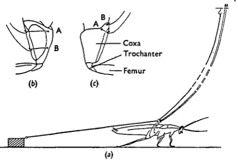
In these preparations recordings were made simultaneously from the coxal levator and depressor muscles while the animal was walking, or running, over a smooth sur-face. The animal was prevented from moving outside an area 2 ft in diameter by attaching a fine light thread to a steel ring inserted through the dorsal cuticle of the metathoracic segment and to an elevated support in the centre of this area (Fig. la). The recording electrodes, which were 50 fim copper wires insulated except at the ends,
Pre-amp.
[image:3.451.105.346.218.383.2](a)
Fig. 1. Experimental arrangement for recording activity in coxal levator and depressor muscles during walking, (a) The animal's movements were restricted to an area 2 ft in diameter by at-taching a fine thread to a ring inserted through the dorsal cuticle of the metathoracic segment and to an elevated support in the centre of this area. The recording leads ran parallel to this thread and were also attached to the ring before being fixed to the coxa. Retraction of the metathoracic legs was resisted by allowing the animal to drag a weight, as shown, or by adding a weight to the animal's back. (6) Ventral and (c) dorsal views of the coxa showing the arrangement of the electrodes for recording the activity in coxal depressor and levator muscles respectively.
176 K. G. PEARSON
Two methods were used to increase the resistance to retraction of the metathoracrai legs. The first, shown in Fig. 1 (a), was to make the animal drag a lead weight (weighing between 1 and 2 g) by a thread attached to the ring on its back. The second method was to add the weight directly to the animal's back. For this second method, the ring had an additional vertical projection on to which the weight was attached.
3. Partially de-qfferented preparations
Recordings were taken from nerves 6Br4 and 5 r i b after removal of all sensory information from the legs. T h e techniques used for exposing and recording from these nerves and the procedure for de-afferentation have been described elsewhere (Pearson & lies, 1970).
4. Identification of motor units
Nerve potentials. In previous studies various motor axons in nerves 6Br4 and 5 0
have been identified and labelled according to the amplitudes of the extracellularly recorded action potentials and discharge patterns (Pearson & Bergman, 1969; Pearson & lies, 1970, 1971; lies & Pearson, 1971). Axons 5 and 6 in nerve 6Br4 innervate the posterior coxal levator muscle 182 and produce slow graded contractions. Axon Ds in
nerve 5r 1 innervates the coxal depressor muscles 177D and 177E and also produces graded contractions. Nerve 5 r 1 also contains a fast axon, Df, which produces twitch
contraction in muscles 178,179 and in parts of muscles 177D and 177E. Although other axons have been identified in the previous studies, these are the only four considered in the present investigation.
Muscle potentials. Since axon Ds is the only slow axon to muscles 177D and 177E,
the corresponding muscle potentials elicited by activity in this axon were readily observed. The amplitude of the extracellularly recorded junctional potentials was usually in the range of 0-2-0-6 mV, depending on the exact location of the recording electrode. Often this potential showed marked facilitation with an increase in the dis-charge rate, but at very high rates the amplitude was reduced by partial summation of the junctional potentials.
RESULTS
1. Partially de-afferented nerve cord
In an earlier study (Pearson & lies, 1970) it was reported that in headless animals, and in preparations in which the meta-mesothoracic connectives were severed, re-ciprocal burst activity persisted in metathoracic levator and depressor motoneurones after removal of all sensory input from the legs. The frequency of the reciprocal activity varied from 0-5 to 5 cyc/sec. As Hoyle (1970) has pointed out, the highest frequency of 5 cyc/sec is considerably less than the maximum frequency of leg move-ments of about 24 cyc/sec seen in running animals (Delcomyn, 1971 a). The failure of Pearson and lies to observe these correspondingly high frequencies of reciprocal motoneuronal activity could have been due either to de-afferentation or to the removal of anterior segments, or to a combination of both. To determine whether the maximum frequency depended on sensory input, recordings were made from nerves 5 r 1 b and 6Br4 in twelve animals from which all sensory input from leg receptors had been removed but which were not decapitated.
(a)
•niimaiiitu 1
Fig. 2. Reciprocal activity in coxal levator and depressor motor axons after removal of all sen-sory input from leg receptors. Top traces, records from levator nerve 6Br4; bottom traces, records from depressor nerve 5 r 1 b. (a) Reciprocal activity elicited by stimulation of the ipsi-latcral cercus. Interaction of the action potentials during levator bursts does not allow identi-fication of the action potentials although the first spike in each burst is from axon 5. The small and large spikes seen during the depressor bursts arise from activity in axons D8 and D(
respectively. (6) Spontaneously generated reciprocal activity. The two spikes in the first levator burst are from axons 5 and 6 (the spike from axon 6 being larger). Only axon D8 discharges
during the depressor bursts. Note that the maximum discharge rate of axon D, occurred at the beginning of the burst. Time scale: (a) 80msec; (6) 200 msec.
Stimulation of the ipsilateral cercus in these partially de-afferented preparations often elicited high-frequency reciprocal activity (Fig. 2 a). These periods of reciprocal activity usually did not last for more than five cycles. The maximum observed fre-quency of the reciprocal activity was 15 cyc/sec (Fig. 2 a), which was much higher than that observed in partially de-afferented headless preparations, but still less than the maximum frequency of leg movements seen in rapidly running animals. The repetition rate of 15 cyc/sec was observed in only one of twelve preparations. Recip-rocal activity could not be evoked in six other preparations, while in the remaining five the maximum frequency was between 8 and 12 cyc/sec. Periods of reciprocal
178 K. G. PEARSON
activity sometimes occurred spontaneously, but in contrast to the evoked responses thU frequency was low (Fig. 26).
The intensity of the levator bursts during high-frequency reciprocal activity was variable and not related in any obvious manner to the intensity of activity in axon Ds
or to the frequency of the reciprocal activity. Sometimes the levator bursts were very intense (Fig. 2 a) and interaction of the action potentials from different axons pre-vented any measurement of the exact activity patterns in the various levator moto-neurones. It was clear, however, that apart from axons 5 and 6 discharging during these bursts, a number of other axons were also active. At other times only axons 5 and 6 were active during these bursts, while occasionally only axon 5 discharged. There was less variability in the intensity of activity in axons Ds and Df during high-frequency
reciprocal activity. Axon Ds usually discharged at rates between 300 and 400 impulses/
sec throughout the depressor bursts (Fig. za). Axon Df almost always became active
during these bursts and fired a number of times within each.
When the frequency of the reciprocal activity was lower, the burst activity in axons 5 and 6 was reciprocal with that in axon Ds (Fig. 26). This low-frequency
pattern of reciprocal activity was similar to that reported by Pearson & lies (1970) for headless de-afferented preparations. Other similarities with the results of Pearson & lies (1970) were that the maximum activity in axon Ds occurred immediately after the
levator burst (Fig. zb), and occasionally the activity in axon Ds was not maintained
between the levator bursts.
The durations of the levator bursts usually varied from 20 to 200 msec. However, there were occasions when the levator-burst duration was longer (> 0-5 sec). In the earlier study (Pearson & lies, 1970) long-duration bursts were not associated with sequences of reciprocal activity in which more than four cycles occurred, whereas in this study the long-duration levator bursts did sometimes occur in such sequences. Characteristically, these long-duration levator bursts were very intense and also associated with extremely intense activity (350 impulses/sec) in the antagonistic depressor motor axon Ds.
A characteristic feature of the activity patterns recorded in non-decapitated partially de-afferented preparations was the high rate of activity in axon Ds. This axon was
usually continuously active between periods of reciprocal activity sometimes discharg-ing at rates greater than 100/sec for tens of seconds. This intense activity in axon Ds was
not seen before de-afferentation. Thus de-afferentation apparently releases a facilatory influence, or removes inhibition, from the depressor motoneurone Ds, but does not
necessarily prevent the generation of reciprocal activity patterns. In six of twelve preparations neither evoked nor spontaneous reciprocal patterns of activity occurred. In these preparations axon Ds usually discharged continuously at rates between 50 and
200 impulses/sec.
2. Freely walking animals
Reciprocal activity. To determine whether the patterns of motoneuronal activity
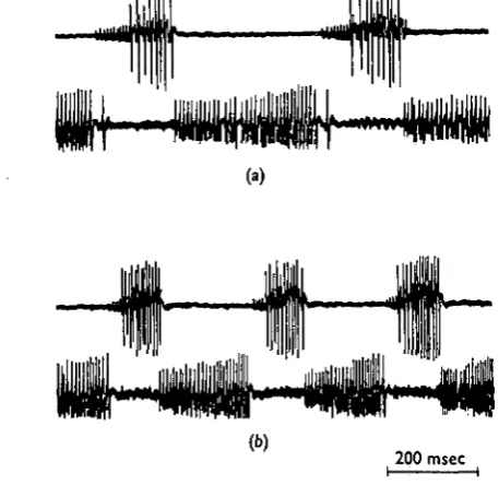
77E (Pearson & lies, 1971). To monitor the activity in these motoneurones, recording electrodes were placed in muscles 182C and 177D (Fig. 1 b, c). Examples of the recorded potentials during walking at two different speeds are shown in Fig. 3. The two muscle potentials recorded from muscle 182C corresponded to activity in axons 5 and 6. This conclusion was reached from the following two observations. First, after recording levator activity in a walking preparation, the animal was pinned ventral side up on a cork board without displacement of the recording electrodes and the connec-tives between the meso- and metathoracic ganglia were cut. In this preparation axons 5 and 6 discharge in bursts and it is rare to observe the larger axons firing during these
(a)
<*•)
[image:7.451.101.330.199.421.2]200 msec I : 1
Fig. 3. Reciprocal activity recorded in coxal levator and depressor muscles during walking at two different speeds. Top traces, records from levator muscle 182C; bottom traces, records from depressor muscle 177D. The small and large junctional potentials recorded in the levator muscle correspond to activity in axons 5 and 6 respectively. The single junctional potential recorded in the depressor muscles corresponds to activity in axon De. Note that with
an increase in walking speed there is an increase in the rate of discharge in all motor axons.
bursts. Correspondingly, the two muscle potentials persist in this restrained prepara-tion and, moreover, the patterns of activity are similar to those of axons 5 and 6 ob-served from nerve recordings (Pearson &Iles, 1970). Secondly, the relative amplitude of the smaller spike was considerably larger when the recording electrode was placed in muscle 182 D as compared to that recorded from muscle 182C, whereas the amplitude of the larger spike was about the same in each. This observation strongly indicates that the smaller spike arises from activity in axon 5 since muscle 182D has a larger fraction of its fibres innervated by axon 5, while axon 6 innervates the same fraction of fibres in both muscles 182C and 192D (Pearson & Bergman, 1969).
180 K. G. PEARSON
have been active, but the corresponding muscle potentials may have been too small be observed using the present techniques. Thus it appears that rhythmic movements of the femur relative to the coxa at rates less than about io cyc/sec are produced by reciprocal bursts of activity in axon Ds (giving extension movements) and axons 5 and
6 (giving flexion movements). All three of these axons can be classified as slow (Pearson & Bergman, 1969; Pearson & lies, 1971; lies & Pearson, 1971), but even so, high-frequency bursts can give rise to strong and rapid contractions which are required to produce femur movements at rates as high as 10 cyc/sec.
When the animal is running rapidly so that the legs move at a rate higher than 10 eye/ sec, axon Df and a single levator axon are recruited (Fig. 4). This additional levator
motor axon recruited at high running speeds has not yet been classified, but is un-doubtedly a fast axon supplying either the main levator muscle 181 or muscle 182.
100 msec
Fig. 4. High-frequency reciprocal activity in single fast axons to coxal levator and depressor muscles during rapid running. Top trace, record from levator muscle 182C; bottom trace, record from depressor muscle 179. Running was initiated by pinching the tarsus at the instant indicated by the initial artifact in each record. The single spike recorded from the depressor muscle 179 corresponds to activity in axon D(.
Only activity in a fast axon could produce contraction of sufficient rapidity to give femur flexion movements lasting less than 25 msec. The functions of the other fast axons to the levator muscles have not yet been clearly established although at least two of these are active during flight (unpublished observations). Axon Df usually discharges
only once per cycle when the animal is running rapidly. This is unlike the pattern seen in sequences of high-frequency reciprocal activity in the partially de-afferented nerve cord where it discharges a number of times each cycle even when the cycle time is longer than ioo msec (compare the activity of axon Df in Figs. 2(a), 4).
There was no evidence in the records taken from rapidly running animals that one set of motoneurones becomes continuously active. At all times the activity in slow and fast axons remained reciprocal. This is in contrast with the results of an earlier study by Ewing & Manning (1966) in which it was reported that at high running speeds the slow motoneurones supplying the flexor and extensor tibiae muscles become continuously active and the fast flexors drive the extensors. Hoyle (1964) also reported driving of either flexor or extensor tibiae muscles of the locust when the frequency of leg move-ment was about 5 cyc/sec. Subsequently, however, Usherwood, Runion & Campbell (1968) have shown a clear reciprocal activity pattern in extensor and flexor tibiae motoneurones at this rate of leg movement.
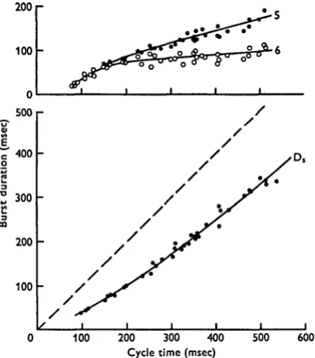
Burst durations. Fig. 5 shows, for a typical preparation, the relationship between the
Tnsec) the burst durations of axons 5 and 6 were very similar and increased rapidly as the cycle time increased. For cycle times longer than about 200 msec the burst duration of axon 6 remained fairly constant at about 80 msec while that of axon 5 continued to increase, but more slowly. For cycle times greater than about 500 msec, axon 6 did not discharge during the levator bursts. The burst durations of axon Ds increased to a
much greater extent than those of axons 5 and 6 with increases in cycle time. At all walking speeds the intervals between the bursts in axon Ds were slightly longer
on average than the burst duration of axon 5. This observation reflects the fact that in normal walking, as well as in restrained de-afferented preparations, there is usually no overlap of activity in axon 5 and axon Ds. The interburst intervals for axon Ds
there-fore give an approximate measure (slight over-estimate) of the burst duration in axon 5.
200 r
100
0
500
400
300
200
100 CO
[image:9.451.101.330.218.478.2]0 100 200 300 400 500 600 Cycle time (msec)
Fig. s. Variation in the burst durations of levator motor axons 5 and 6 (top) and depressor motor axon D, (bottom) with cycle time. Cycle time was measured from the beginning of one levator burst to the beginning of the next. The interrupted line in the bottom graph has a slope of one and the difference between this line and the line through the data points measured along the ordinate gives an approximate measure of the burst duration of motor axon 5.
The variation in burst duration in axon Ds for an animal which had a very large range
of walking speeds is shown in Fig. 6. This figure shows clearly that for slow walking the major variable is in the duration of the depressor burst length, this varying from 35 msec to I-I sec, while that of axon 5 (as indicated by the interburst interval) varied from 50 msec to 170 msec. The ratios for the burst duration of axon 5 to axon Ds
therefore varied from 1-4 to 0-16.
l 82 K. G. PEARSON
I
observations of Delcomyn (1971a) for the leg protraction and retraction phases 0 walking.
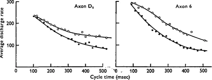
Discharge rates. Because of the interaction of muscle potentials evoked by activity
in axons 5 and 6 it was impossible to accurately determine the variation in discharge rate in axon 5 throughout its burst for different walking speeds. This was not so for axon 6. When records were taken from muscle 182C the excitatory junctional potentials from axon 5 were often considerably smaller than those from axon 6, thus allowing the pat-tern of activity in axon 6 to be accurately determined. Since axon Ds was the only active
axon to the depressor muscles for leg movements at less than 10/sec, there was no difficulty in measuring the pattern of activity of this motoneurone.
1200
1000
~ 750
C o
500
250
250 500 750 Cycle time (msec)
1000 1250
Fig. 6. Variation in the burst duration of motor axon D, with cycle time for an animal displaying a wide range of walking speeds. The line through the origin has a slope of one and the difference between this line and the line through the data points measured along the ordinate gives an approximate measure of the burst duration of motor axon 5.
The discharge rates of axons 6 and Ds were usually fairly constant throughout most
of their bursts, although the discharge rate in axon 6 generally increased at the begin-ning of the burst (Fig. 3). When the animal was walking slowly, maximum activity in axon Ds often occurred at the end of the burst (Fig. 3), although sometimes this
activity was not appreciably greater than that during the rest of the burst. This charac-teristic of long-duration depressor bursts in walking animals is the most noticeable difference from the long depressor bursts in partially de-afferented preparations where the maximum frequency occurs at the beginning of the burst (Fig. 2b). Sometimes in slowly walking animals the bursts in axon Ds showed a decline in activity after
dis-charging at a high rate near the beginning of the bursts, but this decline was followed by an increase near the end. Axon Ds discharged at a fairly uniform rate for shorter
The average discharge rates of axons Ds and 6 increased with decreasing cycle time
as shown in Fig. 7. The minimum discharge rates for axons D3 and 6 were both about
80 impulses/sec. For long cycle times, axon 6 did not discharge and femur flexion was produced by a burst of activity in axon 5 alone. Therefore, axon 6 was recruited as the cycle time decreased to less than about 500 msec. The maximum observed discharge rates of axons Ds and 6 were between 250 and 300, and 300 and 350 impulses/sec
respectively, these occurring when the cycle time was less than 100 msec, i.e. when the leg movements occurred at frequencies greater than 10 cyc/sec.
Effect of resistance to leg retraction. If the motor output patterns during walking were
entirely centrally generated, and thus independent of sensory feedback, these patterns
300
£?200
100
Axon Ds Axon 6
100 200 300 400 500 *G . 100 Cycle time (msec)
[image:11.451.48.394.216.347.2]200 300 400 500
Fig. 7. Variation of the average discharge rate of axon D, (left) and axon 6 (right) with cycle time. • , Unloaded animal; O, same animal dragging a weight of i •$ g. Cycle time was divided into bins of 20 msec width and each point is the mean of average discharge rates determined for all bursts with cycle times between t and t + 20 msec.
would not be expected to be changed by any procedure leading to an alteration in the sensory feedback from leg receptors. Two methods were used to change the sensory feedback, the first being to add a weight to the animal's back above the third thoracic segment, while the second was to allow the animal to drag a weight attached by a fine thread to the cuticle above the third thoracic segment. Both methods of loading the animal result in an increased resistance to retraction of the metathoracic legs, and qualitatively the same effects were produced by each.
The most obvious effect of increasing the resistance to retraction was an increase in the average discharge rates of axons Ds and 6 throughout their bursts for a given cycle
time (Fig. 7). The increase in the average discharge rate declines as the cycle time decreases so that at stepping speeds of a little greater than 10 cyc/sec the increase in load had no appreciable effect on the discharge rates of the two motoneurones. The progressive decline of the reflex effect on axon Ds with decreasing cycle time suggests
that the reflex effect diminishes as walking speed increases. Thus central influences on to axon Dg probably dominate in the rapidly running animal. The increase in the
dis-charge rate of axon 6 was generally more marked than that of axon Ds, particularly at
184 K. G. PEARSON
Another more natural method of increasing the resistance to leg retraction was t ™ allow the animal to walk up an inclined surface. Under this condition, the extension movement of the metathoracic leg must be more powerful in order to carry the animal up the slope. An increase in activity in axons 6 and Ds was also apparent under this
condition.
Apart from causing increases in the discharge frequency of axons 6 and Ds, an
in-crease in load also led to a slight dein-crease in the duration of the levator bursts (Fig. 8). This decrease in burst duration corresponded to a decrease in the interburst interval of axon Ds. The decrease in levator burst duration was most obvious in slowly
walking animals.
•r> 200 r
I 100
200 400 Cycle time (msec)
600
Fig. 8. Decrease in levator burst duration with an increase in resistance to leg retraction. • , No load; O, dragging weight of 1-5 g. Each point is the mean duration of bursts in axon 5 for cycle times between t and t + 20 msec.
Fig. 9. Abrupt increase in discharge rate of axon Ds (arrow) with a sudden increase in resistance
to retraction of the metathoracic leg from which recordings were being taken.
These reflex effects could be due to local reflex pathways in each leg, reflex pathways from other legs, an increase in the central drive to the rhythm-generating system, or various combinations of these. It is difficult to conceive of experiments to separate these three possibilities. However, two observations do make it appear that local reflex pathways are at least partially responsible for modifying the activity of axon Ds.
If phasic reflex influences from receptors in the metathoracic legs can modify the motor output, then a sudden change in the resistance to retraction would be expected to produce an abrupt increase in the activity of axon Ds. This effect is shown in Fig. 9.
Here the animal began to drag a weight during the retraction phase of the leg from which recordings were taken and correspondingly there was a marked increase in activity (arrow). The second observation indicating that local reflex effects influence the activity of axon Ds is shown in Fig. io, where activity in axon Ds was recorded
Speeds the prothoracic and metathoracic legs step at the same time (Delcomyn, 1971 a), which results in a sudden increase in load supported by the mesothoracic leg. Cor-respondingly, there was often a marked increase in the rate of discharge of the meso-thoracic axon Ds during stepping of the pro- and metathoracic legs. This effect was
not observed in all animals and was more apparent when the animal was loaded.
Receptors. An attempt was made to identify the receptors responsible for the
pre-viously described reflex effects by observing whether these effects remained after removal of the sensory input from various groups of leg receptors. Almost the entire
[image:13.451.62.402.182.243.2]I 1
Fig. 10. Increased discharge rate of mesothoracic axon D8 during protraction of the ipsilateral
metathoracic leg. Top record, metathoracic axon D,; bottom record, mesothoracic axon DB.
Protraction of the metathoracic leg is indicated by the silent periods in the top record.
1 1 1 1 1 1 i 1 1 1 1 i 1 iuli|i!mti
I I I ! I I ! I I I : I
OS sec
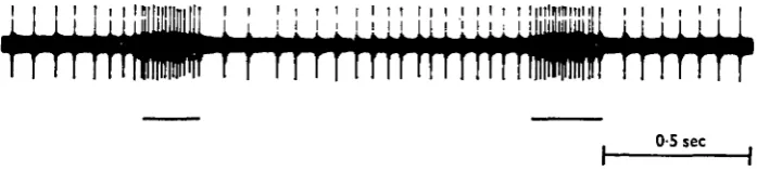
Fig. 11. Increased discharge rate of axon D8 elicited by pressure to the trochanter (horizontal bars).
afferent input from the legs to the thoracic ganglia is via nerves 3 b and 5 (Pipa & Cook, 1959). Afferent axons arising from coxal chordotonal organs and hair plates are con-tained in nerve 3 b, while nerve 5 contains afferents from the campaniform sensilla of the trochanter and femur, and from the hairs and spines of the femur, tibia and tarsus. Cutting nerve 3 b of both metathoracic legs did not abolish rhythmic walking movements in these legs, and although no analysis was made using cinemagraphic techniques these movements appeared to be coordinated in the normal manner with the remaining four legs. However, the movement of the metathoracic legs after cutting nerve 3 b was somewhat abnormal. The exact nature of this abnormality does not concern us here for the observation relevant to the present investigation was that an increase in load still produced an increase in activity of motoneurones 6 and Ds.
Moreover, these reflex effects were not abolished by removal of the tarsus and destruc-tion of the tibial afferents in both metathoracic legs (tibial afferents were destroyed by pushing a thick wire up the tibia). Thus, if the receptors giving rise to the reflex effects observed with an increase in load are located wholly within the metathoracic legs, then they must be located in the coxa, trochanter or femur, or combinations of these. Alternatively, or in addition, the reflex effects may arise from receptors in other legs. This final possibility has not yet been tested.
[image:13.451.56.405.289.368.2]186 K. G. PEARSON —
shown in Fig. 11. Since these receptors are orientated so as to respond to changes irP cuticle strain during leg retraction (Pringle, 1961), an increase in resistance to retrac-tion probably leads to a greater excitaretrac-tion and consequently to an increase in activity in motoneurone Ds. However, this cannot be accepted unreservedly because recordings
have not yet been made from the afferents of the campaniform sensilla during walking, so it is not known whether the activity in these afferents does increase with an increase in resistance to leg retraction.
The reflex effects of other receptors in the femur and coxa have not yet been deter-mined, so at present it is impossible to speculate on their possible function in the reflex regulation of rhythmic leg movements during walking.
DISCUSSION
1. Central program and reflex control
There has been considerable uncertainty in the past regarding the extent to which rhythmic leg movement during insect walking is centrally controlled, and the impor-tance of peripheral feedback in regulating, or even producing, various phases of leg movement (Pringle, 1961; Wilson, 1966). One of the clear results of this investigation, together with those obtained in an earlier study (Pearson & lies, 1970), is that, for the cockroach, reciprocal patterns of activity can be generated in motoneurones sup-plying the metathoracic coxal levator and depressor muscles in the absence of all sensory input from leg receptors (Fig. 2). However, the central program generating these reciprocal patterns is not entirely responsible for producing normal motoneuronal activity during walking, for there are differences in the activity patterns seen in par-tially de-afferented and freely walking animals (compare Fig. 2 with Figs. 3 and 4). These differences are most obvious in the discharge patterns of the depressor moto-neurones Ds and Df. Motoneurone Ds is always continuously active throughout the
intervals between levator bursts in the walking animal, often discharging maximally near the end of the burst. In contrast to this, the activity of Ds in de-afferented
pre-parations often was not maintained between the levator bursts and was always maxi-mal at the beginning of the burst. Another difference was seen for short cycle times. Here, motoneurone Df discharged a number of times per cycle in de-afferented
pre-parations (Fig. 2), whereas in running animals Df rarely discharged more than once per
cycle even when the cycle times were less than those observed in the de-afferented preparations in which Df was active (Fig. 4). A third difference is that the maximum
frequency of reciprocal activity seen in running animals was about 20 cyc/sec, while that in de-afferented preparations was usually less than 15 cyc/sec.
activity patterns seen in partially de-afferented preparations are related to walking is that during periods of reciprocal activity in these preparations the levator burst duration remains relatively constant for large variations in cycle time, usually being in the range of 100-200 msec, but being less than this for very short cycle times. The magnitude and relative constancy of the levator burst durations are very similar to those seen in normal walking animals (Fig. 5). From these observations, then, it is concluded that the reciprocal activity patterns generated in partially de-afferented preparations reflect the activity of a central program controlling rhythmic leg move-ments during walking. The differences in the activity patterns in de-afferented and walking animals must therefore be due to influences of sensory input from leg receptors in the walking animal.
Sensory input could influence the central program in one of two ways. The first is that afferent input from certain leg receptors could tonically affect the central program, and the second, which probably occurs concomitantly with the first, is that phasic information in the feedback sensory signals produces cycle-to-cycle changes in the motor activity, the magnitude of which depends on the magnitude of the phasic sensory signal. It is difficult to separate clearly these two effects, but a number of observations indicate that both types influence the central program controlling rhythmic leg movements in the cockroach.
In a partially de-afferented preparation motoneurone Ds is extremely active and often
discharges for long periods of time at rates between 100 and 200 impulses/sec. This type of activity is not seen before cutting nerve 5, so it can be concluded that afferents in this nerve exert a net tonic inhibitory effect on motoneurone Ds and/or that these
afferents release a tonic facilitory effect on to motoneurone Ds. The excitability of
motoneurone Df is also increased after cutting nerve 5 since this motoneurone is much
more readily activated by cereal stimulation in de-afferented preparations. Conversely, de-afferentation results in a decrease in the excitability of the levator motoneurones. In some de-afferented preparations levator bursts never occurred, while in others it was more difficult to elicit these bursts as compared to the ease with which they can be generated (either spontaneously or by stimulation of leg or cereal receptors) in restrained intact animals. Thus de-afferentation leads to a facilitation of depressor motoneurones and a decline in the excitability of levator motoneurones. This tonic sensory influence would explain at least two of the differences seen in the reciprocal activity patterns in de-afferented and normal walking animals, namely the increased excitability of axons Ds and Df in de-afferented preparations and the lower maximum
frequency of reciprocal activity in de-afferented preparations. The latter effect would result from a decrease in excitability of the system producing the levator burst activity (see §3 below).
Apart from the sensory input having a tonic influence on the central program, there is evidence that the motor activity can be modified in each cycle by phasic sensory input from leg receptors (Figs. 9, 10). The function of this phasic input will now be considered.
2. Reflex modulation of motoneurone Ds
Two observations in the current investigations strongly indicate that phasic sensory feedback from leg receptors modulate the activity of motoneurone Ds during the leg
etraction phase of walking. First, a sudden increase in load results in an immediate
188 K. G. PEARSON
increase in activity (Fig. 9), and second, an increase in the fraction of the body weighf carried by the mesothoracic leg during stepping in the ipsilateral pro- and metathoracic legs often results in an increased activity in motoneurone Ds of the mesothoracic
segment (Fig. 10). The evidence that the receptors responsible for these reflex effects are the trochanteral campaniform sensilla comes from the observations (1) that the reflex effects persist after cutting nerve 3 b and destroying the tibial and tarsal receptors, and (2) that there is a strong excitatory reflex pathway from trochanteral campaniform sensilla to motoneurone Ds (Fig. 11). This evidence is by no means
conclusive, but it does demonstrate that if the reflex effects seen on motoneurone Ds
with an increase in load are the result of increased activity in receptors of that leg, then it is highly likely that the receptors responsible are the trochanteral campaniform sensilla. A major possibility, which has not been excluded by these experiments, is that afferent inputs from other legs are partially responsible for the reflex effects produced by an increase in resistance to leg retraction. With this possibility in mind, the following discussion proceeds on the assumption that local reflex pathways from the trochanteral campaniform sensilla have a strong phasic excitatory effect on moto-neurone Ds.
The reflex pathway from the trochanteral campaniform sensilla to the depressor motoneurone constitutes a positive feedback pathway during walking. These receptors are excited by strains in the cuticle. Thus during leg retraction, which is in part produced by activity in motoneurone Ds, these receptors will be excited and produce a
further excitation in motoneurone Ds. This excitatory reflex pathway therefore
facili-tates the activity in the depressor motoneurone Ds and will contribute to the
main-tenance of activity in this motoneurone throughout the leg retraction phase. The existence of this effect can therefore account for the different patterns of activity in motoneurone Ds seen in partially de-afferented and walking animals. In the former the
maximum activity is at the beginning of the burst and there is a progressive decline in activity throughout the burst (Fig. 2b), while in the latter the activity is well maintained throughout the burst and often maximal at the end of the burst (Fig. 3).
Apart from functioning to maintain activity in motoneurone Ds throughout leg
retraction, the reflex pathway would provide a mechanism for compensating for any variations in load, as first suggested by Pringle (1961). For example, when an animal is walking up a smooth slope, a stronger extension movement is required compared to that required to move the animal along a flat surface. Correspondingly, there is an increase in the frequency of axon Ds for a given burst length. Under these conditions
the cuticle is presumably more stressed so as to produce greater activity in the cam-paniform sensilla and hence a greater facilitation of activity in motoneurone Ds.
Another example of where this reflex would function to compensate for load variations is when the animal is walking over an uneven surface. This positive feedback pathway is in some respects analogous to feedback from primary spindle afferents in mamma-lian systems, where for jaw, intercostal and leg musculature this feedback is maximal during contractions of the homonymous muscle (Critchlow & Euler, 1963; Severin, Orlovskii & Shik, 1967; Taylor & Davey, 1968) and functions to compensate for varia-tions in load (Euler, 1966; Lundberg, 1969).
The effect of an increase in load on the activity of motoneurone Ds progressively
Campaniform sensilla on to this motoneurone decrease as the walking speed increases. The possibility of decreased reflex control during running was suggested by Wilson (1966) from observations on rapidly running animals. However, Wilson (1965) has also demonstrated that reflex effects on to extensor tibiae motoneurones are apparent when the leg is passively moved at frequencies as high as 20/sec. Thus, the possibility exists that reflex control operates even at the highest running speeds. The reflex effects observed by Wilson (1965) may have been elicited from receptors other than the campaniform sensilla so these results do not necessarily conflict with the conclusion of this investigation that the reflex effects from the campaniform sensilla diminish as walking speed increases. A major difficulty in the functional interpretation of Wilson's results is that it is not known whether these reflex effects operate in a normal walking animal. It is possible that the properties of different reflex pathways are altered in walking animals compared to the properties observed in quiescent preparations. For mammalian systems there is evidence suggesting that the efficacy of the ia inhibitory pathway to antagonistic motoneurones is altered during walking (Lundberg, 1969).
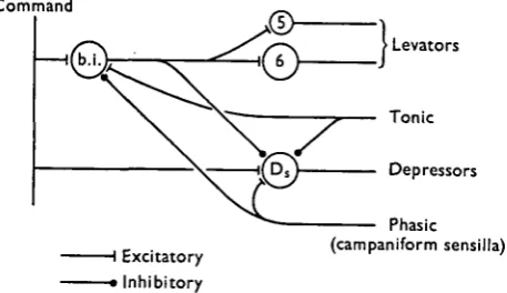
3. Model
Since there is no evidence that the reciprocal patterning depends on connexions between the motoneurones (Pearson & lies, 1970), the simplest model for describing these patterns in de-afferented preparations is one in which a bursting interneurone, or a bursting interneuronal network, simultaneously excites the levator motoneurones and inhibits the depressor motoneurones, and it is assumed that a command input
Command
> Levators
Tonic
Depressors
I Excitatory > Inhibitory
[image:17.451.113.342.368.500.2]Phasic (campaniform sensilla)
Fig. 12. Hypothetical scheme for describing the observed discharge patterns of levator motor axons s and 6 and depressor motor axon DB. See text for details.
190 K. G. PEARSON
motoneurone Ds. The phasic input from the campaniform sensilla is also postulate?!^
to inhibit the bursting interneuronal network (see below). At present, the receptors responsible for the tonic effect have not been identified, although tarsal leceptors could in part be involved since these are known to inhibit the activity in motoneurone Ds and to produce bursts of activity in the levator motoneurones (unpublished
observations).
An unexpected finding was that an increase in the resistance to leg retraction produced an increase in activity in the motoneurones producing leg protraction, i.e. the levators (Fig. 7 right), as well as producing a decrease in the burst durations of these moto-neurones (Fig. 8). The simplest explanation for these findings is that the interneuronal burst generator is inhibited by the phasic input from the campaniform sensilla (Fig. 12). Assuming that the command input remains unchanged, an increase in resistance to retraction would lead to a decrease in the net excitatory input to this generator during leg retraction and hence to an increase in the interval between levator bursts. There would not necessarily be any changes in the levator burst intensity or duration, because during protraction the inhibitory input from the campaniform sensilla would be removed and the excitatory command input to the interneuronal burst generator would be the same as that had retraction not been resisted. Therefore, the effect of an increase in resistance to retraction would be to increase the cycle time but not to change the intensity and duration of the levator bursts, which is equivalent to a shift in the relationships between intensity and cycle time (Fig. 7 right) and between duration and cycle time (Fig. 8) towards longer cycle times. Thus, for a given cycle time, the levator burst intensity will be increased and the duration decreased. The increase in cycle time would explain an unpublished observation that an increase in the resistance to retraction always leads to a decrease in the walking speed.
What are the functional implications of the increase in the discharge rate and the decrease in discharge duration of the levator motoneurones with an increase in load? The behavioural effect of these changes in the characteristics of the levator burst would be to give a stronger and more rapid stepping movement. The most obvious advantage of this change in behaviour would be that when, for example, the animal is walking up a steep incline, the tarsi would be in contact with the ground for a larger fraction of each cycle, thus increasing the time all six legs are simultaneously in contact with the ground. This effect would tend to minimize the average increase in load carried by each leg.
4. Comparison with other systems
In this final section we wish first to examine whether the model proposed in Fig. 12 has any features which could be common with systems producing rhythmic leg move-ments in other insects, and secondly, to discuss the function of phasic sensory feedback in insect motor control.
indicate that sensory input from leg receptors is of extreme importance in controlling rhythmic leg movements. This is not to say, however, that a central pro-gram does not exist. In fact, many of the observations made on walking locusts are compatible with the model shown in Fig. 12. The findings which indicate the im-portance of sensory input in controlling rhythmic leg movements in these animals are that slowing and consequently discoordination of leg movements results from the removal of either the femoral chordotonal organ or the receptors in the tibia and tarsus (Usherwood et al. 1968; Usherwood & Runion, 1970). Moreover, the intensity of acti-vity in both flexor and extensor motoneurones is reduced by removal of these receptors. Both these sets of observations could be explained by a removal of tonic facilitatory sensory input to the bursting interneuronal network. Removal of this excitatory input would increase the interburst duration and decrease the intensity of the flexor bursts generated by this network. The consequence of a decrease in burst intensity would be less inhibition to the extensor motoneurones. This could account for Usherwood and Runion's observation that after removal of tibial and tarsal receptors the extensor motoneurones are not completely inhibited during leg flexion. The decrease in in-hibitory input to the extensor motoneurone would also reduce the effect of post-inhibitory excitation and consequently result in a less intense extensor burst.
We now turn to a brief discussion on the function of phasic sensory input in the control of rhythmic movements. The effect of sensory input in the control of rhythmic movements has been studied in the following systems: locust flight (Wilson & Gettrup, 1963), cricket song (Kutsch & Huber, 1970), and ventilatory movements in a number of different insects (Miller, 1966; Farley & Case, 1968; Mill, 1970). A comparison of the results from these investigations with those of the present study leads to the following generalization. Phasic information in fedback signals becomes increasingly important in controlling the motor output as there is an increase in the probability of variations in load from cycle to cycle. For cricket song there is very little likelihood of unexpected variations in load from cycle to cycle, and correspondingly the motor output patterns are almost entirely independent of sensory input (Kutsch & Huber, 1970). Similarly, for a locust flying through a uniform medium where load variations from cycle to cycle would be expected to be small, the phasic information in fedback sensory signals from wing stretch receptors is irrelevant (Wilson & Gettrup, 1963). This is not true for ventila-tory movements of the abdomen however. Recently, Farley & Case (1968) and Mill (1970) have demonstrated that phasic input from abdominal receptors can modify the motor output patterns. The exact function of this phasic feedback has not yet been established, but one possibility is that it compensates for any variations in load which could occur because the abdomen is in different positions from one cycle to the next. Finally, we have seen in the present study that phasic reflex effects on to the depressor motoneurone D8 could be extremely important in compensating for cycle-to-cycle load
192 K. G. PEARSON
SUMMARY
1. The activity in identified motor units supplying the coxal levator and depressor muscles of the cockroach have been recorded in intact freely walking animals and in preparations after removal of all sensory input from leg receptors.
2. Reciprocal activity in levator and depressor motoneurones can be evoked, or occurs spontaneously, in the partially de-afferented preparations, thus indicating the existence of a central locomotory rhythm generator.
3. The reciprocal activity patterns recorded in the same motoneurones in intact freely moving animals are not identical to those recorded in partially de-afferented preparations. Thus, the production of normal rhythmic leg movements depends to some extent on sensory input from leg receptors, this input probably exerting tonic and phasic effects on the central rhythm generator.
4. An increase in the resistance to leg retraction during normal walking results in an increase in discharge rate of the levator and depressor motoneurones. This observation further demonstrates that rhythmic leg movements are not exclusively centrally con-trolled. The receptors responsible for this reflex effect are probably the trochanteral campaniform sensilla.
I would like to thank Doctors R. B. Stein and A. V. Holden for their helpful criticisms of this paper. This work was supported by a grant from the Canadian Medical Research Council.
REFERENCES
CARBONELL, C. S. (1947). The thoracic muscles of the cockroach, Periplaneta americana. Smithson.
Misc. Collns 107 (2), 1-23.
CRITCHLOW, V. & EULER, C. v. (1963). Intercostal muscle spindle activity and its y-motor control.
J. Physiol., Lond. 168, 820-847.
DELCOMYN, F. (1971a). The locomotion of the cockroach, Periplaneta americana. J. exp. Biol. 54, 443— 52.
DELCOMYN, F. (19716). The effect of limb amputation on locomotion in the cockroach, Periplaneta
americana. J. exp. Biol. 54, 453-72.
EULER, C. V. (1966). The control of respiratory movement. In Breathlessness (ed. J. B. L. Howell and E. J. M. Campbell), pp. 19-32.
EWING, A. W. & MANNING, A. (1966). Some aspects of the efferent control of walking in three cockroach species. J. Insect Physiol. 12, 1115-18.
FARLEY, R. D. & CASE, J. F. (1968). Sensory modulation of ventilative pacemaker output in the cock-roach, Periplaneta americana. J. Insect Physiol. 14, 591-601.
HOY, R. R. & WILSON, D. M. (1969). Rhythmic motor output in leg motor neurons of the milkweed bug, Oncopeltus. Fedn Proc. 28 (2), 588.
HOYLE, G. (1964). Exploration of neuronal mechanisms underlying behaviour in insects. In Neural
Theory and Modelling (ed. R. F. Reiss), pp. 346—76. Stanford University Press.
HOYLE, G. (1970). Cellular mechanism underlying behaviour - neuroethology. Adv. Insect Physiol. 7, 349-444.
HUGHES, G. M. (1952). The coordination of insect movements. I. The walking movements of insects.
J. exp. Biol. 29, 267-84.
HUGHES, G. M. (1957). The coordination of insect movements. II. The effect of limited amputation and the cutting of commissures in the cockroach, Blatta orientalis. J. exp. Biol. 34, 306-33.
HUGHES, G. M. (1965). Locomotion: Terrestrial. In The Physiology of Insecta, vol. 2, pp. 227-54. New York and London: Academic Press.
ILES, J. F. & PEARSON, K. G. (1971). Coxal depressor muscles of the cockroach and the role of peri-pheral inhibition. J. exp. Biol. (in the Press).
KUTSCH, W. (1969). Neuromuscular activity in three cricket species during various behavioural patterns.
PLUTSCH, W. & HUBBR, F. (1970). Central versus peripheral control in cricket stridulation. Z. vergl.
Physiol. 67, 140-59.
LUNDBERG, A. (1969). Reflex control of stepping. Nansen Memorial Lecture to Norwegian Academy of
Sciences and Letters.
MILBURN, N. S. (1963). Sensitivity of cockroach campaniform sensilla to adrenergic drugs. Am. Zool. 3,
5i3-MILL, P. J. (1970). Neural patterns associated with ventilatory movements in dragon fly larvae. .7. exp.
Siol. 52, 167-76.
MILLER, P. L. (1966). The regulation of breathing in insects. Adv. Insect Physiol. 3, 279-344. PEARSON, K. G. & BERGMAN, S. J. (1969). Common inhibitory motoneurones in insects. ,7. exp. Biol. 50,
44S-73-PEARSON, K. G. & ILES, J. F. (1970). Discharge patterns of coxal levator and depressor motoneurones of the cockroach, Periplaneta americana. J. exp. Biol. 52, 139-65.
PEARSON, K. G. & ILES, J. F. (1971). Innervation of the coxal depressor muscles of the cockroach,
Periplaneta americana. J. exp. Biol. 54, 215-32.
PIPA, R. L. & COOK, E. F. (1959). Studies on the hexapod nervous system. I. The peripheral distribution of the thoracic nerves of the adult cockroach, Periplaneta americana. Ann. ent. Soc. Am. 52 (6), 695-710.
PRINGLE, J. W. S. (1938a). Proprioception in insects. II. The action of the campaniform sensilla on the legs. J. exp. Biol. 15, 114-31.
PRINGLE, J. W. S. (19386). Proprioception in insects. III. The function of the hair sensilla at the joints.
J. exp. Biol. 15,
467-73-PRINGLE, J. W. S. (1939). The motor mechanism of the insect leg. J. exp. Biol. 16, 220-31. PRINGLE, J. W. S. (1940). The reflex mechanism of the insect leg. J. exp. Biol. 17, 8-17.
PRINGLE, J. W. S. (1961). Proprioception in arthropods. In The Cell and the Organism, ed. J. A. Ramsay and V. B. Wigglesworth.
SEVERIN, R. V., ORLOVSKII, G. N. & SHIK, M. L. (1967). Work of the muscle receptors during controlled locomotion. Biophysics 12, 575-86.
TAYLOR, A. & DAVEY, M. R. (1968). Behaviour of jaw muscle stretch receptors during active and passive movements in the cat. Nature, Lond. 220, 301—2.
USHERWOOD, P. N. R. (1962). The nature of 'fast' and 'slow' contractions in the coxal muscles of the cockroach. J. Insect Physiol. 8, 31-52.
USHERWOOD, P. N. R. & RUNION, H. I. (1970). Analysis of the mechanical responses of metathoracic extensor tibiae muscles of the free-walking locust. J. exp. Biol. 52, 39-58.
USHERWOOD, P. N. R., RUNION, H. I. & CAMPBELL, J. I. (1968). Structure and physiology of a chordotonal
organ in the locust leg. J. exp. Biol. 48, 305-23.
WENDLER, G. (1966). The coordination of walking movements in arthropods. Symp. Soc. exp. Biol. no. 20. Nervous and hormonal mechanisms of integration, pp. 229-50.
WILSON, D. M. (1965). Proprioceptive leg reflexes in cockroaches. J. exp. Biol. 43, 397-409. WILSON, D. M. (1966). Insect walking. A. Rev. Ent. 11, 103-22.
WILSON, D. M. (1968). The nervous control of insect flight and related behaviour. Adv. Insect Physiol. 5, 289-338.
WILSON, D. & GETTRUP, E. (1963). A stretch reflex controlling wing beat frequency in the grasshopper.
J. exp. Biol. 40, 171-85.