_____________________________________________________________________________________________________ 29(2): 1-9, 2019; Article no.JAMMR.47751
ISSN: 2456-8899
(Past name: British Journal of Medicine and Medical Research, Past ISSN: 2231-0614, NLM ID: 101570965)
The Prognostic Implication of CD49d Expression by
Flow Cytometry and Trisomy 12 Detection by
Fluorescent
in situ
Hybridization in Chronic
Lymphocytic Leukemia
Eman Mosaad Zaki
1, Sahar Abd Allah El-Gammal
2, Noha Gaber Sayed
1,
Mayada Fawzy Sediek
3and Asmaa Ahmed Mohamed
1*1Department of Clinical Pathology, South Egypt Cancer Institute, Assiut University, Egypt. 2
Department of Clinical Pathology, Faculty of Medicine, Assiut University, Egypt.
3
Department of Medical Oncology, South Egypt Cancer Institute, Assiut University, Egypt.
Authors’ contributions
This work was carried out in collaboration among all authors. Author AAM designed the study, performed the statistical analysis, wrote the protocol and wrote the first draft of the manuscript. Authors EMZ and NGS managed the analyses of the study. Authors SAAEG and MFS managed the literature searches. All authors read and approved the final manuscript.
Article Information
DOI: 10.9734/JAMMR/2019/v29i230064 Editor(s): (1) Dr. Syed Faisal Zaidi, Department of Basic Medical Sciences, College of Medicine, King Saud Bin Abdulaziz University-HS, National Guard Health Affairs, King Abdulaziz Medical City, Kingdom of Saudi Arabia. Reviewers: (1) Anil Tombak, Mersin University, Medical Faculty Hospital, Turkey. (2)Mohammaad Nadeem Khan, Bastar University, India. Complete Peer review History:http://www.sdiarticle3.com/review-history/47751
Received 26 December 2018 Accepted 13 March 2019 Published 20 March 2019
ABSTRACT
Chronic lymphocytic leukemia (CLL) is the most common chronic lympho‑proliferative disorder.
This study done to detect the level of cluster of differentiation (CD)49d in CLL patients by flow cytometry and its correlation with the prognosis (survival) and with (trisomy12) detected by fluorescent in situ hybridization (FISH).
Methods: Clinico - hematological profiles done to fourty CLL patients. CD49d tested by flow cytometry and trisomy12 was detected by FISH.
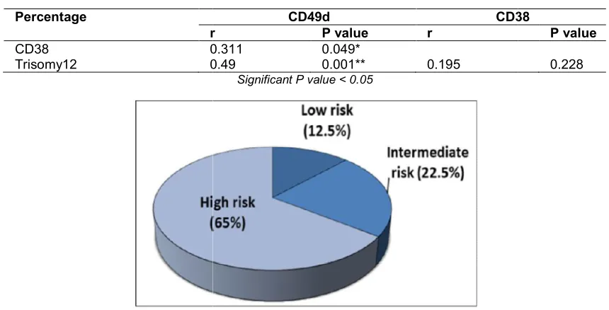
Results: CLL patients classified according to modified Rai staging system into: low risk 12.5%, intermediate risk 22.5% and high risk 65%. CD49d and trisomy12 positivity were detected in 29
Zaki et al.; JAMMR, 29(2): 1-9, 2019; Article no.JAMMR.47751
patients (72.5%) and 22 patients (55%), respectively. There was a significant positive correlation between the percentage of trisomy12 and of CD49d cells in CLL patients (P =0.034). And also, between CD49d and CD38 (P =0.034). On the other hand, there was no significant relation between both CD49d and trisomy12 expression and modified Rai staging system.
As regard to overall survival (O.S) and disease free survival (DFS), both CD49d, trisomy12 positive cases were associated with shorter disease free, and overall survivals compared to the negative cases.
Regarding to the relation between the use of combination of fludarabine, cyclophosphamide, and rituximab (FCR) as a standard treatment in CLL and OS and DFS of patients in our study, we found that FCR account for the better outcome associated with its use.
Conclusion: CLL B-cell membrane expression of CD49d as measured by flow cytometry is a powerful prognostic parameter in patients with CLL. Its positive correlation with the trisomy12 and CD38 and the association of both CD49d and trisomy12 with short survival times indicate that they may have roles in the prognosis of CLL.
Keywords: Chronic lymphocytic leukemia; prognosis; CD49d; trisomy12.
1. INTRODUCTION
Chronic lymphocytic leukemia (CLL) defined as a
lymphoproliferative disorder, composed by
monomorphic round B-lymphocytes involving peripheral blood (PB), bone marrow (BM) and lymphoid organs [1]. CLL is one of the most common types of leukemia in the Western world, however, infrequent in the Eastern. In Egypt, CLL was the most common subtype of leukemias, the National Cancer Registry reported over 80% of lymphoid leukemias are CLL [2]. It is the most common types of leukemia diagnosed in adult.
CD49d is a surface molecule that binds to the β-integrin CD29 to form very late antigen-4
(VLA-4), the expression of which promotes
microenvironment-mediated proliferation of CLL leukemic cells and identifies a subgroup of patients characterized by progressive course and short survival [3]. It should be noted that the expression of CD49d correlates with some other prognostic factors. Specifically, with unmutated IGHV, CD38 and ZAP70 with the major cytogenetic lesions such as trisomy12.Moreover, trisomy12 CLL cases were characterized by the higher mean fluorescence intensity levels of CD49d compared with cases belonging to the other cytogenetic categories, probably facilitated through a NOTCH1 or methylation-mediated mechanism [4].
The presence of cytogenetic abnormalities is a hallmark of CLL, and has historically been best studied by interphase fluorescence in situ hybridization (FISH) [5]. Trisomy12 is the third most common cytogenetic abnormality identified by fluorescence in situ hybridization (FISH). In some reports, trisomy 12 CLL carry an
intermediate prognostic risk, while other reports
suggest a certain degree of clinical
heterogeneity, with a higher incidence of second malignant neoplasms and Richter transformation [6].
The aim of the present study was to detect the level of expression of CD49d in CLL patients by flow cytometry and its correlation with the
prognosis and with trisomy12detected by
fluorescent in situ hybridization (FISH).
2. PATIENTS AND METHODS
The study was included 40 CLL patients (22 men and 18 women; age range 38-81 years). These patients were presented to South Egypt Cancer Institute Assiut University hospital in the period between December 2015 and July 2017. The study was approved by the Institutional Review Board of Faculty of Medicine, Assiut University. An informed written consent was taken from of all cases.
All patients were subjected to:
History taking and clinical examination.
Complete blood picture.
Bone marrow examination
Immunophenotyping: analysis was done by
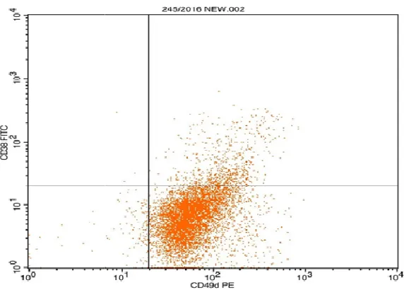
Fig. 1. CLL case with positive CD49d and CD38
Immunophenotyping diagnosis of our CLL patients was done according to scoring system [7]. Fig. 1 illustrate a CLL case with positive CD49d.
Cytogenetic study: The
detect the presence and level of
expression of trisomy 12 by
in situ hybridization (FISH) by using Alpha
Satellite 12 plus probe (Cytocell,
Catalogue No: REF LPH069), labelled in red, which recognized the centromeric repeat sequence D12Z3. FISH procedures were performed as usual, after Preparation of mitotic cells from short
cultures, slide preparation, Pre
denaturation, denaturation, hybridization
and final post-hybridization washes.
Analysis was done by a fluorescent microscope (Carl Zeiss AxioSkop 2 Mot FL). The images were captured throug Leica CW 4000 camera assembled to a computer having FISH software (Carl Zeiss/Cytovision, Axiovision control 3.1). Slides showing more than 50% cells with fluorescent dots were selected for analysis. Then at least 200 cells were counted. In a normal cell, 2 red signals should be observed. While in a cell with trisomy12, there should be 3 red signals.
2.1 Statistical Analysis
Statistical calculation was performed with Statistical Package for Social Sciences (SPSS) software (version 16.0; SPSS Inc, Chicago, Ill).
Zaki et al.; JAMMR, 29(2): 1-9, 2019; Article no.
Fig. 1. CLL case with positive CD49d and CD38
Immunophenotyping diagnosis of our CLL patients was done according to scoring . Fig. 1 illustrate a CLL case with
test aims to
detect the presence and level of
expression of trisomy 12 by fluorescence in situ hybridization (FISH) by using Alpha
Satellite 12 plus probe (Cytocell,
Catalogue No: REF LPH069), labelled in red, which recognized the centromeric repeat sequence D12Z3. FISH procedures were performed as usual, after Preparation itotic cells from short-term blood
cultures, slide preparation,
Pre-denaturation, Pre-denaturation, hybridization
hybridization washes.
Analysis was done by a fluorescent microscope (Carl Zeiss AxioSkop 2 Mot FL). The images were captured through a Leica CW 4000 camera assembled to a computer having FISH software (Carl Zeiss/Cytovision, Axiovision control 3.1). Slides showing more than 50% cells with fluorescent dots were selected for analysis. Then at least 200 cells were counted. In a ll, 2 red signals should be observed. While in a cell with trisomy12, there should be 3 red signals.
Statistical calculation was performed with Statistical Package for Social Sciences (SPSS)
Chicago, Ill).
3. RESULTS
CLL patients classified according to modified Rai staging system into: low risk 12.5%, intermediate risk 22.5% and high risk 65% as in
The expression of CD49d on malignant lymphocytes was detected in 29 patients (72.5%). However, the expression of CD38 on malignant lymphocytes was detected in 21 patients (52.5%) as in Fig. 3.
As regard to trisomy 12, 22 patients were trisomy12 positive (55%).
As regard to correlation between trisomy12, CD49d and CD38; there was a signif
relation between trisomy12 and CD49d
expression on CLL group (P value = 0.001**) (r =0.49), a significant relation between CD38 and CD49d in the studied CLL group. (P value = 0.05*) (r =0.311). On the other hand, there was no significant relation between trisomy and CD38 in the studied CLL group as in
addition, there was no significant relation between modified Rai staging system and the presence of both CD49d and trisomy12 as in Table 2.
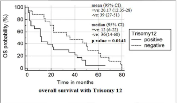
Concerning the relation between the overall survival (OS) and the disease free survival (DFS) of the studied CLL group and the percentage of trisomy12 and CD49d expression, there was a significant relation between OS and both CD49d
(P value= 0.0192) and tr
value=0.0141) as in Table 3 & Figs. 4, 5. As
; Article no.JAMMR.47751
CLL patients classified according to modified Rai staging system into: low risk 12.5%, intermediate risk 22.5% and high risk 65% as in Fig. 2.
The expression of CD49d on malignant lymphocytes was detected in 29 patients However, the expression of CD38 on malignant lymphocytes was detected in 21
As regard to trisomy 12, 22 patients were
As regard to correlation between trisomy12, CD49d and CD38; there was a significant
relation between trisomy12 and CD49d
expression on CLL group (P value = 0.001**) (r =0.49), a significant relation between CD38 and CD49d in the studied CLL group. (P value = 0.05*) (r =0.311). On the other hand, there was ween trisomy and CD38 in the studied CLL group as in Table 1. In addition, there was no significant relation between modified Rai staging system and the presence of both CD49d and trisomy12 as in
Concerning the relation between the overall survival (OS) and the disease free survival (DFS) of the studied CLL group and the percentage of trisomy12 and CD49d expression, there was a significant relation between OS and both CD49d
(P value= 0.0192) and trisomy12 (P
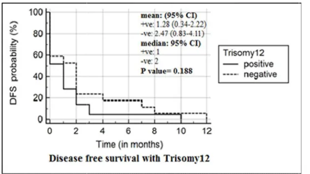
regard to DFS, there was a significant relation between CD49d and DFS (P value=0.0190). However, there was no significant relation between trisomy12 and DFS (P value=0.1882) as in Table 3 & Figs. 6, 7.
4.DISCUSSION
In this study, CLL patients were classified according to modified Rai staging system into: low risk 12.5 %, intermediate risk 22.5% and high risk 65% in their first presentation.
In this study, 29 patients (72.5%) were CD49d positive and 11 patients (27.5%) were CD49d negative according to the use of cut off value of 30%. This result is approximately close to the
Table 1. The relation between CD49d, CD38 and Trisomy12
Percentage
r
CD38 0.311
Trisomy12 0.49
Fig. 2. The distribution of CLL patients, according to modified Rai staging system
Fig. 3. Expression of CD49d and CD38 on malignant lymphocytes in CLL patients Zaki et al.; JAMMR, 29(2): 1-9, 2019; Article no.
regard to DFS, there was a significant relation between CD49d and DFS (P value=0.0190). However, there was no significant relation between trisomy12 and DFS (P value=0.1882)
In this study, CLL patients were classified according to modified Rai staging system into: low risk 12.5 %, intermediate risk 22.5% and high
In this study, 29 patients (72.5%) were CD49d 11 patients (27.5%) were CD49d negative according to the use of cut off value of 30%. This result is approximately close to the
results found by Hendy et al. who reported that, CD49d was positive in (75%) of all studied CLL cases [8]. Other studies done by Benedetti et al. and Wesam et al. found that, CD49d expression was positive in (37.7%) and (55.3%) of cases
respectively [9,10]. These variations in
expression of CD49d in CLL patients between our results and previous studies may be d the variability of the sample size and the possible ethnic variations between the studied groups.
Remarkably, the finding of differential CD49d expression in CLL is an older discovery than anticipated. In 1996, it had already been demonstrated that CD49d expression in CLL is variable, with higher expression of CLL samples of advanced (Rai III, IV) than early stages
Table 1. The relation between CD49d, CD38 and Trisomy12
CD49d CD38
r P value r
0.311 0.049*
0.49 0.001** 0.195
Significant P value < 0.05
distribution of CLL patients, according to modified Rai staging system
Fig. 3. Expression of CD49d and CD38 on malignant lymphocytes in CLL patients
; Article no.JAMMR.47751
results found by Hendy et al. who reported that, CD49d was positive in (75%) of all studied CLL Other studies done by Benedetti et al. and Wesam et al. found that, CD49d expression was positive in (37.7%) and (55.3%) of cases
. These variations in
expression of CD49d in CLL patients between our results and previous studies may be due to the variability of the sample size and the possible ethnic variations between the studied groups.
Remarkably, the finding of differential CD49d expression in CLL is an older discovery than anticipated. In 1996, it had already been demonstrated that CD49d expression in CLL is variable, with higher expression of CLL samples
early stages [11].
CD38
P value
0.228
distribution of CLL patients, according to modified Rai staging system
Fig. 4. Kaplan –Meier curves show the relation between overall survival (OS) and pati
Fig. 5. Kaplan-Meier curves show the relation between overall survival (OS) and
Table 2. The relation between the modified Rai staging system and CD49d and trisomy12
Percentage
Low(N = 5) ±SD
CD49d 38.94±35.27
Trisomy12 6.02 ± 4.42
Table 3. The relation between the overall survival (OS) and the
the studied CLL patient’s group and the percentage of trisomy12 and CD49d expression
Trisomy12 Positive
trisomy12 NO (22)
Negative trisomy12 NO (18)
OS (months) 20.17± 7.82 39 ± 12
DFS (months) 1.28 ± 0.92 2.47 ± 1.64
Significant P value < 0.05; OS:
Zaki et al.; JAMMR, 29(2): 1-9, 2019; Article no.
Meier curves show the relation between overall survival (OS) and pati and without trisomy12
Meier curves show the relation between overall survival (OS) and patients with and without CD49d.
Table 2. The relation between the modified Rai staging system and CD49d and trisomy12
Grade
Intermediate(N = 9) ±SD High(N =26) ±SD
41.46±31.72 60.52±32.62
8.87 ± 5.71 6.95 ± 4.37
Significant P value < 0.05
Table 3. The relation between the overall survival (OS) and the disease free survival (DFS) of the studied CLL patient’s group and the percentage of trisomy12 and CD49d expression
Trisomy12 p. value CD49d Negative
trisomy12 NO (18)
Positive CD49d NO (29)
Negative CD49d NO (11)
39 ± 12 0.0141* 20.9 ± 6.25 41.51±15.97
2.47 ± 1.64 0.1882 1.0 ± 0.56 3.18 ± 2.38
Significant P value < 0.05; OS: Overall survival; DFS: Disease free survival
; Article no.JAMMR.47751
Meier curves show the relation between overall survival (OS) and patients with
patients with
Table 2. The relation between the modified Rai staging system and CD49d and trisomy12
P. value (N =26) ±SD
0.20 0.48
disease free survival (DFS) of the studied CLL patient’s group and the percentage of trisomy12 and CD49d expression
P. value
41.51±15.97 0.0192*
Fig. 6. Kaplan-Meier curves show the relation between the disease free survival (DFS) and patients w
Fig. 7. Kaplan-Meier curves show the relation between disease free survival (DFS) and patients
Within the same concept, our study also revealed that, higher expression of CD49d was associated with advanced disease stage but these results were statistically insignificant, this may be due to low number of our studied cases. This also matched to the results of Wesam E Elderiny, who found that higher levels of CD49d in advanced stages [10].
As regard to correlation between CD49d and CD38 Zucchetto and colleagues were the first in 2006 that reported the strong association of CD38 and CD49d expression on CLL cells using both parameters as categorical variables These findings were found to be in harmony with our study results where we found that, there was
Zaki et al.; JAMMR, 29(2): 1-9, 2019; Article no.
curves show the relation between the disease free survival (DFS) and patients with or without CD49d expression
Meier curves show the relation between disease free survival (DFS) and patients with or without trisomy12
Within the same concept, our study also revealed that, higher expression of CD49d was associated with advanced disease stage but these results were statistically insignificant, this may be due to low number of our studied cases. This also sults of Wesam E Elderiny, who found that higher levels of CD49d in advanced
As regard to correlation between CD49d and CD38 Zucchetto and colleagues were the first in 2006 that reported the strong association of CD38 and CD49d expression on CLL cells using both parameters as categorical variables [12]. be in harmony with our study results where we found that, there was
a significant positive correlation between CD49d and CD38 expression in CLL patients. In addition, this is in agreement with Kamel et al. and Buggins et al. [13,14].
By using Kaplan-Meier curves, patients with CD49d positivity had shorter survival and disease free survival than those negative for CD49d (using 30% as cut off level for CD49d positivity). This is in agreement with Gattei et al.
who reported that, when ana
retrospectively, CLL patients with
positive tumor cells revealed significantly shorter treatment-free and overall survival than patients with <30% CD49d positivity [15]. A prospective analysis indicated that an alternative cut
; Article no.JAMMR.47751
curves show the relation between the disease free survival (DFS) and
Meier curves show the relation between disease free survival (DFS) and patients
a significant positive correlation between CD49d and CD38 expression in CLL patients. In addition, this is in agreement with Kamel et al.
Meier curves, patients with CD49d positivity had shorter survival and disease free survival than those negative for CD49d (using 30% as cut off level for CD49d positivity). This is in agreement with Gattei et al.
who reported that, when analyzed
retrospectively, CLL patients with ≥30% CD49d-positive tumor cells revealed significantly shorter
Zaki et al.; JAMMR, 29(2): 1-9, 2019; Article no.JAMMR.47751
of 45% CD49d expression might be superior to the 30% level [16]. Following these first reports, the prognostic relevance of increased CD49d expression was rapidly and unequivocally confirmed by several groups, using the 30% cut-off level. All of them found that, CD49d expression consistently identifies a subgroup of CLL characterized by poor outcome in their study [3,17,18,19].
Concerning trisomy 12 in our CLL patients, there was a higher incidence of trisomy 12 than that previously thought, (20%) according to WHO 2008 [20]. 55% of our study group were found to be positive for trisomy 12. Other studies including the incidence of trisomy 12 in CLL patients revealed under estimation of these results in comparison to our results. Dal Bo et al., Bulian et al. and Alp. reported that, trisomy 12 was found in 13.2%, 15-20% and between 16% and 35% respectively [21,22,23]. A much lower frequency of trisomy12 (13.2%) reported by Dal Bo et al. On reviewing their study, we found that the majority (50%) of CLL patients who were included in that study had early-stage disease. In contrast, only 12.5% (5/40) of the patients included in our study were at low risk group in the modified Rai staging system, with the majority (65%) were at high risk groups.
As regard to the relation between the staging of the disease and the incidence of the trisomy 12 finding in CLL patients we found that, there was no significant relation between trisomy 12 positivity and staging system. This is in agreement with Alp [23].
On the other side Witzig et al. stated that, there was an increased frequency of trisomy 12 in patients with more advanced Rai stages [24].
In contrast, there was a significant relation between trisomy 12 positivity and CD49d in our study. This is in agreement with Gooden et al., Baumann et al .and Riches et al. they found that a higher CD49d expression was frequently linked to trisomy 12 positive cases in their study [4,25,26]. On the other hand more recently Bullian et al. stated that, despite the high frequency of NOTCH1 and BIRC3mutations and of CD49d and CD38 overexpression, these markers failed to convey a prognostic risk in trisomy12 CLL, while there is a peculiar clinical relevance of IGHV mutations in tris12 CLL patients [22].
In this study, patient’s group with trisomy12 positivity had shorter survival times than those
without trisomy12. This is in agreement with Juliusson et al., Bulian et al. and González-Gascón y Marín et al. they found that overall survival was shorter in patients with high trisomy12 expression in comparison to those with low trisomy12 expression [22,27,28]. These results found to be contradirectory to results reported by Döhner et al. who their prospective trials suggests that overall survival in trisomy12
positive cases was favorable despite
progression-free survival may be shorter [29].
5. CONCLUSION
CLL B-cell membrane expression of CD49d as measured by flow cytometry is a powerful prognostic parameter in patients with CLL. Its positive correlation with the trisomy12 and CD38 and the association of both CD49d and trisomy12 with short survival times indicate that they may have roles in the prognosis of CLL.
CONSENT
An informed written consent was taken from of all cases.
ETHICAL APPROVAL
The study was approved by the Institutional Review Board of Faculty of Medicine, Assiut University.
COMPETING INTERESTS
Authors have declared that no competing interests exist.
REFERENCES
1. Scarfò L, Ferreri AJM, Ghia P. Chronic
lymphocytic leukaemia. Critical Reviews in Oncology/Hematology. 2016;104:169-182.
2. Ibrahim AS, Khaled HM, Mikhail NN,
Baraka H, Kamel H. Cancer incidence in Egypt: Results of the national population-based cancer registry program. J Cancer Epidemiol. 2014;437971.
3. Majid A, Lin TT, Best G, Fishlock K,
Zaki et al.; JAMMR, 29(2): 1-9, 2019; Article no.JAMMR.47751
4. Riches JC, O'donovan CJ, Kingdon SJ,
Mcclanahan F, Clear AJ, Neuberg DS, Werner L, Croce CM, Ramsay AG, Rassenti LZ, Kipps TJ, Gribben JG. Trisomy 12 chronic lymphocytic leukemia cells exhibit upregulation of integrin signaling that is modulated by NOTCH1 mutations. Blood. 2014;123:4101-10.
5. Hernandez JA, Rodriguez AE, Gonzalez
M, Benito R, Fontanillo C, Sandoval V, Romero M, Martin-Nunez G, De Coca AG, Fisac R, Galende J, Recio I, Ortuno F, Garcia JL, De Las Rivas J, Gutierrez NC, San Miguel JF, Hernandez JM. A high number of losses in 13q14 chromosome band is associated with a worse outcome and biological differences in patients with
B-cell chronic lymphoid leukemia.
Haematologica. 2009;94:364-371.
6. Strati P, Abruzzo LV, Wierda WG, O'brien
S, Ferrajoli A, Keating MJ. Second cancers and Richter transformation are the leading causes of death in patients with trisomy 12
chronic lymphocytic leukemia. Clin
Lymphoma Myeloma Leuk. 2015;15:420-7.
7. Bain BJ, Barnett D, Linch D, Matutes E,
Reilly JT, BSOH. General haematology
task force of the British Committee for
Standards in Haematology: Revised
guideline on immunophenotyping in acute
leukaemias and chronic
lympho-proliferative disorders. Clin Lab Haematol. 2002;24:1-13.
8. Hendy O, El Shafie M, Allam M, Motalib T,
Khalaf F, Gohar S. The diagnostic and prognostic value of CD38 and CD49d
expressions in chronic lymphocytic
leukemia. The Egyptian Journal of
Haematology. 2016;41:70.
9. Benedetti D, Tissino E, Pozzo F, Bittolo T,
Caldana C, Perini C, Martorelli D, Bravin V, D'agaro T, Rossi FM, Bomben R, Santinelli E, Zaja F, Pozzato G, Chiarenza A, Di Raimondo F, Del Poeta G, Rossi D, Gaidano G, Dal Bo M, Gattei V, Zucchetto A. NOTCH1 mutations are associated with
high CD49d expression in chronic
lymphocytic leukemia: link between the NOTCH1 and the NF-kappaB pathways. Leukemia; 2017.
10. Wesam E, Elderiny LI. CD49d and CD26
are independent prognostic markers for disease progression in patients with chronic lymphocytic leukemia. Journal of Leukemia, 03; 2015.
11. Eksioglu-Demiralp E, Alpdogan O, Aktan
M, Firatli T, Ozturk A, Budak T, Bayik M,
Akoglu T. Variable expression of CD49d antigen in B cell chronic lymphocytic leukemia is related to disease stages. Leuk Off J Leuk Soc Am Leuk Res Fund UK. 1996;10:1331–1339.
12. Zucchetto A, Bomben R, Dal Bo M, Bulian
P, Benedetti D, Nanni P, Del Poeta G, Degan M, Gattei V. CD49d in B-cell chronic lymphocytic leukemia: Correlated expression with CD38 and prognostic
relevance. Leukemia. 2006;20:523-5;
author reply 528-9.
13. Kamel AM, El-Sharkawy NM, Osman RA,
Abd Fattah EK, Noshokaty E, Abd El-Hamid T, Kandeel EZ. Adhesion molecules expression in CLL: Potential impact on clinical and hematological parameters. J Egypt Natl Canc Inst. 2016;28:31-7.
14. Buggins AG, Levi A, Gohil S, Fishlock K,
Patten PE, Calle Y, Yallop D, Devereux S. Evidence for a macromolecular complex in poor prognosis CLL that contains CD38, CD49d, CD44 and MMP-9. Br J Haematol. 2011;154:216-22.
15. Gattei V, Bulian P, Del Principe MI,
Zucchetto A, Maurillo L, Buccisano F, Bomben R, Dal-Bo M, Luciano F, Rossi FM, Degan M, Amadori S, Del Poeta G. Relevance of CD49d protein expression as overall survival and progressive disease prognosticator in chronic lymphocytic leukemia. Blood. 2008;111:865-873.
16. Shanafelt TD, Geyer SM, Bone ND,
Tschumper RC, Witzig TE, Nowakowski GS, Zent CS, Call TG, Laplant B, Dewald GW, Jelinek DF, Kay NE. CD49d expression is an independent predictor of overall survival in patients with chronic
lymphocytic leukaemia: A prognostic
parameter with therapeutic potential. Br J Haematol. 2008;140:537-46.
17. Rossi D, Zucchetto A, Rossi FM, Capello
D, Cerri M, Deambrogi C, Cresta S, Rasi S, De Paoli L, Bodoni CL, Bulian P, Del Poeta G, Ladetto M, Gattei V, Gaidano G. CD49d expression is an independent risk factor of progressive disease in early
stage chronic lymphocytic leukemia.
Haematologica. 2008;93:1575-9.
18. Nuckel H, Switala M, Collins CH, Sellmann
L, Grosse-Wilde H, Duhrsen U, Rebmann V. High CD49d protein and mRNA expression predicts poor outcome in
chronic lymphocytic leukemia. Clin
Immunol. 2009;131:472-80.
19. Rossi D, Bodoni CL, Zucchetto A, Rasi S,
Zaki et al.; JAMMR, 29(2): 1-9, 2019; Article no.JAMMR.47751
M, Gattei V, Gaidano G. Low CD49d expression and long telomere identify a chronic lymphocytic leukemia subset with highly favourable outcome. Am J Hematol. 2010;85:619–622.
20. Swerdlow SH, Campo E, Harris NL, Jaffe
ES, Pileri SA, Stein H, Thiele J, Vardiman J. WHO classification of tumours of haematopoietic and lymphoid tissues. IARC Press, Lyon; 2008.
21. Dal Bo M, Bulian P, Bomben R, Zucchetto
A, Rossi FM, Pozzo F, Tissino E, Benedetti D, Bittolo T, Nanni P, Cattarossi I, Zaina E, Chivilo H, Degan M, Zaja F, Pozzato G, Chiarenza A, Di Raimondo F, Del Principe MI, Del Poeta G, Rossi D, Gaidano G, Gattei V. CD49d prevails over the novel
recurrent mutations as independent
prognosticator of overall survival in chronic lymphocytic leukemia. Leukemia. 2016;30: 2011-2018.
22. Bulian P, Bomben R, Bo MD, Zucchetto A,
Rossi FM, Degan M, Pozzo F, Bittolo T, Bravin V, D'agaro T, Cerri M, Chiarenza A, Chaffee KG, Condoluci A, D'arena G, Spina M, Zaja F, Pozzato G, Di Raimondo F, Rossi D, Poeta GD, Gaidano G, Shanafelt TD, Gattei V. Mutational status of IGHV is the most reliable prognostic marker in trisomy 12 chronic lymphocytic leukemia. Haematologica. 2017;102:e443-e446.
23. Alp MY. Association of TGF-1 gene
polymorphism with chronic lymphocytic
leukemia. International Journal of
Hematology and Oncology. 2015;25:12-18.
24. Witzig TE, Borell TJ, Herath JF, Tefferi A,
Li CY, Jenkins RB. Detection of trisomy 12
by FISH in untreated B-chronic
lymphocytic leukemia: Correlation with stage and CD20 antigen expression intensity. Leuk Lymphoma. 1994;14:447-51.
25. Gooden CE, Jones P, Bates R,
Shallenberger WM, Surti U, Swerdlow SH,
Roth CG. CD49d shows superior
performance characteristics for flow
cytometric prognostic testing in chronic lymphocytic leukemia/small lymphocytic lymphoma. Cytometry B Clin Cytom; 2016.
26. Baumann T, Delgado J, Santacruz R,
Martinez-Trillos A, Rozman M, Aymerich M, Lopez C, Costa D, Carrio A, Villamor N, Montserrat E. CD49d (ITGA4) expression is a predictor of time to first treatment in
patients with chronic lymphocytic
leukaemia and mutated IGHV status. Br J Haematol. 2016;172:48-55.
27. Juliusson G, Oscier D, Juliusson G,
Gahrton G, Oscier D, Fitchett M, Ross F, Brito-Babapulle V, Catovsky D, Knuutila S, Elonen E, Lechleitner M, Tanzer J, Schoenwald M, Castoldi GL, Cuneo A, Nowell P, Peterson L, Kay N. Cytogenetic findings and survival in B-cell chronic lymphocytic leukemia. Second IWCCLL Compilation of Data on 662 Patients. Leuk Lymphoma. 1991;5(Suppl 1):21-5.
28. González-Gascón Y, Marín I,
Hernández-Sánchez M, Rodríguez-Vicente AE, Sanzo C, Aventín A, Puiggros A, Collado R, Heras C, Muñoz C, Delgado J, Ortega M, González MT, Marugán I, De La Fuente I, Recio I, Bosch F, Espinet B, González M, Hernández-Rivas JM, Hernández JÁ. A high proportion of cells carrying trisomy 12 is associated with a worse outcome in patients with chronic lymphocytic leukemia. Hematological Oncology. 2016;34:84-92.
29. Dohner H, Stilgenbauer S, Benner A,
Leupolt E, Krober A, Bullinger L, Dohner K, Bentz M, Lichter P. Genomic aberrations
and survival in chronic lymphocytic
leukemia. The New England Journal of Medicine. 2000;343:1910–1916.
_________________________________________________________________________________
© 2019 Zaki et al.; This is an Open Access article distributed under the terms of the Creative Commons Attribution License (http://creativecommons.org/licenses/by/4.0), which permits unrestricted use, distribution, and reproduction in any medium, provided the original work is properly cited.
Peer-review history: