Scholarship@Western
Scholarship@Western
Electronic Thesis and Dissertation Repository
7-9-2012 12:00 AM
Effect of Head Movement on Sound Localization in Real and
Effect of Head Movement on Sound Localization in Real and
Simulated Cochlear Implant Users
Simulated Cochlear Implant Users
Kassandra-Anne H. Birtch The University of Western Ontario Supervisor
Dr. E. A. Macpherson
The University of Western Ontario
Graduate Program in Health and Rehabilitation Sciences
A thesis submitted in partial fulfillment of the requirements for the degree in Master of Science © Kassandra-Anne H. Birtch 2012
Follow this and additional works at: https://ir.lib.uwo.ca/etd
Part of the Speech and Hearing Science Commons
Recommended Citation Recommended Citation
Birtch, Kassandra-Anne H., "Effect of Head Movement on Sound Localization in Real and Simulated Cochlear Implant Users" (2012). Electronic Thesis and Dissertation Repository. 634.
https://ir.lib.uwo.ca/etd/634
This Dissertation/Thesis is brought to you for free and open access by Scholarship@Western. It has been accepted for inclusion in Electronic Thesis and Dissertation Repository by an authorized administrator of
(Spine title: Sound Localization in Real and Simulated Cochlear Implantees)
(Thesis format: Monograph)
by
Kassandra-Anne H. Birtch
Graduate Program in Health and Rehabilitation Sciences
A thesis submitted in partial fulfillment of the requirements for the degree of
Master of Science
The School of Graduate and Postdoctoral Studies The University of Western Ontario
London, Ontario, Canada
ii
THE UNIVERSITY OF WESTERN ONTARIO School of Graduate and Postdoctoral Studies
CERTIFICATE OF EXAMINATION
Supervisor
______________________________ Dr. Ewan Macpherson
Supervisory Committee
______________________________ Dr. Vijay Parsa
______________________________ Dr. Susan Scollie
Examiners
______________________________ Dr. Margaret Cheesman
______________________________ Dr. Susan Scollie
______________________________ Dr. Jack Scott
The thesis by
Kassandra-Anne H. Birtch
entitled:
Effect of Head Movement on Sound Localization in Real and
Simulated Cochlear Implant Users
is accepted in partial fulfillment of the requirements for the degree of
Master of Science
iii
Abstract
Cochlear implant (CI) users’ limited ability to use acoustical cues for sound localization causes left/right confusions and front/back reversals. Head movement is beneficial in reducing these errors in acoustically hearing listeners. This study investigated the effect
of head movement on localization throughout 360o of azimuth for both real and simulated
CI users. Listeners in a bilateral electro-acoustic (CI with ipsilateral hearing aid)
simulation derived the greatest head movement benefit in reducing front/back reversals. Left/right confusions were reduced in simulations with matched bilateral stimulation. Sensitivity to both timing and level cues for sound localization was correlated with sound localization performance without head movement for simulated device users. Sensitivity to timing cues was correlated with sound localization performance with head movement cues for simulated device users. Simulations of bilateral CI and bimodal users’ (CI with contralateral hearing aid) listening predicted real users’ sound localization performance, binaural sensitivity and head movement patterns.
Keywords
iv
Acknowledgments
Many thanks to Dr. Ewan Macpherson for his encouragement throughout the
development of this project and without whose MATLAB expertise I could not have done this work. I would also like to thank my advisory committee members, Drs. Vijay Parsa and Susan Scollie, as well as clinical audiologist Dr. Kim Zimmerman, for sharing their contributions to this project.
To my parents, thanks for helping me get to this point both in life and in my studies. To my friends, I’m so glad you were there when I needed to unwind, that in and of itself was a great support. To Vytas, a million socks! To Linh and Teresa, thanks for being there to celebrate the mini-milestones and to talk through the rough patches. To all of you: without your support, understanding and love this would have been a much harder process.
v
Table of Contents
CERTIFICATE OF EXAMINATION ... ii
Abstract ... iii
Acknowledgments... iv
Table of Contents ... v
List of Abbreviations ... ix
List of Tables ... x
List of Figures ... xi
List of Appendices ... xiv
Chapter 1 ... 1
1 Introduction ... 1
1.1 Sound Localization Cues in the Horizontal Plane ... 2
1.1.1 Interaural Time Difference (ITD) – A Cue to Lateral Position ... 2
1.1.2 Interaural Level Difference (ILD) – A Cue to Lateral Position ... 3
1.1.3 Spectral Cues – A Cue to Front/Back Location ... 4
1.1.4 Head Movement – Resolving Ambiguous Information About Front/Back Location ... 4
1.2 Monaural Localization in the Horizontal Plane ... 7
1.3 Restoring a Sensation of Hearing with Cochlear Implants (CI) ... 10
1.4 Device Features Impacting Sound Localization Cues ... 12
1.4.1 Continuous Interleaved Sampling (CIS) ... 12
1.4.2 Fine-Structure Processing (FSP) ... 14
1.4.3 Automatic Gain Control (AGC)... 15
1.4.4 Microphone Placement ... 15
vi
Implant (BiCI) Users... 17
1.5.2 Sensitivity to Interaural Time Differences (ITD) in Bimodal (CI+HA) Users ... 20
1.5.3 Sensitivity to Interaural Level Differences (ILD) in Bilateral Cochlear Implant (BiCI) Users... 22
1.5.4 Sensitivity to Interaural Level Differences (ILD) in Bimodal (CI+HA) Users ... 22
1.6 Abilities of Cochlear Implant (CI) Users to Localize in the Horizontal Plane ... 23
1.6.1 Unilateral Cochlear Implantation (UCI) and Sound Localization ... 23
1.6.2 Bimodal Fitting (CI+HA) and Sound Localization ... 24
1.6.3 Bilateral Cochlear Implantation (BiCI) and Sound Localization ... 26
1.6.4 Who Benefits More, the Bilateral Cochlear Implant (BiCI) User or the Bimodal (CI+HA) User? ... 27
1.6.5 Bilateral Electro-acoustic Stimulation (BiEAS) and Sound Localization... 30
1.6.6 Electro-acoustic Stimulation with Contralateral Hearing Aid (EAS+HA) and Sound Localization ... 32
1.7 Simulating Cochlear Implants... 33
1.8 Filling the Gaps ... 34
1.9 Objectives and Hypotheses ... 35
Chapter 2 ... 37
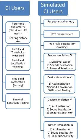
2 Methods ... 37
2.1 Participants ... 37
2.1.1 General Inclusion Criteria ... 37
2.1.2 General Exclusion Criteria ... 37
2.1.3 Simulated Cochlear Implant (CI) Users ... 38
vii
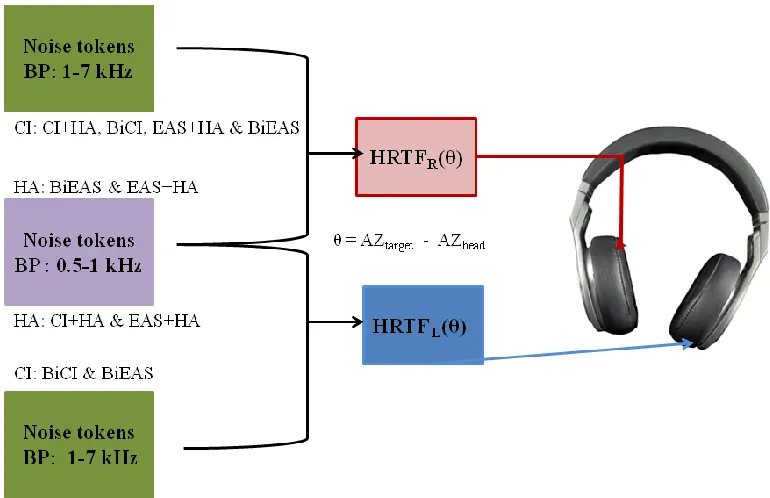
2.2.1 Generating Noise Tokens ... 43
2.2.2 Modifying Individualized Head-Related Transfer Functions (HRTFs) .... 45
2.2.3 Device Simulations ... 48
2.3 Procedures ... 50
2.4 Experimental Conditions ... 54
2.4.1 Sound Localization Tests ... 54
2.4.2 Binaural Testing ... 57
2.5 Analysis Methods... 58
2.5.1 Simulated Cochlear Implant (CI) Users ... 58
2.5.2 Real Cochlear Implant (CI) Users... 64
Chapter 3 ... 66
3 Results ... 66
3.1 Performance on Tests of Sound Localization in Simulated Cochlear Implant (CI) Users ... 66
3.1.1 Visual Inspection of Azimuth and Lateral Angle Plots ... 66
3.1.2 Measures Derived for Analysis ... 71
3.1.3 Statistical Analysis of Front/Back Error Rates ... 72
3.1.4 Statistical Analysis of Left/Right Error Rates ... 77
3.1.5 Statistical Analysis of Lateral Angle Gain ... 79
3.1.6 Statistical Analysis of Lateral Angle Bias ... 81
3.1.7 Statistical Analysis of Response Scatter ... 83
3.2 Performance of Real Cochlear Implant (CI) Users ... 85
3.2.1 Descriptive Analysis of Front/Back Error Rates ... 89
3.2.2 Descriptive Analysis of Left/Right Error Rates and Lateral Angle Bias .. 93
viii
3.3 Performance on Tests of Binaural Sensitivity in Simulated Cochlear Implant
(CI) Users ... 97
3.3.1 Predicting Simulated Cochlear Implant (CI) Users’ Sound Localization Performance without Head Movement Cues ... 100
3.3.2 Predicting Simulated Cochlear Implant (CI) Users’ Sound Localization Performance with Head Movement Cues ... 100
3.4 Comparing Sound Localization Performance of Real and Simulated Bilateral Cochlear Implant (BiCI) and Bimodal (CI+HA) Users ... 103
3.4.1 Measures of Sound Localization ... 103
3.4.2 Head Movement Behaviour in the Free Movement Condition ... 108
4 Discussion ... 121
4.1 Simulated Cochlear Implant (CI) Users Performance ... 121
4.1.1 Sound Localization Tests ... 121
4.1.2 Binaural Sensitivity Tests ... 125
4.2 Real Cochlear Implant (CI) Users’ Sound Localization Performance ... 128
4.3 Representing Bilateral Cochlear Implant (BiCI) and Bimodal (CI+HA) Users’ Performance through Simulation ... 130
4.4 What do Cochlear Implant (CI) Simulations tell us About Real Users’ Performance? ... 134
4.5 Improvements in Cochlear Implant (CI) Devices that might Improve Sound Localization Performance ... 136
4.6 Future Research ... 139
References ... 141
Appendices ... 148
ix
List of Abbreviations
ACE – Advanced Combination Encoder
AGC – Automatic gain control
BiCI – Bilateral cochlear implant
BiEAS- Bilateral electro-acoustic stimulation
BiHA – Bilateral hearing aids
BTE – behind-the-ear or placed above the helix
CI – Cochlear implant
CIS – Continuous interleaved sampling
CI+HA – Cochlear implant paired with contralateral hearing aid
DAI – Direct audio input
EAS+HA – electro-acoustic stimulation paired with contralateral hearing aid
FSP – fine-structure processing
GET – Gaussian-enveloped tone
HA – Hearing aid
HRTF – Head-related transfer function
ILD – Interaural level difference
ITD – Interaural time difference
ILD – JND – Interaural level difference just-noticeable difference
ITD-JND – Interaural time difference just-noticeable difference
JND – Just-noticeable difference
LED – light emitting diode
pps – pulses per second
RMS – root mean square
SPEAK – Spectral Peak
SNHL – Sensorineural hearing loss
UCI – Unilateral cochlear implant
x
List of Tables
Table 1: Hearing history for UCI Users ... 39
Table 2. Hearing history for CI+HA users. ... 40
Table 3. Hearing History for BiCI Users. ... 41
Table 4. Description of the devices worn by all real CI users at the time of testing. ... 42
Table 5. Stimulus presentation level for all real CI users. ... 53
Table 6. Front/back error for real cochlear implant users across head movement conditions. ... 90
Table 7. Left/right error rates for real cochlear implant users across head movement conditions. ... 90
Table 8. Lateral angle gain for real cochlear implant users across head movement conditions. ... 90
Table 9. Lateral angle bias for real cochlear implant users across head movement conditions. ... 91
xi
List of Figures
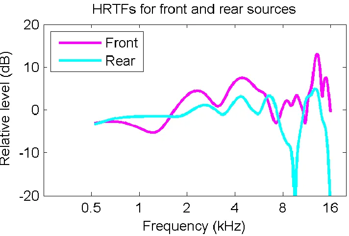
Figure 1. Head-related transfer functions measured at the right ear of one listener for sounds presented from front (magenta) and rear (teal) locations . ... 5
Figure 2. The Cone of Confusion and iso-interaural difference bands. ... 6
Figure 3. Differentiating front from rear sound sources using head movement cues. ... 8
Figure 4. Block diagram of an n-band continuous interleaved sampling (CIS) processor. ... 13
Figure 5. Diagram of the automatic gain control (AGC) in the cochlear implant (CI). . 16
Figure 6. Sequence of steps used to process the individual head-related transfer functions (HRTFs) measured to generate the device simulation assessed. ... 44
Figure 7. Measured head-related transfer functions (HRTFs) for the left and right ears and hearing aid (HA) and cochlear implant (CI) device simulation outputs. ... 47
Figure 8. Modified HRTFs applied to the noise tokens in each of the device simulations. ... 49
Figure 9. Flowchart describing the procedure of assessing both real (left) and simulated (right) CI users on the experimental tasks. ... 50
Figure 10. Diagram of the head sweep performed in the controlled movement condition. ... 56
Figure 11. Sample data illustrating the analysis of front/back errors in azimuth plots... 59
Figure 12. Sample data illustrating the analysis of left/right errors in azimuth plots. .... 61
Figure 13. Sample lateral angle data illustrating good localization performance. ... 62
xii
for listener L061 across both head movement conditions and device simulations. ... 67
Figure 16. The relationship between response azimuth and target azimuth is presented for listener L061 across head movement conditions and device simulations. ... 68
Figure 17. Front/back error rates for normally hearing listeners both in the normally hearing condition and device simulations when no head movement cues are available. . 74
Figure 18. Front/back error rates across simulation and head movement conditions. .... 75
Figure 19. Left/right error rates across simulation and head movement conditions. ... 78
Figure 20. Lateral angle gain for both simulation and head movement conditions. ... 80
Figure 21. Lateral angle bias for both simulation and head movement conditions. ... 82
Figure 22. Response scatter for both simulation and head movement conditions. ... 84
Figure 23. Azimuth scatter plots for real CI users across head movement conditions. .. 86
Figure 24. Lateral angle scatter plots for real CI users across head movement conditions. ... 87
Figure 25. Comparison of real and simulated CI users’ front/back error rates. ... 92
Figure 26. Comparison of real and simulated CI users’ left/right error rates. ... 94
Figure 27. Comparison of real and simulated CI users’ lateral angle bias. ... 95
Figure 28. Comparison of real and simulated CI users’ lateral angle gain. ... 96
Figure 29. Comparison of real and simulated CI users’ response scatter. ... 98
Figure 30. Binaural sensitivity performance for both real and simulated CI users. ... 99
xiii
binaural sensitivities (ILD-JND or ITD-JND) across real and simulated CI users. ... 102
Figure 33. Head movement tracks for listener L061 in the BiCI device simulation. .. 109
Figure 34. Head movement tracks for listener L061 in the CI+HA device simulation. 110
Figure 35. Head movement tracks for listener L038 (real CI+HA user). ... 111
Figure 36. Head movement tracks for listener L067 (real BiCI user). ... 112
Figure 37. Comparison of real and simulated CI users’ initial direction of movement. 114
Figure 38. Comparison of real and simulated CI users’ total head movement. ... 116
Figure 39. Comparison of real and simulated CI users’ average head position. ... 118
xiv
List of Appendices
Appendix A: Ethics Approval ... 148
Appendix B. Summary of Studies Assessing Sound Localization in UCI Users ... 149
Appendix C. Summary of Studies Assessing Sound Localization in CI+HA Users .... 154
Appendix D. Summary of Studies Assessing Sound Localization in BiCI Users ... 156
Appendix E. Summary of Studies Assessing Binaural Sensitivity in BiCI Users ... 159
Chapter 1
1
Introduction
Problem
Cochlear implantation is used to restore a sensation of hearing to individuals with severe-to-profound hearing impairment. The restoration of speech perception in cochlear implant (CI) users is well documented; however, less is known about the benefits of CIs in sound localization. Sound localization benefits acquired by CI users are highly dependent on the type of device they are using. There are four different methods for fitting an individual who has bilateral, severe-to-profound hearing impairment that involve a cochlear implant component, these include: i) bilateral cochlear implantation (BiCI); ii) cochlear implantation paired with a contralateral hearing aid (CI+HA); iii) electro-acoustic stimulation (i.e. a hearing aid and cochlear implant in the same ear; EAS) paired with contralateral hearing aid (EAS+HA); and bilateral electro-acoustic
stimulation (BiEAS). Each of these fitting methods could affect the availability of cues necessary for sound localization differently for several reasons, including: the frequency range represented in the electrical signal, how that frequency range is processed and how the stimulation differs between ears. However, it is likely that all of these methods remove spectral information important for differentiating front/back location because of the speech processor in the CI and the placement of the microphone on the CI. To date this issue is not well understood, although it would be expected that CI users might derive benefits in tests of sound localization if head movement cues were available to them. Head movement benefit is expected because acoustically listening individuals derive benefits from head movement and because head movement provides additional
The following sections include background information about the basic principles of
sound localization (Section 1.1), monaural localization in unilateral hearing loss (Section
1.2), binaural and monaural fitting of CI devices (Section 1.3) and how CI speech
processing strategies work (Section 1.4). Each of these sections provides a framework
for discussing:
i) What might be expected when localizing with one CI only (Section 1.6.1);
ii) How sensitivity to cues necessary for sound localization might differ
among different populations of CI users (Section 1.5);
iii) How well different populations of CI users would be expected to localize
(Section 1.6);
iv) And how sensitivity to binaural cues might explain sound localization
performance in CI users (Section 1.6).
1.1 Sound Localization Cues in the Horizontal Plane
Binaural hearing is essential for localization of sounds in the horizontal plane. In order to localize sound sources, higher auditory centers analyze and compare the acoustic input at each ear (for review: Middlebrooks & Green, 1991). Analysis of the differences in the signals arriving at each ear provides two cues to sound localization: interaural time difference (ITD) and interaural level difference (ILD). In addition, spectral cues arising from interactions between the acoustic signal and the listener’s body can be used to distinguish front from rear sound sources in the horizontal plane. Lastly, head movement provides a cue to resolve ambiguous interaural difference cues and/or resolve front/back confusions when spectral cues are unavailable. Head movement cues are derived from the auditory system’s ability to track changes in ITD and/or ILD as the head moves relative to the sound source. In order to localize accurately, all of these cues are important; therefore, a more detailed discussion of each of these cues follows.
1.1.1
Interaural Time Difference (ITD) – A Cue to Lateral Position
midline are equidistant from the pinnae making ITD equal to zero; whereas sound sources placed more laterally have larger ITDs. While this information provides cues to the lateral angle, the side on which the sound source is located is determined by which ear receives the leading signal. Sensitivity to ITD is best at low-frequencies because the auditory nerve is unable to reliably track differences in the fine-structure of the waveform for mid- to high-frequency stimuli (i.e. stimuli greater than ~1200 Hz, Zwislocki & Feldman, 1956; Newton & Hickson, 1981). Interaural time differences are the predominant cue used when localizing wideband noise in the horizontal plane (Macpherson & Middlebrooks, 2002). There are three types of ITD cues, which are derived from different components of the acoustic signal: i) onset ITD, available in the envelope; ii) offset ITD, also available in the envelope; and iii) on-going ITD, available in both the fine-structure and envelope depending on the nature of the stimulus (i.e. if the envelope is unmodulated or modulated). These different cues refer to the point at which the temporal disparity between acoustic signals is analyzed – e.g. the onset of the
stimulus (onset ITD), the offset of the stimulus (offset ITD) or throughout the duration of the stimulus (on-going ITD; Blauert, 1983). The relative strength of these cues is
dependent on specific features of the stimulus (Freyman, Zurck, Balakrishnan & Chiang, 1997); however, evidence suggests that the most salient of the three is on-going ITDs (Macpherson & Middlebrooks., 2002). On-going ITDs are likely the most salient of the three ITD cues because the auditory system tracks temporal information once per cycle of the waveform for low frequencies, as opposed to once either at the beginning or end of the stimulus for either onset or offset-ITD cues respectively.
1.1.2
Interaural Level Difference (ILD) – A Cue to Lateral Position
Interaural level difference is different from ITD in that it is the disparity in amplitude between the acoustic signals arriving at each of the ears is analyzed by the auditory system. Once the acoustic signal arrives at the head, it is attenuated due to the head shadow effect; the presence of the head attenuates the acoustic signal as it travels to the ear that is furthest from the sound source. The attenuation of the acoustic signal is spectrally dependent because the head shadow effect is most effective for high
less able to bypass the head). Therefore, ILDs are greatest for high-frequency sounds (Newton & Hickson, 1981; Yost & Dye, 1988). As was the case with ITD cues, ILDs are largest for sound sources located laterally and are equal to 0 dB when the sound source is on the midline. Also, left/right location is differentiated by which ear acquires the most intense input.
1.1.3
Spectral Cues – A Cue to Front/Back Location
Spectral cues are derived from the interaction between the acoustic signal and the pinnae, head and shoulders. These interactions filter and modify the acoustic signal. As shown in figure 1, predictable spectral changes occur which can be used by the listener as cues to localization (Middlebrooks & Green, 1991). Spectral cues result from both
constructive and destructive interference, which can increase or decrease the amplitude of the signal, respectively. The amount of constructive or destructive interference is
spectrally dependent because filtering characteristics of the pinna and external auditory canal are greatest for high frequencies. Consequently, spectral cues are most salient at high frequencies. Evidence from studies evaluating spectral cues indicates that these cues provide directional information specifying the front/back position of a stimulus (Merhgardt & Mellert, 1977; Langendijk & Bronkhorst, 2002; Zhang & Hartmann, 2010). The presence of peaks and notches in the HRTFs in the 4 to 10 kHz band reduces front to back errors while the presence of peaks and notches in the HRTFs in the 10 to 12 kHz band reduces back to front errors (Hebrank & Wright, 1974). Therefore, localization of band-passed stimuli containing only low-frequency energy below 4 kHz is associated with poor front/back localization (Best et al., 2011). Findings from Hebrank and Wright (1974) indicate that front-back discrimination in the median plane is most effective for wideband stimuli.
1.1.4
Head Movement – Resolving Ambiguous Information About
Front/Back Location
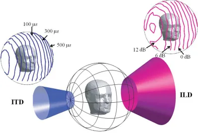
Figure 2. The Cone of Confusion and iso-interaural difference bands.
angle and therefore all positions in space with this lateral angle are known as cones of confusion (Fig. 2). Each cone of confusion represents infinite positions with the same lateral angle; consequently, each cone of confusion represents infinite possible front/back confusions. Although spectral cues provide a cue to front/back position, these cues may not be available (for example, in stimuli that lack frequencies above 4 kHz) and in these cases additional means for resolving front/back confusions are needed. These additional cues are derived from dynamic integration of changing ITD and/or ILD information while the head and/or body are moving.
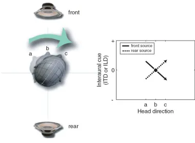
In studies of localization in the horizontal plane, front-back confusions have been shown to be reduced when head movement cues are available (Wightman & Kistler, 1999; Zahorik, Bangayan, Sundareswaran, Wang & Tam, 2006; Iwaya, Suzuki & Kimura, 2003, Macpherson & Middlebrooks, 2002; Macpherson, Cumming & Quelch, 2011). Wallach (1939, 1940) suggested that head movement cues might resolve ambiguous ITD and ILD cues. In principle, as the head moves, the auditory system processes changing ITD and/or ILD information provided that an individual is sensitive to ITD and/or ILD. Therefore, the auditory system tracks changes in interaural difference information in order to identify the location of sound sources that are otherwise ambiguous. For example, as the head moves from left to right for a sound source located in the front, the intensity of the stimulus and lead in the stimulus at the right ear would decrease; if the sound source was located in the rear, and the listener’s head moves from left to right, the intensity of the stimulus and the lead in the stimulus at the right ear would increase (Fig. 3). Studies investigating the effect of head movement on sound localization in normally hearing and unaided hearing impaired listeners indicate that head movement reduces the number of front-back confusions (for review: Best et al., 2011).
1.2 Monaural Localization in the Horizontal Plane
As discussed in section 1.1., binaural hearing is important for localizing sound sources in
localization in the horizontal plane. Studies of monaural localization have investigated the abilities of: monaurally impaired listeners (Newton et al., 1981; Slattery III &
Middlebrooks, 1994), normally hearing listeners with a plugged/muffed ear (Slattery III & Middlebrooks, 1994; Wightman & Kistler, 1997) and normally hearing listeners undergoing monaural simulations virtually (Wightman & Kistler, 1997). In experiments of monaural hearing in which plugs and/or muffs are used, results suggest that these methods are ineffective models of unilateral sensorineural hearing loss (USNHL; for review see Wightman & Kistler, 1997). These methods likely distort rather than attenuate the acoustic signal at the blocked ear because it is extremely difficult to completely attenuate low-frequency sounds. A common finding among studies of
differences in the results across studies may be better addressed by studies that consider the nature of the hearing loss, the spectral content of the stimuli and whether or not overall loudness cues are removed or reduced when drawing conclusions about localization behaviour in populations with USNHL.
1.3 Restoring a Sensation of Hearing with Cochlear
Implants (CI)
Sensorineural hearing loss (SNHL) is a condition in which varying degrees of acoustic information reach higher auditory processing centers because acoustic information cannot be transmitted to the auditory center of the brain because of impairment in either the inner hair cells or a defect in the acoustic nerve. This condition may be congenital or acquired in nature, and can occur both unilaterally and bilaterally. Depending on the severity of the loss, several treatment options are available for restoring bilateral hearing in those whose hearing impairment is caused by an inability to convert the acoustic signal to a neural impulse. The options including CI device fitting include: i) BiCI; ii) CI+HA; iii) BiEAS; and iv) EAS+HA.
ii) one ear is still available for alternative treatments should the outcome of cochlear implantation be poor; iii) there is less discomfort and/or risk is associated with implanting one ear only; and iv) acoustic sensitivity to low-frequencies is preserved in the aided ear, which improves speech perception in noise and pitch perception (McDermott, 2010). In cases where CI+HA is chosen as the preferred method of fitting, it is more likely that the individual will be implanted in the worst ear and that a HA is fitted to the better one. Electro-acoustic stimulation (EAS) combines electric stimulation in the high frequencies and acoustic amplification in the low frequencies to restore hearing to patients with both mild-to-moderate low-frequency hearing loss and severe-to-profound high-frequency loss in the same ear. This technology is young relative to CIs and HAs, and is used as an intermediary between the two for patients who would not benefit from the use of HAs exclusively. The prevalence of EAS use relative to CI or HA use is unknown; however, 250 EAS users have been involved in studies investigating the preservation of residual hearing following implantation (Talbot & Hartley, 2008). In addition, this technology also provides access to low-frequency acoustic information that typical CI users do not have access to. The benefits acquired by patients using EAS are highly dependent upon the degree of residual low-frequency hearing preserved following implantation (von Ilberg Baumann, Kiefer, Tillein & Adunka, 2011). Individuals fitted with BiEAS or EAS+HA have primarily been assessed on tasks of speech and music perception; to date no studies have investigated the effects of either of these configurations on sound
localization (von Ilberg et al., 2011).
Despite the fact that SNHL typically occurs bilaterally, many individuals currently wear a CI unilaterally without any device present in the contralateral ear. The number of
1.4 Device Features Impacting Sound Localization Cues
As mentioned in the previous section, CIs can restore a sensation of hearing to
individuals with severe-to-profound hearing loss. Cochlear implants consist of two major parts: i) the internal component (i.e. the electrode array, magnet and antenna); and ii) the external component (i.e. the battery pack, coil, microphone and speech processor). The speech processor transduces acoustic information picked up by the microphone into an electrical signal that is then transmitted to the internal component. The electrical signal is then used to modulate biphasic pulse trains that are emitted by the intracochlear
electrodes to excite neural tissue. Several different processing strategies are available on the market, each of which processes acoustic stimuli differently. The specific parameters of these programs vary across companies (i.e. Med-El, Cochlear Americas and Advanced Bionics etc.); however, the basic principles are similar. For the purposes of this review, two general program types will be discussed: Continuous Interleaved Sampling (CIS) and Fine-structure processing (FSP). In addition, two features of a CI will be discussed because they may hinder sound localization performance. These two features are the automatic gain control circuitry (AGC) and the placement of the microphone. These two features are also present in HA devices. Consequently, any of the effects of AGC and/or microphone placement on localization cues are also present in the signal presented to the aided ear for CI+HA, EAS+HA and BiEAS users.
1.4.1
Continuous Interleaved Sampling (CIS)
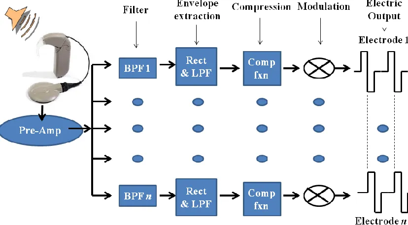
The CIS strategy processes sound in four stages (Fig. 4). In the initial processing stage,
the acoustic signal is picked up by the microphone and filtered by a number ofbandpass
filters into severalchannels. The second stage in processing involves the extraction of
the envelope signal in each of these channels. Envelope extraction might be performed through half-wave rectification or through the application of a Hilbert Transform depending on the specific strategy. The third stage in processing uses a compressive function to compress the acoustic dynamic range in each channel onto a more narrow electric dynamic range. This function is used in order to achieve near normal loudness growth via the electrical output of the CI because its electrical dynamic range is
Figure 4. Block diagram of an n-band continuous interleaved sampling (CIS) processor. The CIS-like strategies uses a pre-amplifier (Pre-amp.) to attenuate high amplitude frequencies in the acoustic signal. Following the pre-amplifier the acoustic input undergoes multiple channels of processing (filter). Each channel includes stages of bandpass filtering (BPF), envelope detection, compression, and modulation. The envelope detectors generally use a rectifier (Rect.) and lowpass filter (LPF). For example, rectifiers might include Hilbert Transform or a half-wave rectifier. The resulting output pulse trains are shown on the right. The outputs of the multipliers are
directed to intracochlear electrodes (Electrode-1 to Electrode-n) via a transcutaneous link
dB. Therefore, the acoustic dynamic range needs to be compressed onto the smaller electric dynamic range. Lastly, the modified envelope signal in each of the channels is used to modulate biphasic pulse trains at each individual stimulating electrode. Most strategies used today are based on the CIS processing strategy, and therefore process the acoustic stimuli in a similar fashion. These CIS and CIS-like strategies are all similar in that they are envelope based strategies; thus, they convert the amplitude modulation of
the acoustic signal’s envelope in n channels into an electric signal presented over m
electrodes (i.e. the number of channels and electrodes is program specific). Variants on this strategy are available from each of the cochlear implant companies. Examples of
CIS-like strategies are HiRes (Advanced Bionics), n-of-m (Med-El), Advanced
Combination Encoder (ACE; Cochlear Americas), HDCIS (Med-El) and Spectral Peak (SPEAK; Cochlear Americas). The most notable difference between these processing strategies has to do with the number of channels available in the electric output.
Processing strategies like HDCIS and HiRes reproduce all of the filtered channels in the
electrical signal. In contrast, processing strategies like ACE, n-of-m, and SPEAK only
stimulate a subset of electrodes that correspond to a specific number of channels that are
selected based on their relative amplitudes. ACE and n-of-m select the number of
channels based on a fixed number while SPEAK does so adaptively. Therefore, even though the deletion of low-amplitude information in the electric outputs of these strategies may reduce masking levels across the electrode array and reduce channel interaction, it likely also creates additional issues for CI users with respect to interaural
difference sensitivities (see Section 1.5).
1.4.2
Fine-Structure Processing (FSP)
Fine-structure processing strategies are a modification of CIS. The fine-structure
preservation of fine-structure in the waveform is done by tracking the positive-zero crossings over a set low-frequency range. The low-frequency information may be conveyed on the three (FSP) or four most apical channels (FS4; Arnoldner, Riss,
Brunner, Durisin, Baumgartner & Hamzavi, 2007; Riss, Arnoldner, Baumgartner, Kaider & Hamzavi, 2008). In FSP, the fine-structure processing channels track the positive-zero crossings between 70-350 Hz. Therefore, FSP encodes the periodicity of the waveform at frequencies between 70-350 Hz and conveys this information on up to three of the most apical channels (i.e. low-frequency channels) at any given time. In contrast, FS4 encodes fine-structure information for frequencies between 70-1000 Hz on the four most apical or low-frequency channels at all times (K. Twitchell, personal communication, Dec. 2010). Consequently, FS4 encodes the periodicity of the waveform over a greater bandwidth than FSP does (70-1000 HZ vs. 70-350 Hz, respectively) and always
maintains this information in the electric output.
1.4.3
Automatic Gain Control (AGC)
The AGC controls the amplification of the acoustic signal before it is filtered into different bands and subsequently compressed (Fig. 5). Activation of the AGC amplifies low-level stimuli prior to any bandpass filtering. Similarly, AGC circuitry in HA serves to increase the amplification of low-intensity sounds. Consequently, the intensity difference between low- and high-level stimuli in the acoustic signal becomes much narrower before additional processing occurs. The AGC circuitry does not function on some CI devices when the direct audio input (DAI) circuitry is being used. As a result, ILD cues might be reduced or distorted when the AGC circuitry is active relative to when stimuli are passed through the DAI circuitry. Lack of coordination between AGC
circuitry for bilateral fittings may result in further distortion of ILD cues.
1.4.4
Microphone Placement
placement of the microphone removes spectral information that might be used to disambiguate front sources from back sources in the electrical output of the CI.
1.5 Sensitivity to Interaural Difference Cues in Cochlear
Implant (CI) Users
Sensitivity to interaural difference cues is a predictor of accurate sound localization performance. Therefore, sensitivity of CI users to both ITD and/or ILD may explain their
performance in sound localization tasks (Section 1.6). Assessment of sensitivity to
binaural cues is only possible in binaural listeners (i.e. BiCI, CI+HA, BiEAS and
EAS+HA users); therefore, a discussion of binaural sensitivity in only these listeners will follow because UCI users do not have access to these cues.
Psychophysical studies use measures of sensitivity like the just-noticeable difference (JND). The JND is defined as the size of the difference between stimuli that the listener is able to detect at a given percent correct which is determined by the experimenter. This measure helps to evaluate ITD and ILD sensitivity in BiCI users.
Several studies have assessed sensitivity to binaural cues in BiCI users (Appendix E). Fewer studies have investigated sensitivity to binaural cues in CI+HA users (Appendix F). While some studies have assessed ITD-JND and ILD-JND, others have investigated methods for optimizing ITD and ILD cues. To date, an assessment of binaural sensitivity in BiEAS and EAS+HA users does not exist. Therefore, sensitivity to binaural cues will only be discussed in the case of BiCI and CI+HA users.
1.5.1
Sensitivity to Interaural Time Differences (ITD) in Bilateral
Cochlear Implant (BiCI) Users
Normally hearing listeners have been shown to have sensitivity to differences in ITD as small as 10 µs (Dunn, Yost, Noble, Tyler & Witt, 2006) whereas BiCI users have ITD-JNDs that are at least an order of magnitude greater than their normally hearing peers (e.g. between ~500 and 5000+ µs; Grantham, Ashmead, Ricketts, Haynes, & Labadie, 2008). Interaural time differences such as these correspond to an ability to differentiate
apart for BiCI users (see Fig. 2.30 in Blauert, 1983; Macpherson & Middlebrooks, 2002). Results from psychophysical studies assessing ITD-JND suggest that several factors affect BiCI users’ sensitivity to ITD, including: i) stimulus characteristics (e.g. pulse and modulation rate; Senn, Kompis, Vischer & Hauser, 2005; van Hoesel, Jones & Litovsky 2009); and ii) place of stimulation (i.e. basal vs. apical stimulation; Long, Eddington, Coburn & Rabinowitz, 2003). The effects of stimulus characteristics on ITD-JND help to explain why it is difficult to compare results across studies of sound localization (Section
1.6.3).
The sensitivity of BiCI users to binaural differences in pulse trains, click trains and modulated noise is likely greater than their sensitivity to other types of stimuli. This is because the speech processor is likely revealing on-going envelope ITDs in the electric
signal (Section 1.6.3 and/or the following paragraph). Comparison across studies
representative of sensitivity in daily life. For example, low rate pulse trains of 200 pps played to a single electrode without simultaneous or interleaved stimulation of any other electrode is not realistic or representative of most CI processing strategies.
It is not surprising that sensitivity to ITD is greater in studies using pulse trains, click trains and modulated noise. This is because all BiCI users in the studies discussed above used CIS or CIS-like speech processing strategies. As was discussed earlier (Section
1.4.1), this strategy extracts the amplitude envelope from the acoustic signal. Therefore, it is likely that pulse trains, click trains and modulated noise would be encoded as
periodic changes in the amplitude of the electric signal which could be extracted by BiCI users as on-going ITDs. From these findings, it might be concluded that stimuli that enabling the extraction of on-going ITD cues (i.e. click trains or modulated noise) are correlated with lower ITD-JNDs relative to stimuli that permit the extraction of onset or offset ITD cues only (i.e. white noise burst or unmodulated noise).
Although poor ITD-JNDs in BiCI users can be explained by extraction of only the amplitude envelope from the acoustic signal, other factors might also be at play.
Distortion of ITD information in the amplitude envelope can occur as a result of: i) jitter in the pulse rate of the electric signal; ii) activation delay of each device; and iii) the removal of low amplitude channels from the electric signal (van Hoesel et al. 2002). First, jitter causes the stimulation pattern of the electrode to vary randomly around the specified stimulation rate. Therefore, it is possible that the random variation in each device prevents the preservation of fine-structure temporal information in the waveform for FSP-like processing strategies and in the envelope for CIS-like processing strategies and also likely introduces ambiguous temporal information in either strategy. Jitter in clinical devices can lead to timing discrepancies of up to ±0.8-ms (van Hoesel et al. 2002).
Second, the activation delay of each device would affect envelope and fine-structure based ITD information. van Hoesel et al. (2002) electrically measured maximal
devices would cause a temporal shift in the overall signal at one ear relative to the other. This asynchrony might distort ITD cues, making them ambiguous or causing bias for a particular direction.
Finally, the number of active filter bands can distort and/or remove ITD information depending on which channels are selected by the processor to be represented in the
electric output. Processing strategies like SPEAK, ACE and n-of-m use an adaptive
approach to determine which channels are maintained in the electric output at any point in time (Wilson, 2006). These strategies generate an electric signal only for the
band-pass filtered (BPF) channels that exceed a pre-determined level (see Section 1.4.1).
Therefore, ITD cues that are present in low-intensity channels would not be encoded in the electrical output. In addition, the channels selected to be presented at each ear may be different; therefore, one ear may receive a signal with timing information that cannot be compared to timing information at the opposite ear because it is not present in the electric signal.
In summary, sensitivity to ITD in BiCI users suggests that their performance on sound localization tasks might be better if transient or modulated stimuli are used. However, even in cases where these stimuli are used, ITD sensitivity is still much poorer in BiCI users than in normally hearing listeners. This is likely due to the reasons previously discussed. For review, these reasons include: i) ITD-sensitivity is reduced by the removal of fine-structure in the electric output due to the extraction of the amplitude envelope; ii) the rate of jitter applied to each clinical device likely distorts ITD cues; iii) asynchronous activation of the clinical processors also likely distorts ITDs; and iv) the number of active filter bands transduced in the electric output can remove ITD cues.
1.5.2
Sensitivity to Interaural Time Differences (ITD) in Bimodal
(CI+HA) Users
tracking of fine-structure in the aided ear only. Relatively few studies have assessed ITD sensitivity in CI+HA users whose CIs are programmed with CIS-like strategies, and to date no studies have investigated ITD sensitivity in CI+HA users whose CIs are
programmed with FSP strategies. Despite the lack of information about ITD sensitivity in CI+HA users several studies have investigated how their ITD sensitivity might be improved (Riss et al., 2008).
Several studies have attempted to optimize ITD sensitivity in CI+HA users via similar methods (e.g. use of a computer interface to directly control both the experimental processor and the hearing aid so that timing between the two devices is co-ordinated). Francart, Brokx and Wouters, (2008a) assessed the effect of (a)synchronous bilateral activation of the auditory nerve through bilateral stimulation of the HA and CI in their study. Given that CI+HA users have two independently operating devices; distortion of interaural differences in their output is likely because the processing times of each of the devices are different. These differences likely cause differences in the timing of
stimulation at each of the cochleae, resulting in the removal or distortion of ITD
information. The findings of Francart, et al. (2008a) support these hypotheses because a mean delay of 1.5-ms applied to the presentation of the electric signal resulted in the perception of a centered image for the CI+HA users assessed. Other studies have found that ITD-sensitivity is not affected by place of stimulation if the carrier frequency of the acoustic signal is equivalent to the modulation frequency of the pulse rate in the electrical signal (Francart, Lensen & Wouters, 2011). Together these findings suggest that in optimal conditions, ITD-sensitivity in CI+HA users is present; however, results from these studies are not applicable to for current CI+HA users for two reasons:
i) most CIs use CIS-like strategies thus leaving only monaural tracking of
fine-structure information possible;
ii) coordinated binaural processors are not available clinically to better
1.5.3
Sensitivity to Interaural Level Differences (ILD) in Bilateral
Cochlear Implant (BiCI) Users
In normally hearing individuals, the most sensitive ILD-JNDs are roughly 1 dB (Yost & Dye, 1988; Dunn et al., 2006). Evidence suggests that BiCI users have comparable ILD-JNDs (~ 2 dB; Grantham, Ricketts, Ashmead, Haynes, & Labadie, 2008; Senn et al., 2005; Laback et al., 2004). All of the studies reviewed have assessed ILD-JND by passing stimuli through CIS and CIS-like processing strategies; therefore, the finding that ILD-JND is low in BiCI users is not surprising because CIS-like strategies encode the amplitude of the waveform. That being said, the AGC and compression of acoustic dynamic range to electric dynamic range may reduce or distort the ILDs available to the listener. Therefore, studies have investigated how ILD is affected by activation of the automatic gain control (AGC) circuitry in the implant (Ricketts, Grantham, D’Haese, Edwards & Barco, 2006; Grantham et al., 2008).
Studies assessing the effect of activating and deactivating the AGC on ILD sensitivity support the hypothesis that an active AGC reduces the dynamic range, because ILD sensitivity was improved with the AGC turned off relative to the AGC turned on (ILD-JND of 1.9 dB vs. 3.8 dB, respectively; Ricketts et al., 2006; Grantham et al., 2008;
Section 1.4.3). These findings suggest that sound localization abilities should be better
correlated with ILD sensitivity when the AGC is inactive; however, correlation between sound localization in the frontal horizontal plane and ILD sensitivity was found both
when the AGC circuitry was active and inactive (r = 0.58 and r = 0.76, respectively;
Grantham et al., 2008). Despite this improvement in ILD sensitivity when the AGC circuitry was off, sound localization was more accurate when the AGC circuitry was active. This suggests that the listeners performed more accurately on tests of localization when listening in their daily configuration and that acclimatization to inactive AGC circuits might lead to improved localization abilities.
1.5.4
Sensitivity to Interaural Level Differences (ILD) in Bimodal
(CI+HA) Users
ILD sensitivity is affected by mismatched place of stimulation between the cochleae. Francart, Brokx and Wouters (2008b) studied the effect on ILD sensitivity of matching place of stimulation between an electrode and a pure tone acoustically presented to the
aided ear using methods described elsewhere (Section 1.5.2; Francart et al., 2011).
Findings from this study indicate that ILD sensitivity is affected by the place of stimulation in the implanted ear. When the pure tone was matched to the place of stimulation of the apical electrode, ILD-JND was approximately 1.7 dB across subjects; however, presentation of the same pure tone and stimulation of the basal electrode resulted in an ILD-JND of approximately 3.7 dB across subjects.
1.6 Abilities of Cochlear Implant (CI) Users to Localize in
the Horizontal Plane
Several studies have investigated the abilities of UCI, CI+HA and BiCI users when localizing sounds in the horizontal plane. The experimental methods and measures used to evaluate these abilities are diverse and will be discussed later (subsections of section
1.6). In general, based on these findings, the ability to accurately localize a sound source
is the poorest in UCI users and the best in BiCI users, leaving CI+HA users somewhere in between.
Although no literature exists to date on the ability of EAS users to localize sounds (von Ilberg et al., 2011), several hypotheses can be made about their abilities based on our knowledge of how typical BiCI and CI+HA users localize, how speech processing strategies function and the sensitivity of BiCI and CI+HA users to interaural difference
cues (for review: Sections 1.1, 1.4, and 1.5). Therefore, the theoretical localization
abilities of BiEAS and EAS+HA users will also be discussed (see Sections 1.6.5 and
1.6.6).
1.6.1
Unilateral Cochlear Implantation (UCI) and Sound
Localization
Studies of sound localization performance in participants with USNHL have shown that
localization with monaural input is possible (Section 1.2). However, in the case of UCI
spectral cues important for localization in listeners with USNHL. Several studies have addressed the question “How well do monaurally implanted adults localize sounds in space?” (Appendix B). In general, findings from these studies indicate that UCI users perform at chance or worse than chance levels. Buhagiar, Lutman, Brinton & Eyles (2004), investigated the ability of UCI users to localize several different types of stimuli in the frontal horizontal plane both with and without reverberation. Of the 17
participants, all listeners performed around chance levels on tests of sound localization
regardless of the stimulus (mean absolute error1 57-61o). However, overall performance
may have been biased by speaker location, because performance was significantly worse
for speakers located between ±54 and ±90o than for central speakers. Similar results
were found by Nava, Bottari, Bonfioli, Beltrame and Pavani (2009). Prelingually and postlingually deafened adults implanted unilaterally were assessed on their ability to localize sounds throughout the horizontal plane. Postlingually deafened adults performed
better than prelingually deafened adults (mean absolute error 48 to 64o vs. 68 to 95o
respectively). Although not all listeners performed below chance levels, performance was poor in all cases. Additionally, most listeners were poor at discriminating front vs. back position (11/14 with greater than 25% of trials front/back confused).
1.6.2
Bimodal Fitting (CI+HA) and Sound Localization
Given that UCI users are poor localizers, several studies have addressed the question: “Does acoustic input from the HA improve or degrade sound localization abilities in CI+HA users?” (Appendix C; Ching, Incerti & Hill., 2004; Seeber, Baumann & Fastl, 2004; Potts, Skinner, Litovsky, Strube & Kuk, 2009). Potts et al. (2004) used CNC words as targets to assess localization in the frontal horizontal plane in CI+HA users. Listeners were tested using both devices simultaneously (CI+HA) and with each device separately (CI only and HA only). Findings show that when subjects listened
1
monaurally, localization errors were frequent and lateral bias was common (i.e. responses were shifted toward the hemifield of the active device). These findings are consistent
with results of studies assessing monaural localization (Section 1.6.1). Performance on
the localization task with binaural input (CI+HA root-mean square (RMS) error2: 39.3
±22.2o) was significantly better than with either device alone (CI RMS error 53.8±
25.15o; HA RMS error: 61.4± 25.85o). When using both devices (CI+HA), RMS error
improved by 22.07o and 14.45o respectively, relative to the use of the HA or CI alone.
Ching et al. (2004) found similar improvement in localization performance when subjects used both devices as opposed to either device alone. Ching et al., (2004) used pulsed pink noise when assessing localization abilities in the frontal horizontal plane. In this study, a statistically significant reduction in RMS error was observed on average when
participants localized binaurally (average RMS error ~3.5o) relative to either the CI-only
(average RMS error ~4.5o) or HA-only conditions (average RMS error ~5o). However,
analysis at the level of the individual showed that only 12 of the 18 participants benefited from CI+HA use relative to either the CI-only or HA-only condition. The findings from Seeber et al. (2004) mirror those of Ching et al. (2009) because intersubject variability was also observed. Seeber et al. (2004) used pulsed Gaussian white noise to assess localization abilities in the frontal horizontal plane. Based on individual performance, three groups were defined. In the first group, one individual, tested bilaterally, performed similarly to normally hearing listeners. The second group demonstrated improvement in performance when localizing binaurally. Finally, a third group showed improved
performance when localizing with the CI only as opposed to binaurally. Therefore, findings across studies assessing localization in CI+HA users suggest that the addition of
a contralateral HA does not degrade localization abilities for a majority of CI+HA users.
However, for some listeners, interference may be occurring when the acoustic input from the HA is added, and these individuals likely are not included in many studies of
2
localization by CI+HA users because they often self-select to be UCI users (Fitzpatrick & Leblanc, 2011).
1.6.3
Bilateral Cochlear Implantation (BiCI) and Sound
Localization
Several studies have addressed the question: “Does a second CI benefit the listener when localizing sounds” (Appendix D). Dunn, Tyler, Oakley, Gantz & Noble, (2008)
compared UCI and BiCI users matched for age at implantation and duration of deafness to address this question. They found that BiCI users performed significantly better than
their matched UCI peers (~20 vs. ~45o RMS error respectively). These findings are not
surprising based on the earlier discussion of UCI users’ poor localization abilities
(Section 1.6.1).
Other studies have assessed the abilities of BiCI users with both devices active or with only one of the two devices active. Grantham, Ashmead, Ricketts, Labadie and Haynes, (2007) tested BiCI users with white noise and speech stimuli presented from speakers
positioned throughout 180o in the frontal horizontal plane. Subjects were tested at two
different time points (5 mo and 15 mo post-activation) in the following conditions: i) right CI alone; ii) left CI alone; and iii) BiCI. There were two important findings. First, results suggest that performance was significantly improved when both devices were active, and that performance was stable over time. Second, performance was quite poor when localization was performed monaurally, and monaural performance actually declined between the first and second visit. Improved performance in bilateral
presentation of stimuli in the frontal horizontal plane. Therefore, these studies did not assess whether BiCI users are able to use spectral cues to differentiate front and back
orientation (for review of spectral cues see Section 1.1.3).
Grantham et al., (2007) also varied the bandwidth of white-noise signals to determine whether or not the spectral content of the stimulus had any effect on the localization abilities of BiCI users. In this experiment, error rates were larger when BiCI users tried to localize low-passed noise (0.1 – 1 kHz) than when they localized high-passed noise (2
– 4 kHz; mean constant error3 of ~28o versus mean constant error of ~15o). These
findings suggest that BiCI users do not have access to ITD cues because they were unable to localize low-passed noise, for which ILDs were minimal.
To date, only one study has assessed the abilities of BiCI users to use spectral cues via presentation of stimuli throughout the horizontal plane (including front/back localization abilities; Laske et al., 2009). In this study, broadband noises were played from speakers
positioned every 30o throughout the horizontal plane. The results of the study were not
described in a way that enables the reader to identify whether there were specific response patterns observed (i.e. was lateral bias observed, were front/back errors a common occurrence etc.), but rather the results describe how accurately the listener was able to identify the sound source (i.e. result described as the mean deviation between
response and target; mean deviation = 57o). Therefore, the only study to date that has
investigated localization abilities of BiCI users throughout the horizontal plane did not describe front/back error rates in its sample population.
1.6.4
Who Benefits More, the Bilateral Cochlear Implant (BiCI)
User or the Bimodal (CI+HA) User?
Restoring a sensation of hearing bilaterally is important for accurate sound localization, but it is difficult to determine whether fitting with BiCI is more effective in restoring
3
localization ability than fitting with CI+HA. In principle, CI+HA fitting may not provide binaural benefits important for sound localization. Specifically, CI+HA users may not have access to either of the binaural cues. In the case of ILD cues, CI+HA users may not have access to this information because the place of stimulation on the basilar membrane is different in each ear. In the aided ear, the place of stimulation on the basilar membrane is apical in nature because the aided ear acquires low-frequency information, whereas in the implanted ear, the place of stimulation on the basilar membrane is basal in nature due to the placement of the electrode. In addition, access to ITD information may be poor for two different reasons. First, the relative difference in the time it takes to process
incoming acoustic information by each device likely distorts ITD cues. Second, CI+HA users are unlikely to acquire benefits because they are unable to tracking on-going ITDs because access to monaural fine-structure in the aided ear is not matched on the CI side. In principle, fitting with BiCI may not restore access to accurate binaural cues either. Given that most speech processors only preserve the envelope of the acoustic signals, BiCI users will have poor, or no, sensitivity to ITD information. In addition, accurate ILD cues may not be available for three reasons. First, the automatic gain control (AGC)
may distort ILD cues in the electric signal (for review: Section 1.4.3). Second, the
compressive function applied to the acoustic signal to generate the electric signal may reduce ILD sensitivity. Third, if each electrode is placed differently in the scala tympani, failure to match place of stimulation between the two ears may prevent comparisons based on level differences between the two ears.
impaired listeners. Second, the type of stimuli used may be a confounding factor. For example, depending on the time delay between pulses in a transient stimulus, the speech processor may capture changes in the amplitude envelope that reveal additional ITD cues. Consequently, the listener may have access to on-going ITD cues found in the envelope of the signal as opposed to its fine-structure when listening to transients, whereas an unmodulated white noise stimulus might only give the listener access to onset and offset ITD cues. Therefore, one would expect that performance on a localization task using transient stimuli would be better than performance on a localization task using white noise stimuli. Generally speaking, a comparison of the results across studies suggests that BiCI users are better at localizing sound sources than CI+HA users. However, in order to effectively assess whether or not fitting with BiCI is better than CI+HA as it pertains to localization, cross-over studies and studies matched by case are needed (Ching et al., 2007).
Seeber et al., (2004), and Ching, van Wanrooy & Dillon, (2007) have presented results which directly compare BiCI and CI+HA users. Seeber et al. (2004) assessed the impact of bilateral device configuration on sound localization abilities. In their study, BiCI users were compared to CI+HA users on their ability to accurately orient to a transient noise presented in the frontal horizontal plane. In both groups, sound localization abilities were improved when tested with binaural stimulation versus monaural stimulation, although inter-subject variability was high. Findings from this study suggest that on average, BiCI users were more accurate in their ability to localize sounds. These findings are supported by those of a cross-over study involving two bimodal users (Ching et al., 2007). These findings showed a significant improvement in the individuals’ ability to localize sounds at seven months following implantation when compared to performance in the CI+HA configuration. These findings also suggest that there is a period of plasticity during which localization improves.
In summary, results from both direct and indirect comparisons of BiCI and CI+HA users suggest that:
1) CI+HA users may not make use of ILD cues as well as BiCI users do because
membrane in each ear. It is likely that CI+HA users have mismatched place of stimulation between the two ears due to the different frequency ranges of the acoustic and electric signals stimulating the cochleae.
2) ITD cues are likely unavailable to CI+HA users because each device has different
processing times therefore, each of the cochleae would be stimulated at different times.
3) Although CI+HA users have monaural access to fine-structure information, the
lack of binaural access to fine-structure information means that they likely do not have access to ITD cues. Therefore, on-going ITD cues are definitely unavailable to BiCI and CI+HA users whose clinical devices are programmed with CIS-like strategies.
4) Simultaneously implanted BiCI users are more likely to be implanted such that
matched place of stimulation along the basilar membrane is optimized. That would make it more likely that they perceive differences in the level of the signal at each ear as being representative of an acoustic signal derived from a single sound source as opposed to two separate sound sources.
5) ILD information can be used by BiCI users despite the distortion of the dynamic
range caused by the AGC and processor envelope compression.
6) ILD cues may be further distorted by the uncoordinated activation of the AGC
circuitry between the CI and HA devices in CI+HA users.
Therefore, evidence to date suggests that sound localization abilities of BiCI users are better than the localization abilities of CI+HA users.
1.6.5
Bilateral Electro-acoustic Stimulation (BiEAS) and Sound
Localization
Bilateral EAS users are different from BiCI users because they are more likely to have access to both ITD and ILD information. For BiEAS users, ITD information would be conveyed in the acoustic signal amplified by the HA component of each device while ILD information would be conveyed in the electric signals produced by the CI
accurately in the horizontal plane than BiCI users given that they have access to
fine-structure information (on-going ITD cues; Section 1.1.1).
Although BiEAS users might make use of fine-structure information, they are likely limited by the independent functioning of each of the two devices. As was the case for BiCI and CI+HA users, BiEAS users may acquire distorted ITD and/or ILD cues. The factors leading to distorted interaural cues in BiEAS users are the same for BiCI users,
and were discussed in sections 1.5 and 1.6.3 therefore, they are only briefly outlined here.
Distortion of binaural cues can be caused by jitter in the pulse rate of the electrical signal (van Hoesel et al., 2002), activation delay of each device (van Hoesel et al., 2002), the number of filter bands preserved in the electric signal (van Hoesel et al., 2002), failure to match for place of stimulation between ears (for example, van Hoesel et al., 1993) and activation of the AGC circuitry (Rickets et al., 2006; Grantham et al., 2008). In addition to these limitations, BiEAS users likely face additional limitations including but not exclusive to: i) the low bandwidth of and differences in residual hearing between ears; and ii) failure to receive a sufficiently loud acoustic signal because perceived loudness of the electric signal might be much greater both between and within each device (i.e. the HA component of an EAS device being too quiet relative to the CI component; von Ilberg et al., 2011). The first limitation could be problematic if fine-structure information in the acoustic signal is not audible to both ears. In such cases, localization would be dependent on ILD cues from the electric signals. The second limitation could also be problematic in two cases. First, if the signal is not balanced for loudness within a device, the acoustic input from the HA might be perceptually non-apparent because the electric input from the CI is perceptually more salient. Consequently, any interaural information carried in the acoustic signal would be unavailable to the listener. Second, if loudness balancing was not sufficient between the two devices, ILD information would be distorted.
the BiEAS user performed well on tests of speech perception in noise (maximum speech reception threshold of -9 dB SNR; Kleine Punte, Vermiere & van de Heyning., 2010). These findings suggest that BiEAS users may be able to use fine-structure information to separate the signal from the noise which is relevant for sound localization because it suggests that BiEAS users may be able to use on-going ITD cues to identify the sound source.
1.6.6
Electro-acoustic Stimulation with Contralateral Hearing Aid
(EAS+HA) and Sound Localization
Users of EAS+HA are likely to have access to ITD and overall loudness information. Dunn, Perreau, Gantz and Tyler (2010) implanted 11 adult EAS candidates and assessed their ability to perform tasks of sound localization. These individuals wore hearing aids bilaterally and were implanted with a hybrid short-electrode CI unilaterally. Assessment of their sound localization abilities at a minimum of 6 months following implantation indicated that performance was better than chance when the tests were performed in the
EAS+HA (average of ~25o RMS error) and HA+HA configurations (average of ~23o
RMS error). Although some individuals performed better than chance in the CI+HA
(average of ~42o RMS error) and EAS (average of ~45o RM error) alone condition, the
group data indicated that performance in the EAS+HA and HA+HA configurations was significantly improved. Individual data suggests relatively little improvement in