R E S E A R C H
Open Access
Function and diversity of P0 proteins
among cotton leafroll dwarf virus isolates
Renan S. Cascardo
1, Ighor L. G. Arantes
1, Tatiane F. Silva
2,3, Gilberto Sachetto-Martins
1, Maité F. S. Vaslin
2and Régis L. Corrêa
1*Abstract
Background:The RNA silencing pathway is an important anti-viral defense mechanism in plants. As a counter defense, some members of the viral familyLuteoviridaeare able to evade host immunity by encoding the P0 RNA silencing suppressor protein. Here we explored the functional diversity of P0 proteins among eight cotton leafroll dwarf virus (CLRDV) isolates, a virus associated with a worldwide cotton disease known as cotton blue disease (CBD).
Methods:CLRDV-infected cotton plants of different varieties were collected from five growing fields in Brazil and their P0 sequences compared to three previously obtained isolates. P0’s silencing suppression activities were scored based on transient expression experiments inNicotiana benthamianaleaves.
Results:High sequence diversity was observed among CLRDV P0 proteins, indicating that some isolates found in cotton varieties formerly resistant to CLRDV should be regarded as new genotypes within the species. All tested proteins were able to suppress local and systemic silencing, but with significantly variable degrees. All P0 proteins were able to mediate the decay of ARGONAUTE proteins, a key component of the RNA silencing machinery.
Conclusions:The sequence diversity observed in CLRDV P0s is also reflected in their silencing suppression capabilities. However, the strength of local and systemic silencing suppression was not correlated for some proteins.
Background
Cotton leafroll dwarf virus (CLRDV) is the causal agent of an economically important cotton (Gossypium hirsutum) disease called cotton blue disease (CBD) [1]. Aphis gossypii-transmitted CBD has been observed in several cotton-producing areas of Central Africa, Asia and South America [2]. CBD symptoms are characterized by stunt-ing, leaf rollstunt-ing, vein yellowstunt-ing, dark-green leaves and small bolls, leading therefore to severe yield losses when aphid populations are not properly controlled. In Brazil, CBD is present in almost all cotton growing fields and the disease was also partially controlled by the application of insecticides to decrease aphid populations and by the use of CBD-resistant cotton cultivars. Since 2006, several resistance breaking CLRDV isolates have been observed throughout the country, producing CBD-like symptoms in formerly resistance cotton lines [3]. Apart from typical CBD symptoms, resistant or susceptible cotton varieties
infected with CLRDV resistance-breaking isolates may also display reddish and withered leaves. Resistance break-ing isolates are now widely distributed in Brazilian cotton growing areas, making the use of insecticide for aphid control compulsory.
The CLRDV genome resembles a typical member of the genus Polerovirus, family Luteoviridae and contains six open reading frames (ORF0 to ORF5) [4]. The genome is divided into two gene-containing portions, separated by an approximately 200 nucleotides inter-genic region. Three open reading frames (ORF3, ORF4 and ORF5) are located in the 3’-end portion of the genome encoding for the structural proteins (capsid, movement and aphid-transmission proteins, respect-ively), while the 5’-end region of the genome encodes replication-related proteins (ORF1 and ORF2) and also a gene (ORF0) encoding the RNA silencing suppression protein P0. In general, the genome sequences of resistance breaking CLRDV isolates are very similar to CLRDV iso-lates from susceptible plants [3]. For example, the degree of sequence identity in all proteins encoded by ORFs 1 to 5 is greater than 93 % between two resistance breaking * Correspondence:[email protected]
1
Department of Genetics, Federal University of Rio de Janeiro, Rio de Janeiro, Rio de Janeiro, Brazil
Full list of author information is available at the end of the article
CLRDV isolates (Ima2 and Acr3) and two non-resistance breaking ones (PV1 and ARG) in cotton plants. However, when the identities among P0 proteins are compared, the diversity is consistently higher, with identity numbers ranging from 85.8 to 86.6 % among the four isolates [3].
The P0 protein from several members of the genera
Polerovirus and Enamovirus, family Luteoviridae, are known to be involved in the suppression of plant’s anti-viral defense mechanisms at variable degrees, depending on the species and isolates [5–11]. P0’s silencing suppres-sion activity is mediated by promoting the destabilization of ARGONAUTE (AGO) proteins, key players in RNA silencing mechanisms [8, 12–15]. In plants, the RNA silencing pathway is triggered by double stranded RNAs (dsRNAs), which are processed by Dicer-like enzymes into small RNAs ranging from 20 to 24 nucleotides [16]. Viral-derived small interfering RNAs (siRNAs) produced during infections are readily recruited by AGO-containing RNA-induced silencing complexes (RISC) and used by the machinery to degrade viral genomic and sub-genomic sequences, being therefore an efficient anti-viral defense mechanism [17]. AGO is an important component of the machinery, since it directly binds to siRNAs and guide RISC to target RNAs. Viral RNA degradation may take place either at locally infected cells or at distal tissues, by the systemic movement of silencing signals [18]. A plethora of evolutionary unrelated viral proteins has evolved to cope with the anti-viral RNA silencing process. The P19 proteins from tombusviruses are one of the best characterized suppressors. P19 proteins are able to bind to sRNAs, preventing their loading into RISC [19]. Similarly, by degrading AGO proteins, P0 proteins are able to suppress the plant’s anti-viral defense, allowing the infection to proceed. P0 proteins probably exert their activity through an F-box-dependent interaction with homologs of the S-phase kinase-related protein 1 (SKP1) ASK1 and ASK2 [20]. SKP1 is a core component of the SKP1/Cullin1/F-box (SCF) family of E3 ubiquitin ligases that mediate the ubiquitination of diverse regulatory and signaling proteins [21]. Point mutations in P0’s F-box motif may abolish its interaction with SKP1 and consequentially decreasing AGO destabilization and viral pathogenesis [8, 9, 11, 20]. However, P0’s activity is insensitive to proteasome inhibitors and the viral pro-tein probably operates by hitchhiking cellular autoph-agy pathways endogenously used to modulate AGO homeostasis [13–15]. This model is supported by the increased accumulation of AGO proteins in the pres-ence of autophagy inhibitors and by its co-localization with autophagic vesicles [14].
Recently, the P0 protein from an Argentinian isolate of CLRDV (P0CL-ARG) has been characterized as a RNA si-lencing suppression protein [10]. The level of both local and systemic silencing suppression observed in P0CL-ARG
seems to be low when compared to other members of the group. Almost no suppression of systemic silencing is observed for P0CL-ARG, in line with what has been pre-viously found for the P0 proteins from other members of the family [5, 10, 22]. However, it’s known that P0 silencing suppression activity can vary even among closely related viruses. For example, European isolates of beet mild yellowing virus vary greatly in their ability to suppress local silencing [7]. Here, the local and systemic silencing suppression activities of P0 proteins from CLRDV isolates collected in different parts of Brazil, including CBD resistant and susceptible cotton varieties, were assessed and compared to P0CL-ARG. Results indi-cate that silencing suppression capabilities are strain-specific and that strength of local and systemic silencing suppression is not correlated in CLRDV P0 proteins.
Results and discussion
Sequence diversity among CLRDV P0 proteins
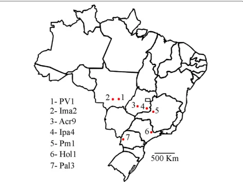
When the genomic sequences of four previously identi-fied CLRDV isolates were compared, significant differ-ences were only observed in the P0 coding sequence [3]. In order to better characterize the sequence diversity of P0 proteins among CLRDV isolates, cotton plants from different varieties, displaying typical or atypical CBD symptoms were collected from different parts of Brazil. Sampled areas covered the main cotton producing areas of the country, most of them with significant geograph-ical distance from each other (Fig. 1). In total, seven Brazilian CLRDV isolates (PV1, Ima2, Acr9, Ipa4, Pm1, Hol1 and Pal3) were analyzed and compared to the Argentinian isolate (ARG) [4] and also to the P0 from an Australian isolate of potato leafroll virus (P0PL-AU) [8] (Table 1). The genomes from two of the Brazilian iso-lates analyzed (PV1 and Ima2) have been previously obtained [3, 23]. Three CLRDV isolates (Acr9, Ima2 and Ipa4) were obtained from known cotton CLRDV-resistant varieties and are therefore treated as resistance-breaking isolates. Two isolates (Ima2 and Pm1) were obtained from plants showing atypical symptoms (Table 1).
sequence should be considered as different species [24]. In this line, the isolates Acr9, Ima2 and Ipa4 should be regarded as a new species associated with CBD. Since P0 sequences are the most variable sequences among pole-roviruses, it has been recently proposed that viruses hav-ing high diversity in this region, but with amino acid identities higher than 90 % in all other proteins should be regarded as genotypes of the same species and not as a different one [25, 26]. This kind of analysis can only be
made for the Ima2 isolate, the only one of the three with the genome fully sequenced [3]. But since the P0 sequences of the three isolates are very similar (Table 2), Acr9 and Ipa4 are probably also new genotypes of CLRDV, as previously stated for Ima2 [3].
The separation of the isolates in two groups is also sup-ported by phylogenetic analysis. It has been shown that, when full genomic sequences are not available, P0-based phylogenies can correctly reconstruct the relatedness Fig. 1Map of Brazil showing sites where cotton plants were harvested for the study. The numbered red dots in the map indicate the different cotton leafroll dwarf virus isolates harvested
Table 1Brazilian isolates of cotton leafroll dwarf virus used in the study
Isolate Locationa G. hirsutumcultivar CBD resistance phenotype Symptoms observed Year
PV1 Primavera do Leste–MT FM966 Susceptible Typical 2004
Acr9 Acreuna–GO CD406 Resistant Typical 2006
Hol1 Holambra–SP Nd Nd Typical 2007
Ima2 Campo Verde - MT IAC25 RMD Resistant Atypical 2009
Ipa4 Ipameri - GO Delta Opal Resistant Typical 2006
Pal3 Palotina - PR CD034928 Nd Typical 2006
Pm1 Patos de Minas - MG Epamig1 Nd Atypical 2007
a
among poleroviruses [25]. A Neighbor-joining tree based on CLRDV P0s groups the isolates Hol1, Pal3 and Pm1 with the two CBD founding members PV1 and ARG, while the isolates Acr9, Ima2 and Ipa4 clearly branch out, forming a well-supported group (Fig. 2). Since the three divergent isolates were all obtained from formerly cotton resistant varieties (Table 1), the silencing suppression ac-tivities from all isolates were then scored and compared.
Suppression of local silencing by CLRDV P0s
Previous data have shown that P0CL-ARG is weak sup-pressor of local silencing when expressed in Nicotiana benthamianaleaves [10]. The suppression assay is based on the Agrobacterium-mediated transient co-expression of mGFP5 and a candidate silencing suppression protein in mGFP5-expressingN. benthamiana leaves (transgenic line 16c) [27]. When no silencing suppression is ob-served, the transiently expressed GFP triggers a strong RNA silencing response that ultimately leads to RNA degradation from both stable and transient transgenes. However, in the presence of a silencing suppression pro-tein, GFP degradation is prevented and both transgenes (stable and transient) are expressed, increasing total GFP levels. The silencing suppression assay can be easily
monitored by hand UV lamps. In order to check whether the sequence diversity observed among the P0s could also reflect variable silencing suppression activities, the P0 from all seven Brazilian isolates and the Argentinian isolate were tested side-by-side in the 16cN. benthamiana
line and compared to P0PL-AU and P19, two known sup-pressors of local silencing. All genes were cloned into the pGWB417 binary vector [28], leading to a 35S-driven expression of Myc-tagged proteins.
As expected, a red patch in the infiltrated area was observed when GFP was expressed in the absence of a silencing suppression protein (Fig. 3). Conversely, strong GFP fluorescence was observed in the presence of P19 or P0PL-AUin all time-points analyzed (Fig. 3). All tested CLRDV P0s displayed obvious RNA silencing suppres-sion activities. When scored based on GFP fluorescence, the levels of silencing suppression activity observed for the CLRDV P0s were similar to P0PL-AU, but significantly lower than the P19 suppressor protein even at 3 days post-infiltration (dpi) (Fig. 3). The observed GFP fluores-cence at 3 dpi correlates well with the accumulation of GFP RNAs when checked by real-time PCR at this time-point in N. benthamiana16c plants (Fig. 4a). GFP RNA levels in GFP/P19-infiltrated plants were approximately 14 times higher than in control mock-infiltrated 16c plants. However, the GFP RNA fold change in P0PL-AUor CLRDV P0-infiltrated plants, varied from only 2 to 6 times the levels obtained in the same control condition (Fig. 4a). At later time-points, GFP fluorescence started to fade at infiltrated areas of P0CL-Acr9, P0CL-Hol1, P0CL-Ima2 and P0CL-Ipa4, indicating that those proteins are weaker suppressors than the other P0s (Fig. 3). The accumulation of the Myc-tagged suppressor proteins in the three bio-logical replicates used for real-time PCR was checked by western blot using anti-Myc antibodies (Fig. 4b). Although all suppressors were expressed from the same vector back-ground (pGWB417) [28], the P19 protein accumulated at levels consistently higher than the P0s. The strong suppression activity observed for P19 in the assay, there-fore, might be correlated with its higher stability in N.
Table 2Percentage of amino acid identity among the viral isolates used in the study
P0CL-Pal3 X
P0CL-Pm1 98.88 X
P0CL-PV1 98.51 98.14 X
P0CL-Hol1 98.51 98.14 97.76 X
P0CL-ARG 98.88 98.51 98.14 98.14 X
P0CL-Ima2 86.24 85.87 86.61 85.5 86.24 X
P0CL-Ipa4 88.1 87.73 87.73 87.36 87.36 96.28 X
P0CL-Acr9 88.1 87.73 87.73 87.36 88.1 95.53 97.02 X
P0PL-AU 18.21 18.21 17.84 18.21 18.58 17.84 18.58 18.58 X
P0CL-Pal3 P0CL-Pm1 P0CL-PV1 P0CL-Hol1 P0CL-ARG P0CL-Ima2 P0CL-Ipa4 P0CL-Acr9 P0PL-AU
benthamianaleaves. Since the P0s accumulated at similar levels, the observed differences in silencing suppression activities for those proteins might be due to functional di-vergence and not expression levels.
The fading phenotypes observed in 16c plants for the suppressor proteins P0CL-Acr9, P0CL-Hol1, P0CL-Ima2 and P0CL-Ipa4were reproduced when the experiment was re-peated in wild type plants (Additional file 1: Figure S1). When transiently expressed alone in wild type plants, the accumulation of GFP was lower than in the presence of the control strong suppressor P19, even at 3 dpi (Additional file 1: Figure S1). At 6 dpi, GFP was almost totally silenced when expressed alone, contrasting to what was observed in the presence of control constructs (P19 or PLP0-AU) or any of the CLRDV P0s. From 6 dpi onwards, as mentioned before, GFP levels started to fade in the presence of P0CL-Acr9, P0CL-Hol1, P0CL-Ima2 and P0CL-Ipa4, also indicating that those proteins are not able to suppress GFP silencing inN. benthamiana leaves for long periods. In all time-points analyzed, GFP accumu-lated at levels expressively higher when co-infiltrated with P19 than in the presence of any other P0, indicating that even the ones able to maintain GFP suppression at later times post-infiltration (P0PL-AU, P0CL-ARG, P0CL-PV1, P0CL-Pal3and P0CL-Pm1) should be regarded as moderate suppressors compared to the control used in the assays.
It has been previously shown that P0s depend on the presence of a F-box-like motif to exert their silencing suppression activity [8, 9, 11, 20]. The hallmark amino acids LPxx(L/I)x10–13P could be found in all CLRDV P0s tested (Additional file 2: Figure S2). However, isoleucine is changed to an amino acid with similar biochemical properties (valine) in the three resistance-breaking CLRDV isolates (Acr9, Ima2 and Ipa4). The ring structure amino acids known to affect local silencing suppression activity of melon aphid-borne yellows virus P0 are also present and conserved among the CLRDV P0s (Additional file 2: Figure S2) [9]. Therefore, the local silencing suppression variability observed among CLRDV P0s is probably associated with alternative functional residues.
Suppression of systemic silencing by CLRDV P0s
Viral siRNAs produced in infected cells may also move systemically through vascular tissues to reach other parts
Fig. 3Suppression of local silencing by cotton leafroll dwarf virus P0s inN. benthamiana16c plants. TransgenicN. benthamianaplants expressing GFP (line 16c) were co-infiltrated withAgrobacterium
of the plants [18]. In the N. benthamiana 16c assay, systemic silencing can be visualized by the appearance of red-silenced areas especially around the veins of newly developed leaves. Eventually, silencing signals may spread throughout the leaves, producing completely si-lenced plants. The systemic silencing suppression activ-ities of all CLRDV P0s were scored and compared to the strong systemic suppressor P0PL-AU [8]. At 16 dpi, 15 out of 20 plants assayed showed systemic silencing when infiltrated with the GFP silencing-trigger construct in the absence of any suppressor protein and almost 100 % of the plants were silenced by 20 dpi (Table 3). As expected, P0PL-AU completely blocked the spread of si-lencing signals as no silenced plants were observed at 16, 20 or 29 dpi. However, the suppression of systemic silencing mediated by the CLRDV P0s varied among the different isolates. In our experimental conditions, only 3 plants out of 20 were silenced at 16 dpi when co-infiltrated with P0CL-ARG (Table 3). The number of si-lenced plants, however, increased to 7 and 10 out of 20 at 20 dpi and 29 dpi, respectively, indicating that P0CL-ARGis
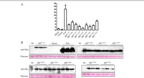
a moderate suppressor of silencing signals (Table 3). This result contrast to what has been previously ob-served for P0CL-ARG, where 8 out 10 plants were already silenced by 15 dpi in the presence of the protein [10]. Since the spread of RNA silencing signals may be Fig. 4Accumulation of GFP mRNA and suppressor proteins in infiltratedN. benthamiana16c leaves at 3 days after infiltration (dpi).aGFP levels in tissues co-infiltrated with cotton leafroll dwarf virus P0 proteins (P0CL-ARG, P0CL-PV1, P0CL-Acr9, P0CL-Hol1, P0CL-Ima2, P0CL-Ipa4, P0CL-Pal3or P0CL-Pm1), potato leafroll virus P0 (P0PL-AU), empty vector or mock-inoculated were detected with real-time PCR. Error bars indicate standard deviation of GFP mRNA in three biological repeats. Normalized value obtained in the mock sample was arbitrary set to 1 and all the other values compared to it. Data was normalized with Ubi3 and EF-1 reference genes. Asterisks indicate values that are statistically different from the control mock construct, with p-values varying from 0.0002 to 0.0478.bWestern blot showing the accumulation of suppressor proteins. Protein extracts from all the three biological replicates from each construct used for real-time PCR were run in SDS-PAGE, transferred to membranes and probed with Myc-tag specific antibodies. Gel loading was observed by Ponceau staining. Non-infiltrated plants (NI) were used as negative controls. Top left panel: accumulation of the control proteins P0PL-AUand tombusvirus P19 and the negative control (infiltrated with the empty vector). Top right panel: accumulation of P0CL-ARG, P0CL-PV1 and P0CL-Acr9proteins. Bottom left panel: accumulation of P0CL-Hol1, P0CL-Ima2and P0CL-Ipa4proteins. Bottom right: accumulation of P0CL-Pal3 and P0CL-Pm1proteins
Table 3Proportion of plants showing systemic silencing at 16, 20 and 29 days post-infiltration (dpi)
Infiltration 16 dpi 20 dpi 29 dpi
GFP + Vector 15/20 19/20 19/20
GFP + P0PL-AU 0/20 0/20 0/20
GFP + P0CL-PV1 0/20 0/20 0/20
GFP + P0CL-ARG 3/20 7/20 10/20
GFP + P0CL-Acr9 5/20 7/20 7/20
GFP + P0CL-Hol1 0/20 1/20 2/20
GFP + P0CL-Ima2 7/20 9/20 11/20
GFP + P0CL-Ipa4 0/20 0/20 0/20
GFP + P0CL-Pal3 0/20 0/20 0/20
influenced by environmental conditions [29–31] and possibly by the number of infiltrated leaves, concentra-tion and strain of Agrobacteriumused in the assay and vector background, the difference in the systemic silen-cing activity observed for P0CL-ARGmight be due to dif-ferent experimental settings.
In line with what has been observed for P0CL-ARG, the P0 proteins P0CL-Acr9and P0CL-Ima2 were also moderate suppressors of systemic silencing (Table 3). However, the P0 proteins from five isolates (PV1, Hol1, Ipa4, Pal3 and Pm1) efficiently blocked the spread of systemic silencing signals, with a silencing suppression activity similar to P0PL-AU (Table 3). During the experiments, 70 % of the plants infiltrated with suppressor proteins P0CL-Pal3 and P0CL-Pm1and 50 % of P0PL-AU-infiltrated ones displayed strong necrotic lesions at late times after infiltration, most of them starting at 10 dpi (data not shown). There-fore, the suppression of systemic silencing by those pro-teins could have also been influenced by the induced cell death. Similar necrotic phenotypes have also been ob-served for P0PL-AU [8] and for the P0s from sugarcane yellow leaf virus and beet western yellows virus [6].
AGO destabilization by CLRDV P0s
The P0 proteins from some members of the family Luteo-viridaeare able to destabilize the expression of AGO pro-teins [8, 11–15]. However, the activity of CLRDV P0 in AGO decay has never been tested. For that, a Myc-tagged version of theArabidopsis thalianaAGO1 protein, known to be involved in several RNA silencing pathways, includ-ing anti-viral defense [32, 33], was transiently expressed via Agrobacterium infiltration in wild type N. benthami-ana leaves in the presence or absence of different P0s or control constructs. The accumulation of AtAGO1-Myc protein was detected by anti-myc antibodies when co-expressed without P0 suppressors or in the presence of P19 (Fig. 5). All P0s, including P0PL-AU, the seven Brazilian
isolates and the Argentinian isolate of CLRDV were able to strongly decrease the AtAGO1-Myc levels when co-expressed. Therefore, the local and systemic silencing differences observed among the isolates might be due to the regulation of still unknown cellular proteins by P0, possibly other AGO members [8], or due to small differ-ences in AGO1 accumulation that are not detected due to western blot resolution limits.
Conclusions
Our results indicated a high diversity among P0 proteins from Brazilian and Argentinian isolates of CLRDV, a virus associated with CBD. All CLRDV P0 proteins ana-lyzed were able to mediate AtAGO1 decay, however, variable silencing suppression activities were observed, probably reflecting their sequence diversity. P0CL-ARG was a moderate silencing suppressor of both local and systemic silencing in our experiments, when compared to the positive control constructs used in the assays (Figs. 3, 4 and Additional file 1: Figure S1). Three proteins (P0CL-PV1, P0CL-Pal3and P0CL-Pm1) were also moderate suppressors of local silencing, but strong suppressors of systemic silen-cing. Four other proteins behaved as weak suppressors of local silencing. Contrasting to control constructs (P19 and P0PL-AU) and to other CLRDV P0s, those four proteins (P0CL-Acr9, P0CL-Ima2, P0CL-Hol1 and P0CL-Ipa4) could not support GFP suppression for long periods when assayed in the mGFP5-expressing N. benthamiana 16c line (Fig. 3) or in wild type plants (Additional file 1: Figure S1). GFP levels clearly started to fade in the presence of those proteins from 6 dpi onwards (Fig. 3 and Additional file 1: Figure S1). However, two of the weak local silencing suppressor proteins (P0CL-Hol1and P0 CL-Ipa4
) were able to almost completely block the spread of systemic silencing signals when assayed in 16c trans-genic lines. Despite of their weak local silencing, P0 CL-Hol1
and P0CL-Ipa4are as strong as the control P0PL-AU
Fig. 5Accumulation of a Myc-tagged version of theA. thalianaAGO1 protein transiently expressed inN. benthamianaleaves in the presence or absence of P0 proteins. Myc-tagged versions of cotton leafroll dwarf virus P0s (P0CL-ARG, P0CL-PV1, P0CL-Acr9, P0CL-Hol1, P0CL-Ima2, P0CL-Ipa4, P0CL-Pal3or P0CL-Pm1) and
potato leafroll virus P0 (P0PL-AU) were used in the assay. Non-infiltrated plants (NI) were used as negative controls and AtAGO1 infiltrated with the empty
protein in suppressing systemic silencing. It’s tempting to speculate therefore that the strength of local and sys-temic silencing suppression activity might be genetically unlinked in P0 proteins. Furthermore, these data indi-cate that the silencing suppression capabilities of the distinct CLRDV P0 proteins are not directly linked to their genetic diversity.
Methods
Plant material and DNA constructs
Gossypium hirsutumplants belonging to at least six cul-tivars (FM966, CD406, CD034928, IAC25 RMD, Delta Opal and Epamig1) were collected in five different States of Brazil (Goiás, Mato Grosso, Minas Gerais, Paraná and São Paulo) (Table 1 and Fig. 1). The Ima2 isolate was collected in Campo Verde – Mato Grosso, but passed through the IAC24 RMD cotton variety at Instituto Matogrossense do Algodão (Primavera do Leste –Mato Grosso) before being sent for analysis [34]. Harvesting and maintenance of plants were performed according to Brazilian rules (MP 2.186-16/2001). Total RNA of all plants were extracted using Qiagen Plant RNA kit and 2.5μg were used to prepare cDNAs with the O5R2 primer (5’-GCAACCTTTTATAGTCTCTCCAAT-3’), which an-neals in the middle of CLRDV ORF5. The ORF0 se-quences from all Brazilian isolates were amplified with primers CLP0_F (5’-CACCATGTTGAATTTGATCATC TGCAG-3’) and CLP0_R (5’-ACTGCTTTCTCCTTCAC-3’) and cloned into pENTRY-D-TOPO (Invitrogen). The ORF0 of the Argentinian isolate of CLRDV [GenBank: NC_014545.1] was synthesized and cloned into a pUC plasmid by the Blue Heron Biotechnology Inc (USA). The Argentinian ORF0 [10] and the P19 coding sequence [35] were amplified with primers CLP0_TOPO_F/CLP0_R and P19_TP_F (5’-CACCATGGAACGAGCTATACAAGGAA ACG -3’)/P19_R (5’-TTACTCGCTTTCTTTTTCGAAG G-3’), respectively, and also cloned into pENTRY-D-TOPO (Invitrogen). All amplifications were performed with the Phusion High Fidelity Polymerase (NEB). Entry vectors containing theArabidopsis thalianaAgo1 coding sequence [TAIR: AT1G48410] and the P0 from the Australian isolate of PLRV [GenBank: D13953.1] were described previously [8].
Genes in entry gateway clones were sequenced in both directions in automated ABI sequencers through dye ter-minator cycle method, using primers annealing in vector sequences. The accession numbers for the new P0 se-quences obtained here are: [GenBank:KR185733] (isolate Acr9), [GenBank:KR185734] (isolate Pm1), [GenBank: KR185735] (isolate Hol1), [GenBank:KR185736] (isolate Ipa4), [GenBank:KR185737] (isolate Pal3). All genes in entry vectors were transferred through LR reac-tions to the binary destination vector pGWB417 [28],
resulting in 35S-driven, Myc-tagged proteins when expressed in plants.
Sequence analysis
Multiple sequence alignments of deduced amino acid se-quences were performed with ClustalW2 (http://www. ebi.ac.uk/Tools/msa/clustalw2/) and phylogenetic recon-structions were performed with the MEGA 4 software [36]. Trees were constructed by the neighbor-joining (NJ) method [37], with the pair-wise deletion option and number of differences matrix.
Agroinfiltration
Agrobacterium tumefaciens, strain GV3101, were infil-trated in Nicotiana benthamiana leaves as described previously [38]. Cells were individually diluted to an optical density of 1.0 at 600 nm before mixing the cul-tures. Leaves were infiltrated in the abaxial surfaces with needleless syringes and the infiltrated plants were incubated in growth chambers with a 16-hour photo-period at 24 °C.
For the local silencing suppression assay, three leaves of 5-weeks-old mGFP5-expressing N. benthamiana plants (wild type or 16c line) [27] were co-infiltrated with equal volumes ofA. tumefaciensharboring plasmids expressing mGFP5 and pGWB417 with or without candidate silen-cing suppressor genes. For the systemic silensilen-cing suppres-sion assay, only one leaf of 3-weeks-oldN. benthamiana
16c plants were co-infiltrated with equal volumes of A. tumefaciens expressing mGFP5 and pGWB417 (negative control) or with pGWB417 expressing candidate silencing suppressor genes. GFP fluorescence was observed under a long-wavelength UV lamp and the number of plants having systemic silencing scored in different time-points.
For the AGO1 destabilization assay, three leaves of 5-week-old wild type N. benthamiana plants were infil-trated with A. tumefaciens harboring plasmids pJL3:P19 [35], pGWB417-AtAGO1-MYC, and pGWB417 with or without candidate silencing suppression genes in a pro-portion of 30 %, 35 % and 35 %, respectively. The infil-trated leaves were collected 4 days after infiltration.
qRT-PCR
instructions. Quantitative PCR reactions were performed in a total volume of 20μL, using 5μL of a 20-fold diluted cDNA. The amplification reactions were performed using the SYBR® Select Master Mix (Applied Biosystems), ac-cording to manufacturer’s instructions. Primers used for GFP were qmGFP5_F3 (5’-AGTGGAGAGGGTGAAGGT GATGC-3’) and qmGFP5_R4 (5’- TCCCTCAGGCATGG CGCTCTT-3’). The genes Ubiquitin3 (Ubi3) and Elong-ation factor-1α(EF-1) were used as reference genes, with primers previously described [39]. Three biological and technical replicates were used for all samples. Quantifica-tion of GFP expression levels was performed using the comparative CT method (ΔΔCT) through the Miner and qBase softwares [40–42]. The t-student test was per-formed to compare the samples.
Western blotting
InfiltratedNicotiana benthamianaleaves were ground in liquid nitrogen and mixed with sample buffer (100 mM Tris [pH 6.8], 20 % glycerol, 4 % SDS, and 0.2 % bromo-phenol blue) containing 10 % β-mercaptoethanol [43]. Samples were then boiled at 90 °C for 10 min, and cen-trifuged for 5 min at 13,000 × g before loading on a gel. Extracts were run in 8 % SDS-PAGE gels for the detec-tion of AtAGO1-Myc and in 12 % SDS-PAGE gels for detecting P19-myc, P0PL-AU-myc and P0CLs-myc with anti-myc antibody (1:2,000; Sigma, clone 9E10), followed by an anti-mouse HRP secondary antibody (1:5,000; Bio-Rad). Antibody–protein interactions were visualized using an en-hanced chemiluminescence detection kit (GE Healthcare) according to the manufacturer’s instructions.
Additional files
Additional file 1: Figure S1.Suppression of local silencing by cotton leafroll dwarf virus P0s inN. benthamianawild type plants.N. benthamiana
plants were co-infiltrated withAgrobacteriumcarrying plasmids to express GFP and candidate suppressor proteins (P0CL-PV1, P0CL-Acr9, P0CL-Hol1, P0CL-Ima2,
P0CL-Ipa4, P0CL-Pal3and P0CL-Pm1). GFP co-expressed with empty vector was
used as negative control. P0CL-ARG, P0PL-AUand P19 were used as
positive controls in the assay. Pictures were taken at 3, 6, 9 and 12 days post-infiltration (dpi). (PNG 4388 kb)
Additional file 2: Figure S2.Amino acid sequence alignment of cotton leafroll dwarf virus P0s. Sequences were aligned with ClustalW2 and conserved amino acids were shaded in black with Mview. The red bar highlights the conserved F-box-like domain. Functional ring structure residues described in Hanet al., 2010 are marked with asterisks. (PNG 1391 kb)
Competing interests
The author(s) declare that they have no competing interests.
Authors’contributions
RSC, GSM, MFSV and RLC conceived and designed the experiments. RSC, ILGA and TFS performed the experiments. MFSV and TFS contributed with plant material. GSM, MFSV and RLC contributed reagents/materials/analysis tools. RSC and RLC wrote the manuscript. All authors read and approved the final manuscript.
Acknowledgements
Authors acknowledge Dr. Jean-Lois Bélot and Dr. Rafael Galbieri, from Instituto Matogrossense do Algodão and Dr. Nelson Suassuna from EMBRAPA Algodão for sending the cotton infected plants. This study is part of the thesis research of RSC in pursuit of his PhD in Genetics at the Genetics Department of the Federal University of Rio de Janeiro. This work was supported by grants from Conselho Nacional de Desenvolvimento Científico e Tecnológico (CNPq) and Fundação Carlos Chagas Filho de Amparo à Pesquisa do Estado do Rio de Janeiro (FAPERJ) to GSM, MFSV and RLC. RSC and ILGA were supported by fellowships from CNPq.
Author details
1Department of Genetics, Federal University of Rio de Janeiro, Rio de Janeiro,
Rio de Janeiro, Brazil.2Department of Virology, Federal University of Rio de Janeiro, Rio de Janeiro, Rio de Janeiro, Brazil.3Present address: Departamento de Biotecnologia, Escola de Engenharia de Lorena, Universidade de São Paulo, Lorena, São Paulo, Brazil.
Received: 28 April 2015 Accepted: 4 August 2015
References
1. Correa RL, Silva TF, Simoes-Araujo JL, Barroso PA, Vidal MS, Vaslin MF. Molecular characterization of a virus from the family Luteoviridae associated with cotton blue disease. Arch Virol. 2005;150:1357–67.
2. Cauquil J. Etudes sur une maladie d’origine virale du cotonnier: la maladie bleue. Cot Fibres Trop. 1977;32:259–78.
3. Da Silva AKF, Romanel E, Silva T da F, Castilhos Y, Schrago CG, Galbieri R, et al. Complete genome sequences of two new virus isolates associated with cotton blue disease resistance breaking in Brazil. Arch Virol. 2015;160:1371–4. 4. Distéfano AJ, Kresic IB, Hopp HE. The complete genome sequence of a virus
associated with cotton blue disease, cotton leafroll dwarf virus, confirms that it is a new member of the genus Polerovirus. Arch Virol. 2010;155:1849–54.
5. Pfeffer S, Dunoyer P, Heim F, Richards KE, Jonard G, Ziegler-Graff V. P0 of beet Western yellows virus is a suppressor of posttranscriptional gene silencing. J Virol. 2002;76:6815–24.
6. Mangwende T, Wang ML, Borth W, Hu J, Moore PH, Mirkov TE, et al. The P0 gene of Sugarcane yellow leaf virus encodes an RNA silencing suppressor with unique activities. Virology. 2009;384:38–50.
7. Kozlowska-Makulska A, Guilley H, Szyndel MS, Beuve M, Lemaire O, Herrbach E, et al. P0 proteins of European beet-infecting poleroviruses display variable RNA silencing suppression activity. J Gen Virol. 2010;91(Pt 4):1082–91.
8. Fusaro AF, Correa RL, Nakasugi K, Jackson C, Kawchuk L, Vaslin MFS, et al. The Enamovirus P0 protein is a silencing suppressor which inhibits local and systemic RNA silencing through AGO1 degradation. Virology. 2012;426:178–87.
9. Han YH, Xiang HY, Wang Q, Li YY, Wu WQ, Han CG, et al. Ring structure amino acids affect the suppressor activity of melon aphid-borne yellows virus P0 protein. Virology. 2010;406:21–7.
10. Delfosse VC, Agrofoglio YC, Casse MF, Kresic IB, Hopp HE, Ziegler-Graff V, et al. The P0 protein encoded by cotton leafroll dwarf virus (CLRDV) inhibits local but not systemic RNA silencing. Virus Res. 2014;180:70–5. 11. Zhuo T, Li YY, Xiang HY, Wu ZY, Wang XB, Wang Y, et al. Amino acid
sequence motifs essential for P0-mediated suppression of RNA silencing in an isolate of potato leafroll virus from Inner Mongolia. Mol Plant Microbe Interact. 2014;27:515–27.
12. Bortolamiol D, Pazhouhandeh M, Marrocco K, Genschik P, Ziegler-Graff V. The Polerovirus F box protein P0 targets ARGONAUTE1 to suppress RNA silencing. Curr Biol. 2007;17:1615–21.
13. Baumberger N, Tsai CH, Lie M, Havecker E, Baulcombe DC. The Polerovirus silencing suppressor P0 targets ARGONAUTE proteins for degradation. Curr Biol. 2007;17:1609–14.
14. Derrien B, Baumberger N, Schepetilnikov M, Viotti C, De Cillia J, Ziegler-Graff V, et al. Degradation of the antiviral component ARGONAUTE1 by the autophagy pathway. Proc Natl Acad Sci U S A. 2012;109:15942–6. 15. Csorba T, Lozsa R, Hutvagner G, Burgyan J. Polerovirus protein P0 prevents
16. Bologna NG, Voinnet O. The diversity, biogenesis, and activities of endogenous silencing small RNAs in Arabidopsis. Annu Rev Plant Biol. 2014;65:473–503.
17. Incarbone M, Dunoyer P. RNA silencing and its suppression: Novel insights from in planta analyses. Trends Plant Sci. 2013;18:382–92.
18. Melnyk CW, Molnar A, Baulcombe DC. Intercellular and systemic movement of RNA silencing signals. EMBO J. 2011;30:3553–63.
19. Lakatos L, Csorba T, Pantaleo V, Chapman EJ, Carrington JC, Liu YP, et al. Small RNA binding is a common strategy to suppress RNA silencing by several viral suppressors. EMBO J. 2006;25:2768–80.
20. Pazhouhandeh M, Dieterle M, Marrocco K, Lechner E, Berry B, Brault V, et al. F-box-like domain in the polerovirus protein P0 is required for silencing suppressor function. Proc Natl Acad Sci. 2006;103:1994–9.
21. Hua Z, Vierstra RD. The cullin-RING ubiquitin-protein ligases. Annu Rev Plant Biol. 2011;62:299–334.
22. Guilley H, Bortolamiol D, Jonard G, Bouzoubaa S, Ziegler-Graff V. Rapid screening of RNA silencing suppressors by using a recombinant virus derived from Beet necrotic yellow vein virus. J Gen Virol. 2009;90:2536–41. 23. De Andrade RRS, Vaslin MFS. SearchSmallRNA: a graphical interface tool for
the assemblage of viral genomes using small RNA libraries data. Virol J. 2014;11:45.
24. Domier LL. Virus Taxonomy, Ninth Report of the International Committee on Taxonomy of Viruses. London: Elsevier/Academic Press; 2012. 25. Lin YH, Gao SJ, Damaj MB, Fu HY, Chen RK, Mirkov TE. Genome
characterization of sugarcane yellow leaf virus from China reveals a novel recombinant genotype. Arch Virol. 2014;159:1421–9.
26. Knierim D, Tsai WS, Maiss E, Kenyon L. Molecular diversity of poleroviruses infecting cucurbit crops in four countries reveals the presence of members of six distinct species. Arch Virol. 2014;159:1459–65.
27. Brigneti G, Voinnet O, Li WX, Ji LH, Ding SW, Baulcombe DC. Viral pathogenicity determinants are suppressors of transgene silencing in Nicotiana benthamiana. EMBO J. 1998;17:6739–46.
28. Nakagawa T, Suzuki T, Murata S, Nakamura S, Hino T, Maeo K, et al. Improved Gateway binary vectors: high-performance vectors for creation of fusion constructs in transgenic analysis of plants. Biosci Biotechnol Biochem. 2007;71:2095–100.
29. Szittya G, Silhavy D, Molnar A, Havelda Z, Lovas A, Lakatos L, et al. Low temperature inhibits RNA silencing-mediated defence by the control of siRNA generation. EMBO J. 2003;22:633–40.
30. Qu F, Ye X, Hou G, Sato S, Clemente TE, Morris TJ. RDR6 has a broad-spectrum but temperature-dependent antiviral defense role in Nicotiana benthamiana. J Virol. 2005;79:15209–17.
31. Kotakis C, Vrettos N, Kotsis D, Tsagris M, Kotzabasis K, Kalantidis K. Light intensity affects RNA silencing of a transgene in Nicotiana benthamiana plants. BMC Plant Biol. 2010;10:220.
32. Qu F, Ye X, Morris TJ. Arabidopsis DRB4, AGO1, AGO7, and RDR6 participate in a DCL4-initiated antiviral RNA silencing pathway negatively regulated by DCL1. Proc Natl Acad Sci U S A. 2008;105:14732–7.
33. Baumberger N, Baulcombe DC. Arabidopsis ARGONAUTE1 is an RNA Slicer that selectively recruits microRNAs and short interfering RNAs. Proc Natl Acad Sci. 2005;102:11928–33.
34. Galbieri R, Cia E, Fuzatto M, Franzon R, Belot J, Dias JC. Transmissibility and cotton genotypes’response to an atypical form of vein mosaic virus disease. Trop Plant Pathol. 2010;35:88–95.
35. Lindbo JA. High-efficiency protein expression in plants from agroinfection-compatible Tobacco mosaic virus expression vectors. BMC Biotechnol. 2007;7:52.
36. Tamura K, Dudley J, Nei M, Kumar S. MEGA4: Molecular Evolutionary Genetics Analysis (MEGA) software version 4.0. Mol Biol Evol. 2007;24:1596–9.
37. Saitou N, Saitou N, Nei M, Nei M. The neighbor-joining method: a new method for reconstructing phylogenetic trees. Mol Biol Evol. 1987;4:406–25. 38. English JJ, Davenport GF, Elmayan T, Vaucheret H, Baulcombe DC.
Requirement of sense transcription for homology-dependent virus resistance and trans-inactivation. Plant J. 1997;12:597–603.
39. Rotenberg D, Thompson TS, German TL, Willis DK. Methods for effective real-time RT-PCR analysis of virus-induced gene silencing. J Virol Methods. 2006;138:49–59.
40. Livak KJ, Livak KJ, Schmittgen TD, Schmittgen TD. Analysis of relative gene expression data using real-time quantitative PCR and the 2(−Delta Delta C(T)) Method. Methods. 2001;25:402–8.
41. Hellemans J, Mortier G, De Paepe A, Speleman F, Vandesompele J. qBase relative quantification framework and software for management and automated analysis of real-time quantitative PCR data. Genome Biol. 2007;8:R19.
42. Zhao S, Fernald RD. Comprehensive algorithm for quantitative real-time polymerase chain reaction. J Comput Biol. 2005;12:1047–64.
43. Laemmli UK. Cleavage of structural proteins during the assembly of the head of bacteriophage T4. Nature. 1970;227:680–5.
Submit your next manuscript to BioMed Central and take full advantage of:
• Convenient online submission
• Thorough peer review
• No space constraints or color figure charges
• Immediate publication on acceptance
• Inclusion in PubMed, CAS, Scopus and Google Scholar
• Research which is freely available for redistribution