International Journal of Research (IJR)
e-ISSN: 2348-6848, p- ISSN: 2348-795X Volume 2, Issue 08, August 2015Available at http://internationaljournalofresearch.org
Bartonella henselae
of the brain; diagnostic challenge
Mohamed A.R. Arbab
1, 2; Sawsan AH AL deaf
2; Lamyaa A.M Hassan
3&
Ahmed M. Hassan
41. Department of surgery, Faculty of Medicine, University of Khartoum.
2. National Centre of Neurological Sciences, Khartoum.
3. School of Medicine, Ahfad Women University, Omdurman.
4. Institute of Endemic Diseases, University of Khartoum.
Correspondence: Mohamed A.R Arbab M.D, Ph.D.
Department of Surgery, Faculty of Medicine, University of Khartoum. P.O Box 13456 Khartoum.
Email: [email protected]
Abstract
A 13 and 35-year-old female patients presented because of fatigability, nocturnal febrile illness,generalized body aches and worsening headache. In the course of the illness both patientsdeveloped worsening headache and attacks of epileptic seizure. Brain MRI imaging showedintraaxial cerebral heterogeneous
irregular masseswith
focalencephalomalacia,cystic component, glial reaction and dystrophic calcification. Histological results of the resected pathology revealed highly vascular mass with features suggestive of Bartonella henselae. In this report, the initial course of the disease was that of febrile illness, a common problem in a tropical country, furthermore the symptoms were not suggestive of cat scratch disease.
Key words: Bartonella henselae; brain;
cerebral mass
Introduction
Bartonella henselae, a bacterial pathogen known to cause central nervous system disease
in humans. It was first reported in 1990 and described as a new species in 1992. The pathogen is mainly carried by cats and can cause in humans wide spectrum of clinical syndromes that include cat scratch disease, bacillary angiomatosis, endocarditis and relapsing bacteremia. Central nervous system involvement has included encephalopathy, myelopathy, meningitis, cerebral arteritis, optic neuritis and radiculopathy (Brazis et al. 1986), (Carithers and Margilet 1991), (Lewis and Tucker 1986), (Pickerill and Milder 1986), (Selby and Walker 1979).
Although about 40% of cats carry the bacteria in their saliva but they do not themselves show manifestations of the disease. Cat scratch disease mostly is a self-limiting disease that resolves without treatment; however, in immune compromised patients it can cause serious illness.In this paper, we report 2cases of young females who had bizarre symptoms that lasted for periods of 8 weeks andone year.
Development of neurologic symptoms
necessitated imaging of the brain which
Field Code Changed
International Journal of Research (IJR)
e-ISSN: 2348-6848, p- ISSN: 2348-795X Volume 2, Issue 08, August 2015Available at http://internationaljournalofresearch.org
revealed intra- axial brain masses with cystic component mimking cerebral neoplasms. The
post-operative diagnosis ofBartonella henselae
was made.
Case 1
A previously healthy 35-year –female developed general fatigability, nocturnal low grade fever, worsening headache, generalized body aches and pains. These symptoms have been going on for one year. One month before presentation to the neurosurgical department, the patient developed neck pain, left ear pain with hyperacuasia and one attack of generalized epileptic seizure. The patient reported in the past history of the illness vague abdominal pains and diarrhea and general ill health that necessitated blood transfusion. The patient denied history of any skin scratching or joints pain.
MRI brain showed a left temporal
heterogeneous irregular mass lesion with focal encephalomalacia, glial reaction and dystrophic calcification(Fig. 1). The patient was operated upon. Left craniotomy flap was reflected. The dura was then opened, this was found adherent to the underlying piaarachnoid. A bright -yellowish firm nodular mass with a tuft of numerous tiny vessels was encountered. The tumor mass was gradually resected and the vascular part secured by coagulation and freed from the Sylvain vessels.
The patient had uneventful post-operative recovery.
Case 2
A 19 years old female presented because of febrile illness that lasted few weeks to be
followed by headache, failing vision and convulsions. The patient was initially been investigated for the febrile illness without
conclusive diagnosis. Development of
neurologic symptoms necessitated brain MRI imaging. The images showed intra-axial thick-walled cystic lesion with perilesional edema (Fig. 1). The patient was subjected to surgery where a thick-walled cystic mass with gliotic white to yellowish content was encountered.
The tumor specimens from both cases 1 and 2 werefixed in neutral formalin saline and stained with Hematoxylin and Eosin, Warthin Starry stain and Masson Fontana and Melan-A for melanin. Aggregates of small black
filamentous structures suggested Bartonella
organisms were confirmed by a mono-clonal
antibody specific for B henselae. It was Mouse
monoclonal antibody: Anti-Bartonella
henselae (Cat Scratch Fever) antibody [H2A10] (ab704) Abcam Call (888) 77-ABCAM (22226. It was obtained from the USA
The cell phenotypes in the lesions were identified by indirect Immunoperoxidase stains that included CD3, CD20, and CD68. IgG and Complement C3a, C3b and C5a were also stained for. CD34 was used for identification of blood vessels.
Results
International Journal of Research (IJR)
e-ISSN: 2348-6848, p- ISSN: 2348-795X Volume 2, Issue 08, August 2015Available at http://internationaljournalofresearch.org
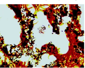
Warthin Starry stain showed small linear organisms in small groups and aggregates. The
features were characteristic of Bartonella
henselae (Fig. 4)
Discussion
In this report, both patients had states of ill health for periods that extended from few weeks up to more than one year. Both patientswere immune competent. The low grade fever, generalized body aches and pains in a tropical country enlist a number of
differential diagnoses. In the two
patientsneither lymph-adenopathy nor skin scratching was reported.Remarkable low hemoglobin in the course of the disease that necessitated blood donation could be attributed to either poor nutritional status of the patient during her illness or could be part of the pathogenesis of the disease.
Emergence of headache, visual deterioration, ear pain, hyperacuasis and convulsive attacks were the early neurologic symptoms that lead ultimately to the diagnosis of the disease.
Bartonella is known as the only genus of bacteria that induces pathological angiogenesis in mammalian host. The mechanism of Bartonella –induced angiogenesis was not well understood. It was found that these bacteria invade human brain vascular pericytes and induces increase pericyte production of
vascular endothelial growth factor
(VEGF)(Varanat et al. 2013).
This can explain the pathologic remarkable vascularity in our cases. Cases of encephalitis or neuroretinitis havebeen reported with Bartonella henselae infection.
Cerebral bacillary angiomatosis has been reported in human immunodeficiency virus-infected patients(Spach et al. 1992).However, the patients in this study were found to be immune-competent.
Of particular clinical importance, in the present
report the Bartonella infection manifested as
cerebral neoplasms as shown in the MRI. Role of infectious agents in tumor genesis has been reported (Hansen et al 2007), (Moss et al 2007).
Vasoproliferative tumors induced by
Bartonellaspecies were reported as benign and can be cured with antibiotic(Rudikoff et al. 1989).
However, in our cases the state of clinical presentation and the absence of supportive
evidence of possibility of B. hensaele disease
gave no chance for therapeutic trail.
In conclusion Bartonella hensaele infection of
the brain can manifest as cerebral neoplasms and do not necessarily present as classical cat
scratch disease.Furthermore the relatively late
presentation of patients with this disease and establishment of cerebral neoplasm call for surgical intervention.
References
[1.]Brazis PW, Stokes HR, Ervin FR. Optic
neuritis in cat scratch disease. J Clin NeurolOpthalmol. 1986; 6:172-174.
[2.]Carithers HA, Margileth AM. Cat
International Journal of Research (IJR)
e-ISSN: 2348-6848, p- ISSN: 2348-795X Volume 2, Issue 08, August 2015Available at http://internationaljournalofresearch.org
[3.]Hansen A, Boshoff C, and Lagos D
(2007). Kaposi sarcoma as a model of oncogenesis and cancer treatment. Expert Rev Anticancer Ther 7:211-220.
[4.]Lewis DW, Tucker SH. Central nervous
system involvement in cat scratch disease. Pediatrics. 1986; 77:714-721.
[5.]Moss SF, Malfertheiner P. (2007)
Helicobacter and gastric malignancies. Helicobacter 12:33-30.
[6.]Pickerill RG, Milder JE. Transvers
myelitis associated with cat scratch disease in an adult. JAMA. 1986; 246:2840-2841.
[7.]Rudikoff D, Phelps RG et al 1989.
Acquired immunodeficiency syndrome-related bacillary vascular proliferation
(epitheloid angiomatosis): rapid
response to erythromycin therapy. Arch Dermatol 125:706-707.
[8.]Selby G, Walker GL. Cerebral arteritis
in cat scratch disease. Neurology. 1979; 29:1413-1418.
[9.]Smith DL. Cat-scratch disease and
related clinical syndromes. Am Fam Physician. 1997; 55:1783-1789.
[10.] Spach DH, Panther LA et al.
Intracerebral bacillary angiomatosis in a
patient infected with human
immunodeficiency virus. Ann Intern Med. 1992; 116:740-742.
[11.] Varanat M, et al. Infection of
human brain vascular pericytes
(HBVPs) by Bartonella henselae. Med
International Journal of Research (IJR)
e-ISSN: 2348-6848, p- ISSN: 2348-795X Volume 2, Issue 08, August 2015Available at http://internationaljournalofresearch.org
A
B
C
Fig 1.
International Journal of Research (IJR)
e-ISSN: 2348-6848, p- ISSN: 2348-795X Volume 2, Issue 08, August 2015Available at http://internationaljournalofresearch.org
AB
Fig 2.
A shows inflammatory cells consisting of lymphocytes and macrophages. Some of the latter had a
clear cytoplasm. In one area there was a focus of necrosis surrounded by chronic inflammatory cells (H&E X40
B The inflammatory cells are positive for LCA (Immune-peroxidase X40).
A B
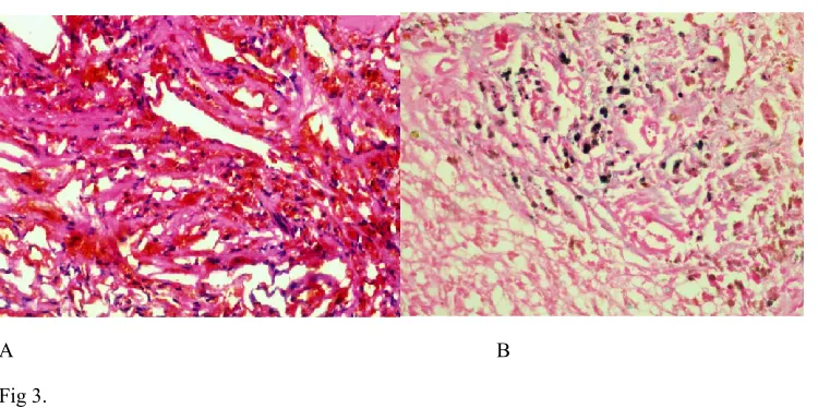
Fig 3.
A lesion shows marked angiomatosis as seen in the figure on the left (H&E X40)
B Many macrophages contained brown granular material (Upper figure. H&E X40)) that was
International Journal of Research (IJR)
e-ISSN: 2348-6848, p- ISSN: 2348-795X Volume 2, Issue 08, August 2015Available at http://internationaljournalofresearch.org