Lung Nodule Classification As Malignant Or
Benign Based On SVM Classifier
T.PoojaDr. Rajesh A
Abstract: Lung cancer is the uncontrolled growth of abnormal cells that start off in one or both lungs, usually in the cells that line the air passages. The abnormal cells do not develop into healthy lung tissue; they divide rapidly and form tumors. Metastasis refers to cancer spread ing beyond its site of origin to other parts of the body. When cancer spreads, it is much harder to treat successfully. Primary lun g cancer originates in the lungs, while secondary lung cancer starts somewhere else in the body, metastasizes, and reaches the lungs. In current medical diagnosis, treatment, and surgery, medical imaging plays one of the most significant roles, since imaging devices such as Computed Tomography (CT), Magnetic Resonance Imaging (MRI), and ultrasound diagnostics yield a great deal of information about diseases and organs. However, radiologists have to analyze and evaluate some medical images comprehensively in a short time, which is an enormous burden. To lighten the burden, computer technology research has been used more often to analyze medical images in recent years. The proposed method which is found to be accurate for tumor detect ion, makes use of Grey Level Co-occurrence Matrix (GLCM). The Support Vector Machine (SVM) classifier classifies the given input stage was normal or diseased and if it is diseased, further it classifies the tumor images into benign (non-cancerous) or malignant (cancerous).
Index Terms: Lung Nodules, Support Vector Machine, Gray Level Concurrence Matrix, Computed tomography, Skewness, Kurtosis Neural networks.
—————————— ——————————
1.
INTRODUCTION
Normal respiration begins by breathing air through the oral cavity and nasal. This air flows down your trachea, that divides into the left and right bronchi, that carry air to every respiratory organ. Once within the respiratory organ, the bronchi divide into smaller tubes referred to as channels and every bronchiole ends with alveoli. The alveoli are to blame for oxygenating the blood for circulation additionally as removing greenhouse emission from the blood. epithelial cell is that the presumably carcinoma to gift as a Pancoast’s neoplasm, that is high within the respiratory organ apex with extension to the chest wall, inflicting shoulder pain that radiates down the nervus ulnaris additionally called epidermoid malignant neoplastic disease. Most common type of lung most cancers makes up 40-50% of all lung cancers most of these cancers originate at the peripheral areas of the lung, like the bronchial mucosa can also begin in scars prompted through fibrosis, effortlessly considered on x-rays two can show up in non-smokers extra common in women, strongly linked to smoking Slow metastasis can occur for the duration of the lung or different physique organs. The most frequent symptom is a chronic and non productive cough breathing problems: shortness of breath accelerated shortness of breath throughout bodily activity, wheezing due to the bronchus being partly obstructed , high pitched respiratory sounds (stridor) changes in phlegm (sputum): increased amount blood in the phlegm (hemoptysis) lung contamination (pneumonia):
widespread lung infections may strengthen the lung contamination may be located in the same region as the tumour.Horner’s syndrome motive recessed eyeball, student constriction, droopy higher eyelid and lowered perspiration on the affected aspect of the face. two This kingdom is introduced on my paralysis of the trunk of cervical sympathetic nerve by such things as a lung tumour. A chest x ray is a painless, non-invasive test that creates images of the constructions inner your chest, such as your heart, lungs, and blood vessels. "Non-invasive" skill that no surgery is done and no devices are inserted into your body. They typically show up mild on a chest x ray. Your lungs, which are crammed with air, normally show up dark. A sickness in the chest that changes how radiation is absorbed also will appear on a chest x ray. Chest x rays have few risks. The quantity of radiation used in a chest x ray is very small. A lead apron may be used to defend certain components of your body from the radiation. CT scans suggests the size, shape, and position of your lungs and different buildings in your chest. Follow up on unusual findings from fashionable chest x rays, Find the cause of lung symptoms, such as shortness of breath or chest pain, find out whether you have a lung problem, such as a tumor, excess fluid around the lungs, or a pulmonary embolism. MRI is like a CT solely it makes use of magnetism as an alternative of x-rays, eliminate all metallic objects, and fill out a screening form, requested to lie down on a simply padded desk that gently glides you into the scanner. Most MRI tests take between 15 to forty-five minutes to entire depending on the physique section imaged and how many pictures are needed.
2. LITERATURE REVIEW
In view of breast cancer, the loss of life rate is turning into elevated. According to WHO (World Health Organization) breast threatening influenced over 1.5 million female constantly by and giant [1]. In 2015; 570,000 girls surpassed out due to the fact of chest unsafe hazard that used to be around 15% of whole passing rate. In 2017 round 252,710 patients of breast dangerous risk are identified and round 40, 610 female perished in America [2]. India is in threat fee with 90,000 sufferers and expanding each yr and passing women is around 40,000 consistently [4]. A CAD system, the usage of
__________________________
PG Scholar, Department of ECE, CVR College of Engineering,
Hyderabad, Telangana.
Associate Professor, Department of ECE, CVR College of
Engineering, Hyderabad, Telangana
E-mail: [email protected]
3607
wave atom seriously change (WAT) algorithm and Support Vector Machine (SVM) [5], is proposed to consider mammography images. During the process, the area of pastime is described earlier than applying the method. Biopsy is an appraised scientific manner in distinguishing bosom malignant growth which is a costly, monotonous and difficult framework. Radiologist recommendation is vital in this stage, once there ought to be an tournament of unsuitable assurance; affected need no longer to do undesirable biopsy [6]. A new robust Global Kernel Fuzzy-C Means (NRGKFCM-F) clustering algorithm [7], the place F refers to kernelized characteristic space. It solves all intermediate troubles making use of Kernel-based Fuzzy C- Means-F (KFCM-F) as a nearby search method procedure. It utilizes the segmentation of Genius MRI image, and then morphological filtering can be formed inevitably to avoid the mis-clustered regions. Mini MIAS informational collections [8] is utilized for appraising the structure. The community implementation is evaluated as affectability, identification & accuracy. Affectability & exactness completed from the structure are 97% and 97.89% separately. A method for segmentation [9] of MRI is primarily based in a hybrid strategy the usage of volumes got with the aid of a method segmentation based totally on Growth Regions as input to an algorithm Parametric Deformable Models. CAD system [10] for recognizing breast cancer in ROIs of digital mammograms. The study additionally investigates the overall performance of the gadget with wave atom radically change and SVM method. The presented results display that wave atom seriously change and SVM are beneficial and effective strategies to distinguish the mammographic photos as normal, benign and malignant.
3. PROPOSED SYSTEM
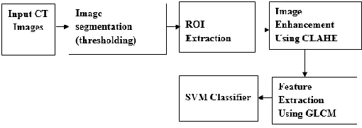
Therefore, the acquired images are first subjected to pre-processing steps that include: 1) segmentation of ROI, 2) edge detection and 3) image enhancement to extract the ROI part of the lung image. Each of the acquired lung images is first subjected to binarization, using a fixed threshold value, to coarsely localize the centre part (ROI) in the image. Some portions of background still appear as connected to the bright regions, predominantly due to uneven illumination. Subsequently, the circle shaped background in the resulting images is eliminated from the thresholding, i.e., based on the low intensity values of the background. The resulting binary mask is used to segment the ROI from the original lung image. The pictures with low distinction and uneven illumination are subjected to picture enhancement the use of CLAHE method. Equalization is applied in my view for all three RGB color spaces. These equalized RGB factors are merged collectively to result the coloration equalized image. CLAHE used to be at the start developed for successfully bettering the low distinction scientific images. A gray level co-occurrence matrix (GLCM) includes records about the positions of pixels having comparable grey degree values. The statistical measures described so a long way are easy to calculate, however do not grant any records about the repeating nature of texture. GLCM includes facts about the positions of pixels having comparable gray level values. Support vector machines are supervised gaining knowledge of models with associated learning algorithms that analyze information and recognize patterns, used for classification the primary SVM takes a set of enter statistics and for every given input, predicts, which of two lessons varieties the input,
making it a non-probabilistic binary linear classifier. From given set of training examples, every marked as belonging to one of two categories, an SVM education algorithm builds a mannequin that assigns new examples into one class or the other. In the proposed technique we are using linear classifier. Best hyper aircraft is the one that represents the biggest separation or margin between the two classes.
3.1 GLCM based Features Extraction
Feature extrication plays a fundamental role in classifying pattern. Gray Level Co occasion Matrix (GLCM) feature is chosen in 0° for entire mammograms. Per users urged to scrutinize the basic minimizing of GLCM. The span of GLCM was chosen from the dark measurement value in an image. For each recipe gave in the conditions, n choose the amount of dim measurement used. The grid part Q (i, j) is the recurrence related with two pixels, secluded by separating the pixel occurred in the area with power i and j. Textual features achieved from GLCM were presented underneath.
Figure 1: Block Diagram of Lung Nodule Classification
Contrast
It evaluates gray dimension extent between reference and the neighborhood pixels, variance in the mammogram is assessed from it.
Correlation
Connection delineates the direct needy of gray value. The correlation value is vast if the mammogram contains direct shape for extensive sum.
where
,
Sum entropy
It is the whole of small scale (nearby) contrasts in the picture. SE
Difference entropy
It demonstrates the variations in small scale contrasts. DE
Where
|i-j| = k, k=0,…n-1
Entropy
difference circulation in a region. It is evaluated from the condition appeared as follows.
N
4. RESULTS AND DISCUSSION
Figure 2: Input Image
Figure 3: GRAYSCALE IMAGE
Figure 4: Histogram of original Image
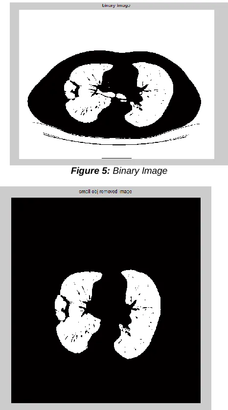
Figure 5: Binary Image
Figure 6: Lung cavities Image
3609
Figure 8: Lung Image-Shape
Figure 9: Tumor Extraction-Thresholding
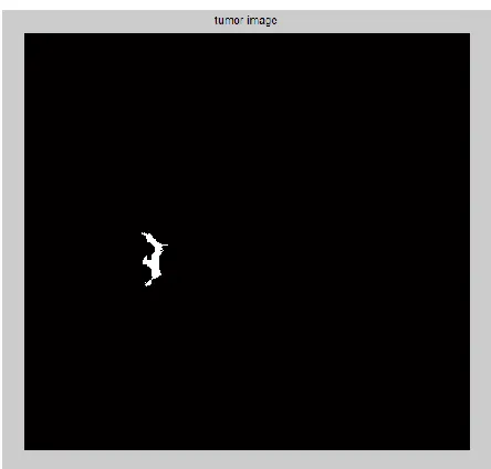
Figure 10: Tumor Image
Figure 11: Output
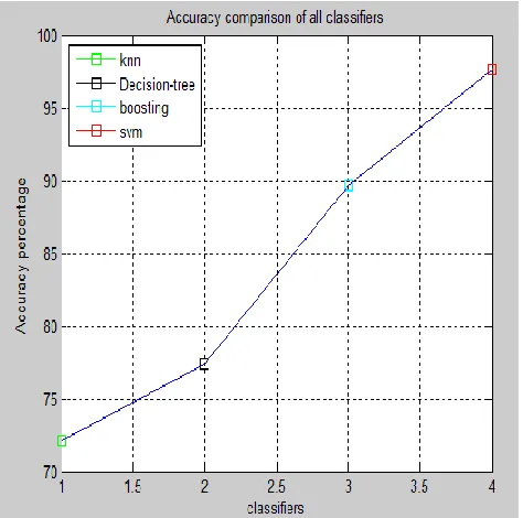
Finally, the performance of the system is evaluated. The following equations in (1) and (2) are to evaluate the correct classification and the incorrect classification of this system.
Figure 12: Comparison Graph
5. CONCLUSION
We proposed a lung cancer classification system which classifies automatically by simultaneously utilizing the normal and cancerous images. We presented GLCM Algorithm and region properties measurement for the feature extraction, which can extract the shape features and SVM Algorithm for classification which achieves much higher accuracy than previously proposed approaches.
ACKNOWLEDGEMENT
First and foremost, the author would like to thank her supervisor, Associate Professor Dr. Rajesh A for providing his valuable advice, helpful guidance and knowledge. The author sincerely wishes to thank to the Head of Department of Electronic Engineering, Professor Dr.K.Lalithendra at CVR College of Engineering,Hyderabad.
REFERENCES
[1] F. Paulin, A. Santhakumaran, Back propagation neural network by comparing hidden neurons: case study on breast cancer diagnosis, International Journal of Computer Applications 2 (June) (2010) 40– 44.
[2] J. Ramesh, K. Gunavathi, et al., Fault classification in phase-locked loops using back propagation neural networks, ETRI, Journal 30 (August (4)) (2008) 546– 554.
[3] Chen C.H., Chang C.K., Tu C.Y., Liao W.C., Wu B.R., Chou K.T., Chiou Y.R., Yang S.N., Zhang G., Huang T.C. Radiomic features analysis in computed tomography images of lung nodule classification. PLoS ONE. 2018;13:e0192002. doi: 10.1371/journal.pone.0192002.
[4] Hu H.H., Ni S.D. Classification of malignant-benign pulmonary nodules in lung CT images using an improved random forest; Proceedings of the 2017 13th International Conference on Natural Computation, Fuzzy Systems and Knowledge Discovery (ICNC-FSKD); Guilin, China. 29–31 July
2017; pp. 2285–2290.
[5] Aggarwal P., Vig R., Sardana H.K. Patient-Wise Versus Nodule-Wise Classification of Annotated Pulmonary Nodules using Pathologically Confirmed Cases. J. Comput. 2013;8:2245–2255.
[6] Mukherjee J., Chakrabarti A., Skaikh S.H., Kar M. Automatic Detection and Classification of Solitary Pulmonary Nodules from Lung CT Images; Proceedings of the 2014 Fourth International Conference of Emerging Applications of Information Technology; Kolkata, India. 19–21 December 2014; pp. 294–299.
[7] Amal A.F., Ali A., Elshazly S., Farag A.A. Feature fusion for lung nodule classification. Int. J. Comput. Assist. Radiol. Surg. 2017;12:1809–1818.
[8] Xie Y.T., Xia Y., Zhang J.P., Feng D.D., Fulham M.J., Cai W.D. Transferable Multi-model Ensemble for Benign-Malignant Lung Nodule Classification on Chest CT; Proceedings of the Medical Image Computing and Computer-Assisted Intervention (MICCAI); Quebec City, QC, Canada. 11–13 September 2017; pp. 656–664.