1556-6811/10/$12.00 doi:10.1128/CVI.00208-09
Copyright © 2010, American Society for Microbiology. All Rights Reserved.
Peptide Microarray-Based Identification of
Mycobacterium tuberculosis
Epitope Binding to HLA-DRB1*0101, DRB1*1501,
and DRB1*0401
䌤
†
Simani Gaseitsiwe,
1,2Davide Valentini,
3Shahnaz Mahdavifar,
1,2Marie Reilly,
3Anneka Ehrnst,
1and Markus Maeurer
1,2*
Department of Microbiology, Tumor and Cell Biology (MTC), Karolinska Institutet, Stockholm, Sweden1; Swedish Institute for Infectious Disease Control (SMI), Stockholm, Sweden2; and Department of
Medical Epidemiology and Biostatistics, Karolinska Institutet, Stockholm, Sweden3
Received 23 May 2009/Returned for modification 30 July 2009/Accepted 6 October 2009
A more effective vaccine againstMycobacterium tuberculosisis needed, and a number ofM. tuberculosisvaccine
candidates are currently in preclinical or clinical phase I and II studies. One of the strategies to selectM.
tuberculosis(protein) targets to elicit a CD8ⴙor CD4ⴙT-cell response is to gauge the binding of candidate peptides to major histocompatibility complex (MHC) class I or class II molecules, a prerequisite for successful
peptide presentation and to expand antigen-specific T cells. We scanned 61 proteins from theM. tuberculosis
proteome for potential MHC class II-presented epitopes that could serve as targets for CD4ⴙT-cell responses.
We constructed a peptide microarray consisting of 7,466 unique peptides derived from 61 M. tuberculosis
proteins. The peptides were 15-mers overlapping by 12 amino acids. Soluble recombinant DRB1*0101 (DR1), DRB1*1501 (DR2), and DRB1*0401 (DR4) monomers were used to gauge binding to individual peptide species. Out of 7,466 peptides, 1,282, 674, and 1,854 peptides formed stable complexes with HLA-DR1, -DR2, and -DR4, respectively. Five hundred forty-four peptides bound to all three MHC class II molecules, 609 bound
to only two, and 756 bound to only a single MHC class II molecule. This allowed us to rankM. tuberculosis
proteins by epitope density.M. tuberculosisproteins contained “hot spots,” i.e., regions with enriched MHC
class II binding epitopes. Two hundred twenty-two peptides that formed MHC class II-peptide complexes had previously been described as exclusively recognized by IgG in sera from patients with active pulmonary
tuberculosis, but not in sera from healthy individuals, suggesting that these peptides serve as B-cell and CD4ⴙ
T-cell epitopes. This work helps to identify not onlyM. tuberculosispeptides with immunogenic potential, but
also the most immunogenic proteins. This information is useful for vaccine design and the development of
future tools to explore immune responses toM. tuberculosis.
CD4⫹T cells play a central role inMycobacterium tubercu-losis-directed cellular immune responses (2, 6, 7, 12). It is most likely that an effective tuberculosis (TB) vaccine would target the expansion of CD8⫹and CD4⫹T cells, which recognizeM. tuberculosis peptides presented by major histocompatibility complex (MHC) class I and class II molecules.
The MHC locus is the most variable gene locus in the human genome, and the variability of MHC class II alleles in different populations is well documented (24). Certain MHC class II alleles have been shown to be associated withM. tuberculosis
infection (1, 11, 15, 16, 23): DRB1*0803 and DQB1*0601 were found to be associated with TB disease progression, develop-ment of drug resistance, and disease severity in Koreans (16). In South Africa, DRB1*1302 and DQB1*0301 to -0304 were apparently associated with active TB compared to control in-dividuals lacking these alleles (23). The prevalence of HLA-DRB1*0401 and HLA-DRB1*0801 was significantly decreased
in Mexican patients with pulmonary TB compared to their prevalence in healthy controls (35).
The association of some MHC class II alleles with “better disease outcome” could be due to the fact that these alleles are “better” at binding and presenting a certain repertoire of pep-tide epitopes to CD4⫹T cells than other alleles. The identifi-cation of peptides binding to molecularly defined MHC class II alleles could therefore represent an important first step in identifying potential targets for TB vaccine design and the development of new diagnostic assays. More recently, De Groot and colleagues used a bioinformatics approach, fol-lowed by validation with functional assays to identify CD4⫹ T-cell epitopes that were used to construct an epitope-based
M. tuberculosisvaccine (5).
Only a fewM. tuberculosis MHC class II binding peptides have been identified so far, and 7% of theM. tuberculosisopen reading frames have been explored for both B-cell and T-cell epitopes (3). We described a peptide microarray assay that allowed us to visualize HIV peptide binding to molecularly defined MHC class II alleles (9). The assay has the major advantage that a high number of candidate peptides can be screened within a short time frame. In the current report, we describe M. tuberculosis peptide binding to the three most frequently encountered MHC class II alleles in different populations; DRB1*0101 (DR1), DRB1*1501 (DR2), and
* Corresponding author. Mailing address: Department of Microbi-ology, Tumor and Cell Biology (MTC), Karolinska Institutet, and the Swedish Institute for Infectious Disease Control (SMI), Nobels Va¨g 18, SE. 17182 Stockholm, Sweden. Phone: 46 84572650. Fax: 46 8337460. E-mail: [email protected].
† Supplemental material for this article may be found at http://cvi .asm.org/.
䌤Published ahead of print on 28 October 2009.
168
on August 17, 2020 by guest
http://cvi.asm.org/
DRB1*0401 (DR4). DR1, DR2, and DR4 exhibit population frequencies of 15.4%, 32.9%, and 20.9% among Caucasians. In the Botswana population, HLA-DRB1*01, -DRB1*02, and -DRB1*04 show population frequencies of 21.7%, 21.3%, and 14.4%, respectively. The candidate test peptides are derived from 61M. tuberculosisproteins that have been tested for IgG and IgA recognition in patients with active pulmonary TB. The data sets contribute to defining “immunogenicity” inM. tuber-culosis candidate target proteins, visualize MHC class II epitope “hot spots,” and allow us to link B-cell targets and potential MHC class II-presentedM. tuberculosisepitopes.
MATERIALS AND METHODS
Mycobacterium tuberculosispeptides.Sixty-oneM. tuberculosisproteins were printed as overlapping peptide (15-amino-acid) stretches on microarray slides, as reported previously (10). Most of these proteins have not been mapped for MHC class II binding, except for antigen 85B, heat shock protein HSPX, and MPT63 (Rv1926c) (20). These data were therefore available for comparative analysis.
The biological functions of the 61 proteins in theM. tuberculosislife cycle have
been addressed in detail previously (10), and an overview is provided in Table 1.
Peptide microarray printing.The peptide microarray slides used in this ex-periment were produced by JPT, Germany. The peptides were synthesized as amino-oxy-acetylated peptides on cellulose membranes in a parallel manner using SPOT synthesis technology (8, 32). The printing process was carried out as reported previously (28), and the slides were stored at 4°C until they were ready for use.
Soluble HLA class II alleles.Three MHC class II alleles, HLA-DRB1*0101 (DR1), -DRB1*1501 (DR2), and -DRB1*0401 (DR4), were supplied by Beck-man Coulter. The process for the production of these alleles has been described in detail elsewhere (29).
Sample processing.HLA-DR monomers were incubated with the peptide microarrays as described previously (9). Briefly, MHC class II monomers were
diluted to a working concentration of 1g/ml using a binding buffer (36 mM
phosphate, 14.4 mM citrate, 0.15% bovine serum albumin [BSA], 0.25% octyl
beta-D-glucopyranoside, 0.02% NaN3, pH 5.5). Three hundred microliters of the
HLA-DR–buffer mixture was incubated with the peptide microarray slide for 48 h at 37°C in a humid chamber. The slides were then washed three times for 5 min each, two times with washing solution (phosphate-buffered saline [PBS] and 0.05% Tween 80), and once with PBS alone. Next, the slides were incubated
for 1 h at room temperature with 300l of a Cy5-labeled monoclonal antibody
(MAb) (clone L243, obtained from Beckman Coulter) diluted to 5g/ml in PBS
to detect stable MHC class II-peptide complexes. The slides were dried by spinning them for 10 s using a slide spinner (Euro Tech, United Kingdom). Two slides were incubated with each monomer, and two slides were incubated with buffer and the detection antibody to identify peptides that were recognized by secondary antibody. These peptides were excluded from analysis.
Data acquisition. (i) Scanning and analysis.Each slide was scanned with the GenPix 4000B microarray scanner (Axon Instruments) at two wavelengths, 532 and 635 nm, and the images were saved in TIFF and JPG formats. Image analysis was performed utilizing the circular feature alignment of the GenePix Pro 5.1 software and the Genepix Array List (GAL) files supplied by JPT, Berlin, Ger-many. Spots with nonuniform foreground or background signals were flagged if
they satisfied the following criteria: {[F635 mean]⬎(1.5⫻[F635 median]){ and
([F635 median]⬎40) or {[B635 mean]⬎(1.5⫻[B635 median])} and ([B635
median]⬎40).
These and other flags assigned by GenePix resulted in four types of spots:
“good” or “nonflagged” spots (labeled as 0⬘), “bad” or “flagged” spots (labeled
as⫺100⬘), not-found spots (labeled as⫺50⬘), and empty spots (labeled as⫺75⬘).
The image from each subarray was saved as a GenePix result (GPR) file, and the median foreground and background intensities for the 635-nm wavelength from individual peptide spots were used in the response analysis. All GPR files were saved in a common folder and imported into R/Bioconductor using the read. GenePix function from the marray R/bioconductor package.
We examined the distribution of the flags (listed above) to monitor the ac-quired data for quality control purposes. This quality control exercise was con-ducted for each of the four groups of slides ([i] slides incubated with buffer only, [ii] DRB1*0101 slides, [iii] DRB1*1501 slides, and [iv] HLA-DRB1*0401 slides), both overall and stratified by the type of feature (control or peptide spots). Visual inspection of the images from the individual subarrays was carried out using the Image function in Bioconductor in order to evaluate
questionable responses that should be excluded from data analysis. For a mea-sure of the strength of the response, we chose the ratio of the median foreground to background (on a log scale). This response index was computed for all spots with background greater than zero, and any spots with zero background were excluded. The data for each of the four groups of slides were arranged in a large matrix, with columns identifying slide, subarray, and block. All the analyses described below used these master data sets.
(ii) Data reduction.Using the distribution of the negative controls to define a cutoff for a “detectable” response, we removed the spots with no detectable response on any slide. The method used to define the cutoff has been described previously (23). Any peptide with a high response on slides incubated with buffer only and the Cy5-labeled MAb L243 was considered a false positive and dis-carded from analysis. After all valid (i.e., unflagged) peptide responses on the buffer slides were normalized using the same linear model as for the negative controls, the cutoff was determined for the definition of a false-positive event.
(iii) Analysis of peptide responses.For each group of slides incubated with soluble recombinant MHC class II molecules, we used the thresholds defined above to exclude from the analysis any peptide that (i) had no detectable re-sponse on any slide or (ii) had a false-positive rere-sponse in at least 10% of replicates. The remaining peptide responses were normalized using a linear model to remove artifacts due to slide, subarray, and block. Since the systematic effects of slide, subarray, and block were removed, we refer to these as the “normalized responses.” For any peptides that were replicated, the normalized values were averaged. Thus, the preprocessed data consist of a list of unique peptides with their normalized values for each slide.
RESULTS
Peptides binding to the three soluble HLA-DR alleles.
Pep-tide microarray slides printed with 7,446 unique pepPep-tides de-rived from 61Mycobacterium tuberculosisproteins were incu-bated with soluble MHC class II monomers, i.e., DRB1*0101, DRB1*1501, and DRB1*0401. The printed peptides were 15-mers overlapping by 12 amino acids. The peptide microarray slides also contained empty spots, which were used as negative control spots, and Cy3 spots, which were used for GAL file alignment.
The reported average index represents a function of both the binding affinity and the off rate of the MHC class II-peptide interaction. Each of these factors contributes to the signal intensity of the antibody that detects properly folded MHC class II-peptide complexes. Table 1 lists the M. tuberculosis
proteins used to screen for MHC class II monomer interaction. The complete list of peptides binding to the MHC class II monomers is provided in Table S1 in the supplemental mate-rial. We observed binding of 1,282, 674, and 1,854 peptides to HLA-DRB1*0101, -DRB1*1501, and -DRB1*0401, respec-tively.
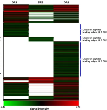
To evaluate the MHC class II-peptide binding pattern for the entire 61M. tuberculosisproteins (and the three MHC class II alleles), we carried out a Pearson centered hierarchical clus-tering analysis (Fig. 1). The peptides are clustered into groups recognized by only one monomer, groups recognized by two monomers, and groups recognized by all three MHC class II monomers. There were more peptides binding to HLA-DRB1*0401 than to HLA-DRB1*1501 and -DRB1*0101.
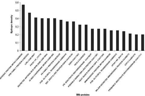
MHC class II epitope densities onM. tuberculosisproteins.
Next, we analyzed M. tuberculosispeptides that bind with a particular index value (i.e., the measure of MHC class II-peptide complex formation) in a reproducible fashion; we set an average index value cutoff of 0.00 in at least 2 of 3 repeats. We then calculated the epitope density of each individualM. tuberculosisprotein, defined as the number of peptides binding to any MHC class II monomer per total number of peptides
on August 17, 2020 by guest
http://cvi.asm.org/
from the respectiveM. tuberculosisprotein. Figure 2 shows the top 20M. tuberculosisproteins with the highest epitope densi-ties; the epitope densities of the entire set of 61M. tuberculosis
proteins are provided in Table 2. The “epitope density value”
provides a good estimate of which proteins are likely to provide epitopes to DRB1*0101, DRB1*1501, and/or DRB1*0401 MHC class II molecules; they are also likely to provide more CD4⫹T-cell epitopes, which may lead to T-cell expansion if
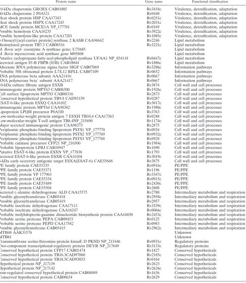
TABLE 1. M. tuberculosisproteins tested for MHC class II peptide binding
Protein name Gene name Functional classification
10-kDa chaperonin GROES CAB01005 Rv3418c Virulence, detoxification, adaptation
60-kDa chaperonin 2 P0A521 Rv0440 Virulence, detoxification, adaptation
Heat shock protein HSP CAA17343 Rv0251c Virulence, detoxification, adaptation Heat shock protein HSPX CAA17245 Rv2031c Virulence, detoxification, adaptation MCE family protein MCE1A YP_177701 Rv0169 Virulence, detoxification, adaptation
Possible hemolysin CAA16235 Rv3922c Virulence, detoxification, adaptation
Possible hemolysin-like protein CAA17201 Rv1085c Virulence, detoxification, adaptation 3-Oxoacyl-关acyl-carrier protein兴synthase 2 KASB CAA94642 Rv2246 Lipid metabolism
Biotinylated protein TB7.3 CAB08316 Rv3221c Lipid metabolism
M. Bovisacyl- coenzyme A synthase gene; U75685 Lipid metabolism
M. Bovismycocerosic acid synthase gene M95808 Lipid metabolism
Putative cyclopropane-fatty-acyl-phospholipid synthase UFAA1 NP_854118 Rv0447c Lipid metabolism
Secreted antigen 85-B FBPB (85B) CAB10044 Rv1886c Lipid metabolism
Alternate RNA polymerase sigma factor SIGF CAB07069 Rv3286c Information pathways Probable 50S ribosomal protein L7/L12 RPLL CAB07109 Rv0652 Information pathways
RNA polymerase beta subunit AAA21416 Rv0667 Information pathways
RNA polymerase beta⬘subunit AAA21417 Rv0667 Information pathways
10-kDa culture filtrate antigen ESXB Rv3874 Cell wall and cell processes
Immunogenic protein MPT63 CAB06500 Rv1926c Cell wall and cell processes
Cell surface lipoprotein MPT83 CAB08316 Rv2873 Cell wall and cell processes Conserved hypothetical protein TB9.8 CAD93159 Rv0287 Cell wall and cell processes
ESAT-6-like protein ESXQ CAA16102 Rv3017c Cell wall and cell processes
Immunogenic protein MPT64 CAA98382 Rv1980c Cell wall and cell processes
Lipoprotein LPQH precursor P0A5J0 Rv3763 Cell wall and cell processes
Low-molecular-weight protein antigen 7 ESXH TB10.4 CAA17363 Rv0288 Cell wall and cell processes Low-molecular-weight T-cell antigen TB8.4NP_215690 Rv1174c Cell wall and cell processes Major secreted immunogenic protein CAA98373 Rv2875 Cell wall and cell processes Periplasmic phosphate-binding lipoprotein PSTS1 YP_177770 Rv0934 Cell wall and cell processes Periplasmic phosphate-binding lipoprotein PSTS2 YP_177769 Rv0932c Cell wall and cell processes Periplasmic phosphate-binding lipoprotein PSTS3 YP_177768 Rv0928 Cell wall and cell processes Probable cutinase precursor CFP21 NP_216500 Rv1984c Cell wall and cell processes
Probable lipoprotein LPRJ CAB10947 Rv1690 Cell wall and cell processes
Putative ESAT-6-like protein ESXN YP_177838 Rv1793 Cell wall and cell processes Secreted ESAT-6-like protein ESXR CAA16104 Rv3019c Cell wall and cell processes 6-kDa early secretory antigenic target ESXA(ESAT-6) CAE55648 Rv3875 Cell wall and cell processes
PE family protein CAE55335 Rv0916c PE/PPE
PPE family protein CAE55371 Rv1196 PE/PPE
PPE family protein YP 177963 Rv3347c PE/PPE
PPE family protein CAE55334 Rv0915c PE/PPE
PPE family protein CAE55489 Rv2430c PE/PPE
PPE family protein CAE55504 Rv2608 PE/PPE
SecretedL-alanine dehydrogenase ALD CAA15575 Rv2780 Intermediary metabolism and respiration Possible glycosyltransferase CAB05418 Rv2958c Intermediary metabolism and respiration Possible glycosyltransferase CAB05419 Rv2957 Intermediary metabolism and respiration Probable isocitrate dehydrogenase CAA17111 Rv3339c Intermediary metabolism and respiration Probable isocitrate dehydrogenase CAA16247 Rv0066c Intermediary metabolism and respiration Probable molybdopterin-guanine dinucleotide biosynthesis protein CAA16030 Rv2453c Intermediary metabolism and respiration Probable serine protease PEPA CAB09453 Rv0125 Intermediary metabolism and respiration Probable serine protease PEPD CAA17582 Rv0983 Intermediary metabolism and respiration Possible glycosyltransferase CAB05415 Rv2962c Intermediary metabolism and respiration
MTB48 AAK31576 Unknown
MTB81 Unknown
Transmembrane serine/threonine-protein kinasE D PKND NP_215446 Rv0931c Regulatory proteins Two-component transcriptional-regulatory protein DEVR NP_217649 Rv3133c Regulatory proteins Conserved hypothetical protein CFP17 CAB01474 Rv1827 Conserved hypotheticals Conserved hypothetical protein TB16.3CAD97060 Rv2185c Conserved hypotheticals Conserved hypothetical protein TB18.5CAD93033 Rv0164 Conserved hypotheticals
Hypothetical protein NP_217139 Rv2623 Conserved hypotheticals
Hypothetical protein NP_217142 Rv2626c Conserved hypotheticals
Iron-regulated conserved hypothetical protein CAB08889 Rv1636 Conserved hypotheticals
Conserved hypothetical protein CAB08634 Rv2629 Conserved hypotheticals
on August 17, 2020 by guest
http://cvi.asm.org/
the appropriate T-cell receptors (TCRs) are present in the TCR repertoire at the time of vaccination or exposure to the nominal target antigen. We then compared the Ag85B and MPT63 peptides that tested positive for MHC class II mono-mer binding with previously published T-cell epitopes (Fig. 3). For MPT63, most of the peptides identified by use of the current approach have been described as CD4⫹ T-cell epitopes.
MHC class II binding peptides represent commonly recog-nized Ig epitopes in sera from patients with pulmonary TB.
We previously identified three patterns of IgG and IgA reac-tivity toM. tuberculosistarget peptides: (i) epitopes that are exclusively recognized in individuals with pulmonary TB (and not in healthy individuals), (ii) epitopes that are recognized in healthy subjects and not in patients with pulmonary TB, and (iii) epitopes that are recognized in both TB patients and healthy controls, but in a differential manner, i.e., either strongly in one group and weakly in the other group or vice versa. Based on the observation that B- and T-cell epitopes can overlap, as defined by the SEREX approach in screening for tumor-specific B- and T-cell responses (20), we tested whether
any MHC class II binding peptide identified in the current report would also serve as targets for an IgG response in sera from patients with acute pulmonary TB. Note that we screened onlyM. tuberculosisepitopes that were commonly recognized (n⫽35/35 patients) in sera from patients with TB and not in any healthy individual (n⫽34) for MHC class II binding. Two hundred twenty-twoM. tuberculosispeptides that bound to any of the three MHC class II monomers were also defined as IgG epitopes in sera from patients with TB (14). Out of these 222 peptides, 33 bound to all three MHC class II monomers, 24 bound to only two monomers, and 165 bound to only a single MHC class II allele. Eighty peptides bound to HLA-DRB1*0101, 52 bound to HLA-DRB1*1501, and 185 peptides bound to HLA-DRB1*0401. These peptides are listed in Table S2 in the supplemental material.
DISCUSSION
Only 7% of the 4,000 open reading frames ofM. tuberculosis
have been explored for B-cell and T-cell epitopes. This is due to the size of theM. tuberculosisgenome, i.e., the number of
FIG. 1. Pearson centered hierarchical clustering analysis ofM. tuberculosispeptides from 61 proteins binding to MHC class II monomers. Peptides binding to only one of the MHC class II alleles can be identified, as well as peptide groups that bind to only two or a to a single MHC class II molecule.Mtb,M. tuberculosis.
on August 17, 2020 by guest
http://cvi.asm.org/
protein targets to be tested, and to the lack of appropriate technology to explore such a massive data set in an affordable manner. This report describes the detailed analysis of anM. tuberculosispeptide microarray using 7,446 overlapping pep-tides from 61 individual M. tuberculosis proteins to identify potential T-cell epitopes that could be presented by three com-mon MHC class II alleles, HLA-DRB1*0101, -DRB1*1501, and -DRB1*0401. Most of the peptides that we identified bound to more than a single MHC class II molecule; only a few peptides bound to only one MHC class II allele. This is not surprising, since MHC class II peptide binding is quite promis-cuous (17, 30), and all MHC class II bindingM. tuberculosis
peptides listed by Blythe and coworkers bound to three or more MHC class II alleles (3).
We identified more M. tuberculosispeptides that bound to HLA-DRB1*0401 than to HLA-DRB1*0101 or -DRB1*1501. HLA-DRB1*0401 was found to be associated with pulmonary TB in Italian patients (31). Thus, whether a broaderM. tuber-culosis peptide epitope presentation by HLA-DRB1*0401 is beneficial or detrimental to mounting a protective anti-M. tu-berculosis-directed CD4⫹T-cell response has to be explored in future studies.
Ranking of M. tuberculosis proteins by epitope densities identified MPT63 Rv1926c and PPE CAE55334/Rv0915c as the 2 of 61M. tuberculosisproteins with the highest epitope densities. Immunization of C57BL/6 mice with MTB41 Rv0915c DNA induced protection againstM. tuberculosis in-fection comparable to the protection induced by
Mycobacte-rium bovisBCG (34), and the cellular immune responses were dominated by CD4⫹T cells. Analysis of T-cell responses was carried out using the Rv0915c protein, and the immune re-sponses were not determined on the peptide level.
Rv1926c, an M. tuberculosis-secreted protein, has recently been shown to be recognized (26) in healthy BCG-vaccinated subjects. Peptides binding to different MHC class II alleles were identified using a virtual matrix-based prediction pro-gram (ProPred). Nine (Rv1926c) peptides predicted to serve as promiscuous CD4⫹T-cell epitopes (24) show significant over-lap with the peptides that we identified as binding to the three MHC class II alleles. This lends support to our approach that MHC class II binding peptide species, defined by the interac-tion of soluble MHC class II molecules and immobilized pep-tides on a microarray chip, serve as CD4⫹T-cell epitopes (9). We were able to match MHC class II bindingM. tuberculosis
epitopes with peptide epitopes that were exclusively recog-nized by IgG from patients with TB (10). These candidate epitopes may represent clinically relevant targets for diagnos-tics. Conversely, MHC class II bindingM. tuberculosispeptides recognized in individuals who have been exposed toM. tuber-culosis but who are protected from development of disease may represent reasonableM. tuberculosis vaccine candidates. The identification of suchM. tuberculosistargets (i.e., exclusive recognition in a clinically well-defined population) in associa-tion with “good immunogenicity,” defined by MHC class II epitope density, is currently under way in our laboratory.
The current study is limited, since we did not test the
iden-FIG. 2. Epitope densities of the top 20M. tuberculosisproteins defined by the number of peptides binding to MHC class II molecules.
on August 17, 2020 by guest
http://cvi.asm.org/
tified HLA-DR binding peptides for CD4⫹T-cell recognition using peripheral blood mononuclear cells (PBMCs) from HLA-DR-matched patients with tuberculosis; corresponding CD4⫹T cells endowed with a clonotypic TCR may not be part of the TCR repertoire in individual patients. The
disadvan-tages of testing peptide-specific T-cell recognition may include the facts that (i) a single cytokine, e.g., interferon, may not reflect the breadth of a CD4⫹T-cell response and (ii) individ-ual peptide species may not be stable and may be quickly degraded in a standard assay gauging intracellular cytokine
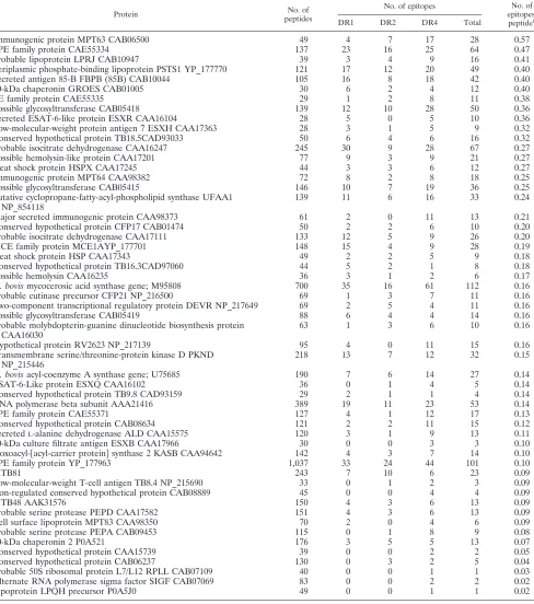
TABLE 2. Number of peptides from each individualM. tuberculosisprotein binding to three MHC class II allelesa
Protein No. of
peptides
No. of epitopes No. of
epitopes/
peptideb
DR1 DR2 DR4 Total
Immunogenic protein MPT63 CAB06500 49 4 7 17 28 0.57
PPE family protein CAE55334 137 23 16 25 64 0.47
Probable lipoprotein LPRJ CAB10947 39 3 4 9 16 0.41
Periplasmic phosphate-binding lipoprotein PSTS1 YP_177770 121 17 12 20 49 0.40
Secreted antigen 85-B FBPB (85B) CAB10044 105 16 8 18 42 0.40
10-kDa chaperonin GROES CAB01005 30 6 2 4 12 0.40
PE family protein CAE55335 29 1 2 8 11 0.38
Possible glycosyltransferase CAB05418 139 12 10 28 50 0.36
Secreted ESAT-6-like protein ESXR CAA16104 28 5 0 5 10 0.36
Low-molecular-weight protein antigen 7 ESXH CAA17363 28 3 1 5 9 0.32
Conserved hypothetical protein TB18.5CAD93033 50 6 4 6 16 0.32
Probable isocitrate dehydrogenase CAA16247 245 30 9 28 67 0.27
Possible hemolysin-like protein CAA17201 77 9 3 9 21 0.27
Heat shock protein HSPX CAA17245 44 3 3 6 12 0.27
Immunogenic protein MPT64 CAA98382 72 8 2 8 18 0.25
Possible glycosyltransferase CAB05415 146 10 7 19 36 0.25
Putative cyclopropane-fatty-acyl-phospholipid synthase UFAA1 NP_854118
139 11 6 16 33 0.24
Major secreted immunogenic protein CAA98373 61 2 0 11 13 0.21
Conserved hypothetical protein CFP17 CAB01474 50 2 2 6 10 0.20
Probable isocitrate dehydrogenase CAA17111 133 12 5 9 26 0.20
MCE family protein MCE1AYP_177701 148 15 4 9 28 0.19
Heat shock protein HSP CAA17343 49 2 2 5 9 0.18
Conserved hypothetical protein TB16.3CAD97060 44 5 2 1 8 0.18
Possible hemolysin CAA16235 36 3 1 2 6 0.17
M. bovismycocerosic acid synthase gene; M95808 700 35 16 61 112 0.16
Probable cutinase precursor CFP21 NP_216500 69 1 3 7 11 0.16
Two-component transcriptional regulatory protein DEVR NP_217649 69 2 5 4 11 0.16
Possible glycosyltransferase CAB05419 88 6 4 4 14 0.16
Probable molybdopterin-guanine dinucleotide biosynthesis protein CAA16030
63 1 3 6 10 0.16
Hypothetical protein RV2623 NP_217139 95 4 0 11 15 0.16
Transmembrane serine/threonine-protein kinase D PKND NP_215446
218 13 7 12 32 0.15
M. bovisacyl-coenzyme A synthase gene; U75685 190 7 6 14 27 0.14
ESAT-6-Like protein ESXQ CAA16102 36 0 1 4 5 0.14
Conserved hypothetical protein TB9.8 CAD93159 29 2 1 1 4 0.14
RNA polymerase beta subunit AAA21416 389 19 11 23 53 0.14
PPE family protein CAE55371 127 4 1 12 17 0.13
Conserved hypothetical protein CAB08634 121 2 2 11 15 0.12
SecretedL-alanine dehydrogenase ALD CAA15575 120 3 1 9 13 0.11
10-kDa culture filtrate antigen ESXB CAA17966 30 0 0 3 3 0.10
3-oxoacyl-关acyl-carrier protein兴synthase 2 KASB CAA94642 142 4 3 7 14 0.10
PPE family protein YP_177963 1,037 33 24 44 101 0.10
MTB81 243 7 10 6 23 0.09
Low-molecular-weight T-cell antigen TB8.4 NP_215690 33 0 1 2 3 0.09
Iron-regulated conserved hypothetical protein CAB08889 45 0 0 4 4 0.09
MTB48 AAK31576 150 4 3 6 13 0.09
Probable serine protease PEPD CAA17582 151 4 3 6 13 0.09
Cell surface lipoprotein MPT83 CAA98350 70 2 0 4 6 0.09
Probable serine protease PEPA CAB09453 115 0 1 8 9 0.08
60-kDa chaperonin 2 P0A521 176 3 5 5 13 0.07
Conserved hypothetical protein CAA15739 39 0 0 2 2 0.05
Conserved hypothetical protein CAB06237 130 0 3 2 5 0.04
Probable 50S ribosomal protein L7/L12 RPLL CAB07109 40 0 0 1 1 0.03
Alternate RNA polymerase sigma factor SIGF CAB07069 83 0 0 2 2 0.02
Lipoprotein LPQH precursor P0A5J0 49 0 0 1 1 0.02
a
DRB1ⴱ0101, DRB1ⴱ1501, and DRB1ⴱ0401.
b
The “epitope density” of each protein.
on August 17, 2020 by guest
http://cvi.asm.org/
production. Not mutually exclusive, anti-M. tuberculosis re-sponses may also be anergic in individuals with active pulmo-nary TB (33, 36). Tetramer-guided analysis may represent a remedy to this problem. However, the fact that some of the identified peptides have been reported previously (3, 26, 27) using functional assays or tetramer-guided analysis (14) sup-ports the validity of the approach reported here. It is also important to note that some of the candidate test peptides might not bind to the HLA-DR alleles in vivo due to differ-ential protein processing and subsequent presentation: peptide processing is dependent on the three-dimensional structure of proteins (18, 21, 22, 25). Future tetramer-guided analysis of PBMCs from patients with TB will aid in determining which peptides are presented in vivo and lead to expansion of anti-gen-specific CD4⫹T cells.
Of note, MHC class II-presented peptides may also drive immunosuppressive immune responses associated with CD4⫹ regulatory T cells (Tregs). This may be considered in rational vaccine design, since Tregs have been associated with the sup-pression of Th1-type immune responses inM. tuberculosis in-fection (13). In addition, instability of the transcription factor Foxp3 may lead to the generation of antigen-specific memory T cells with altered effector properties. This has recently been shown to be true for Tregs with an activated-memory T-cell phenotype, which gave rise to potentially autoreactive effector T cells (37).
We hypothesize that most of the M. tuberculosis peptides
that formed stable complexes with HLA-DRB1*0101, -DRB1*1502, and -DRB1*0401 may serve as CD4⫹ T-cell epitopes and that these peptides could be useful in designing a rational epitope-based vaccine againstM. tuberculosis. We sug-gest that the integrated analysis of IgG-recognized targets from clinically very well-characterized patient cohorts will help make the best choice forM. tuberculosisvaccine targets. (Pep-tide) specific B cells may serve as professional antigen-presenting cells (19) for CD4⫹T cells. Conversely, CD4⫹T cells may provide help for B cells and CD8⫹T cells. Therefore,
M. tuberculosisvaccine target identification should be accom-panied by MHC class I peptide binding analysis, since CD8⫹T cells are instrumental in conferring long-term immune memory in TB (4), particularly in patients with HIV coinfection and decreased CD4⫹T-cell numbers.
ACKNOWLEDGMENTS
The work was supported in part by an EU Marie Curie Fellowship to S.G. and by grants from Vetenskapsrådet and SIDA, Sweden, to M.M.
REFERENCES
1.Amirzargar, A. A., A. Yalda, M. Hajabolbaghi, F. Khosravi, H. Jabbari, N. Rezaei, M. H. Niknam, B. Ansari, B. Moradi, and B. Nikbin.2004. The association of HLA-DRB, DQA1, DQB1 alleles and haplotype frequency in Iranian patients with pulmonary tuberculosis. Int. J. Tuberc. Lung Dis.
8:1017–1021.
2.Beveridge, N. E., D. A. Price, J. P. Casazza, A. A. Pathan, C. R. Sander, T. E. Asher, D. R. Ambrozak, M. L. Precopio, P. Scheinberg, N. C. Alder, M.
FIG. 3. Locations of peptide binding to MHC class II molecules for two selected epitope-rich proteins. The peptides are arranged from the N to the C terminus of the proteins Ag85B (left) and MPT63 (Rv1926c; right), and only positive binding results are shown. These twoM. tuberculosis
proteins have previously been explored for HLA class II binding peptides (3, 14, 26, 27). Peptides that have been previously described as CD4⫹ T-cell epitopes, defined in either functional assays or MHC class II binding or by MHC class II tetramers, are underlined and shown in red.
on August 17, 2020 by guest
http://cvi.asm.org/
Roederer, R. A. Koup, D. C. Douek, A. V. Hill, and H. McShane.2007. Immunisation with BCG and recombinant MVA85A induces long-lasting,
polyfunctional Mycobacterium tuberculosis-specific CD4⫹memory T
lym-phocyte populations. Eur. J. Immunol.37:3089–3100.
3.Blythe, M. J., Q. Zhang, K. Vaughan, R. de Castro, Jr., N. Salimi, H. H. Bui, D. M. Lewinsohn, J. D. Ernst, B. Peters, and A. Sette.2007. An analysis of
the epitope knowledge related to mycobacteria. Immunome Res.3:10.
4.Day, C. L., N. Mkhwanazi, S. Reddy, Z. Mncube, M. van der Stok, P. Klenerman, and B. D. Walker.2008. Detection of polyfunctional Mycobac-terium tuberculosis-specific T cells and association with viral load in
HIV-1-infected persons. J. Infect. Dis.197:990–999.
5.De Groot, A. S., J. McMurry, L. Marcon, J. Franco, D. Rivera, M. Kutzler, D. Weiner, and B. Martin.2005. Developing an epitope-driven tuberculosis
(TB) vaccine. Vaccine23:2121–2131.
6.D’Souza, S., M. Romano, J. Korf, X. M. Wang, P. Y. Adnet, and K. Huygen.
2006. Partial reconstitution of the CD4⫹-T-cell compartment in CD4 gene
knockout mice restores responses to tuberculosis DNA vaccines. Infect.
Immun.74:2751–2759.
7.Endsley, J. J., A. Hogg, L. J. Shell, M. McAulay, T. Coffey, C. Howard, C. F. Capinos Scherer, W. R. Waters, B. Nonnecke, D. M. Estes, and B. Villarreal-Ramos. 2007. Mycobacterium bovis BCG vaccination induces memory
CD4⫹ T cells characterized by effector biomarker expression and
anti-mycobacterial activity. Vaccine25:8384–8394.
8.Frank, R.2002. The SPOT-synthesis technique. Synthetic peptide arrays on
membrane supports—principles and applications. J. Immunol. Methods267:
13–26.
9.Gaseitsiwe, S., D. Valentini, R. Ahmed, S. Mahdavifar, I. Magalhaes, J. Zerweck, M. Schutkowski, E. Gautherot, F. Montero, A. Ehrnst, M. Reilly, and M. Maeurer.2009. Major histocompatibility complex class II molecule-human immunodeficiency virus peptide analysis using a microarray chip.
Clin. Vaccine Immunol.16:567–573.
10.Gaseitsiwe, S., D. Valentini, S. Mahdavifar, I. Magalhaes, D. F. Hoft, J. Zerweck, M. Schutkowski, J. Andersson, M. Reilly, and M. J. Maeurer.2008. Pattern recognition in pulmonary tuberculosis defined by high content pep-tide microarray chip analysis representing 61 proteins from M. tuberculosis.
PLoS ONE3:e3840.
11.Goldfeld, A. E., J. C. Delgado, S. Thim, M. V. Bozon, A. M. Uglialoro, D. Turbay, C. Cohen, and E. J. Yunis.1998. Association of an HLA-DQ allele
with clinical tuberculosis. JAMA279:226–228.
12.Goletti, D., O. Butera, F. Bizzoni, R. Casetti, E. Girardi, and F. Poccia.2006.
Region of difference 1 antigen-specific CD4⫹memory T cells correlate with
a favorable outcome of tuberculosis. J. Infect. Dis.194:984–992.
13.Guyot-Revol, V., J. A. Innes, S. Hackforth, T. Hinks, and A. Lalvani.2006. Regulatory T cells are expanded in blood and disease sites in patients with
tuberculosis. Am. J. Respir. Crit. Care Med.173:803–810.
14.Hohn, H., C. Kortsik, I. Zehbe, W. E. Hitzler, K. Kayser, K. Freitag, C. Neukirch, P. Andersen, T. M. Doherty, and M. Maeurer.2007. MHC class II
tetramer guided detection of Mycobacterium tuberculosis-specific CD4⫹T
cells in peripheral blood from patients with pulmonary tuberculosis. Scand.
J. Immunol.65:467–478.
15.Kettaneh, A., L. Seng, K. P. Tiev, C. Toledano, B. Fabre, and J. Cabane.
2006. Human leukocyte antigens and susceptibility to tuberculosis: a
meta-analysis of case-control studies. Int. J. Tuberc. Lung Dis.10:717–725.
16.Kim, H. S., M. H. Park, E. Y. Song, H. Park, S. Y. Kwon, S. K. Han, and Y. S. Shim.2005. Association of HLA-DR and HLA-DQ genes with susceptibility to pulmonary tuberculosis in Koreans: preliminary evidence of associations with drug resistance, disease severity, and disease recurrence. Hum.
Immu-nol.66:1074–1081.
17.Kobayashi, H., M. Wood, Y. Song, E. Appella, and E. Celis.2000. Defining promiscuous MHC class II helper T-cell epitopes for the HER2/neu tumor
antigen. Cancer Res.60:5228–5236.
18.Landry, S. J.2008. Three-dimensional structure determines the pattern of
CD4⫹T-cell epitope dominance in influenza virus hemagglutinin. J. Virol.
82:1238–1248.
19.Lanzavecchia, A.1996. Mechanisms of antigen uptake for presentation.
Curr. Opin. Immunol.8:348–354.
20.Lee, S. Y., and D. Jeoung.2007. The reverse proteomics for identification of
tumor antigens. J. Microbiol. Biotechnol.17:879–890.
21.Li, H., P. C. Chien, Jr., M. Tuen, M. L. Visciano, S. Cohen, S. Blais, C. F. Xu, H. T. Zhang, and C. E. Hioe.2008. Identification of an N-linked glycosylation in the C4 region of HIV-1 envelope gp120 that is critical for recognition of
neighboring CD4 T cell epitopes. J. Immunol.180:4011–4021.
22.Li, H., C. F. Xu, S. Blais, Q. Wan, H. T. Zhang, S. J. Landry, and C. E. Hioe.
2009. Proximal glycans outside of the epitopes regulate the presentation of
HIV-1 envelope gp120 helper epitopes. J. Immunol.182:6369–6378.
23.Lombard, Z., D. L. Dalton, P. A. Venter, R. C. Williams, and L. Bornman.
2006. Association of HLA-DR, -DQ, and vitamin D receptor alleles and haplotypes with tuberculosis in the Venda of South Africa. Hum. Immunol.
67:643–654.
24.Middleton, D., L. Menchaca, H. Rood, and R. Komerofsky.2003. New allele
frequency database. Tissue Antigens61:403–407.
25.Mirano-Bascos, D., M. Tary-Lehmann, and S. J. Landry.2008. Antigen structure influences helper T-cell epitope dominance in the human immune
response to HIV envelope glycoprotein gp120. Eur. J. Immunol.38:1231–
1237.
26.Mustafa, A. S.2009. Th1 cell reactivity and HLA-DR binding prediction for promiscuous recognition of MPT63 (Rv1926c), a major secreted protein of
Mycobacterium tuberculosis. Scand. J. Immunol.69:213–222.
27.Mustafa, A. S., A. T. Abal, F. Shaban, A. M. El-Shamy, and H. A. Amoudy.
2005. HLA-DR binding prediction and experimental evaluation of T-cell epitopes of mycolyl transferase 85B (Ag85B), a major secreted antigen of
Mycobacterium tuberculosis. Med. Princ. Pract.14:140–146.
28.Nahtman, T., A. Jernberg, S. Mahdavifar, J. Zerweck, M. Schutkowski, M. Maeurer, and M. Reilly.2007. Validation of peptide epitope microarray
experiments and extraction of quality data. J. Immunol. Methods328:1–13.
29.Novak, E. J., A. W. Liu, G. T. Nepom, and W. W. Kwok.1999. MHC class II
tetramers identify peptide-specific human CD4(⫹) T cells proliferating in
response to influenza A antigen. J. Clin. Invest.104:R63–R67.
30.Panina-Bordignon, P., A. Tan, A. Termijtelen, S. Demotz, G. Corradin, and A. Lanzavecchia.1989. Universally immunogenic T cell epitopes: promiscu-ous binding to human MHC class II and promiscupromiscu-ous recognition by T cells.
Eur. J. Immunol.19:2237–2242.
31.Ruggiero, G., E. Cosentini, D. Zanzi, V. Sanna, G. Terrazzano, G. Matarese, A. Sanduzzi, F. Perna, and S. Zappacosta.2004. Allelic distribution of human leucocyte antigen in historical and recently diagnosed tuberculosis
patients in Southern Italy. Immunology111:318–322.
32.Scharn, D., H. Wenschuh, U. Reineke, J. Schneider-Mergener, and L. Germ-eroth.2000. Spatially addressed synthesis of amino- and
amino-oxy-substi-tuted 1,3,5-triazine arrays on polymeric membranes. J. Comb. Chem.2:361–
369.
33.Seitzer, U., K. Kayser, H. Hohn, P. Entzian, H. H. Wacker, S. Ploetz, H. D. Flad, J. Gerdes, and M. J. Maeurer.2001. Reduced T-cell receptor CD3zeta-chain protein and sustained CD3epsilon expression at the site of
mycobac-terial infection. Immunology104:269–277.
34.Skeiky, Y. A., P. J. Ovendale, S. Jen, M. R. Alderson, D. C. Dillon, S. Smith, C. B. Wilson, I. M. Orme, S. G. Reed, and A. Campos-Neto.2000. T cell expression cloning of a Mycobacterium tuberculosis gene encoding a pro-tective antigen associated with the early control of infection. J. Immunol.
165:7140–7149.
35.Teran-Escandon, D., L. Teran-Ortiz, A. Camarena-Olvera, G. Gonzalez-Avila, M. A. Vaca-Marin, J. Granados, and M. Selman.1999. Human leu-kocyte antigen-associated susceptibility to pulmonary tuberculosis: molecu-lar analysis of class II alleles by DNA amplification and oligonucleotide
hybridization in Mexican patients. Chest115:428–433.
36.Weichold, F. F., S. Mueller, C. Kortsik, W. E. Hitzler, M. J. Wulf, D. M. Hone, J. C. Sadoff, and M. J. Maeurer.2007. Impact of MHC class I alleles
on the M. tuberculosis antigen-specific CD8⫹T-cell response in patients
with pulmonary tuberculosis. Genes Immun.8:334–343.
37.Zhou, X., S. L. Bailey-Bucktrout, L. T. Jeker, C. Penaranda, M. Martinez-Llordella, M. Ashby, M. Nakayama, W. Rosenthal, and J. A. Bluestone.2009. Instability of the transcription factor Foxp3 leads to the generation of
patho-genic memory T cells in vivo. Nat. Immunol.10:1000–1007.