Page 1 of 14 Review Article
From the perspective of embryonic tendon development: various
cells applied to tendon tissue engineering
Fangjie Qi1#, Zhantao Deng1#, Yuanchen Ma1, Shuai Wang1, Chang Liu1, Fengjuan Lyu1, Tao Wang1,2, Qiujian Zheng1,2
1Department of Orthopedics, Guangdong Provincial People’s Hospital, Guangdong Academy of Medical Sciences, School of Medicine, South China
University of Technology, Guangzhou Higher Education Mega Centre, Guangzhou 510006, China; 2Centre for Orthopaedic Translational Research, School of Biomedical Sciences, University of Western Australia, Nedlands, Western Australia, Australia
Contributions: (I) Conception and design: F Qi, T Wang, Q Zheng; (II) Administrative support: None; (III) Provision of study materials or patients: Z Deng; (IV) Collection and assembly of data: F Qi; (V) Data analysis and interpretation: F Qi; (VI) Manuscript writing: All authors; (VII) Final approval of manuscript: All authors.
#These authors contributed equally to this work.
Correspondence to: Qiujian Zheng or Tao Wang. Guangdong Provincial Hospital, Guangdong Academy of Medical Sciences, School of Medicine, South China University of Technology, 106 Zhongshan 2nd Rd., Yuexiu Qu, Guangzhou 510080, China. Email: [email protected]; [email protected].
Abstract: There is a high risk of injury from damage to the force-bearing tissue of the tendon. Due to its poor self-healing ability, clinical interventions for tendon injuries are limited and yield unsatisfying results. Tissue engineering might supply an alternative to this obstacle. As one of the key elements of tissue engineering, various cell sources have been used for tendon engineering, but there is no consensue concerning a single optimal source. In this review, we summarized the development of tendon tissue from the embryonic stage and categorized the used cell sources in tendon engineering. By comparing various cell sources as the candidates for tendon regeneration, each cell type was found to have its advantages and limitations; therefore, it is difficult to define the best cell source for tendon engineering. The microenvironment cells located is also crucial for cell growth and differentiation; so, the optimal cells are unlikely to be the same for each patient. In the future, the clinical application of tendon engineering might be more precise and customized in contrast to the current use of a standardized/generic one-size-fits-all procedure. The best cell source for tendon engineering will require a case-based assessment.
Keywords: Tendon; tissue engineering; stem cells; cell types
Submitted Oct 28, 2019. Accepted for publication Dec 06, 2019. doi: 10.21037/atm.2019.12.78
View this article at: http://dx.doi.org/10.21037/atm.2019.12.78
Introduction
Tendons are unique connective tissues that not only connect the muscles and bones, but they are also important for maintaining posture and locomotion through transmitting muscle-contraction force to the skeleton. The tendon is composed predominantly of tenocytes surrounded by extracellular matrices (ECM) such as collagen I fibers, proteoglycans, and glycoproteins. It forms a solid structure and can support high repetitive mechanical loading (1).
However, tendon tissue has poor healing ability and limited regenerative ability; the damage may become irreversible,
and the healing process will be difficult, leading to chronic
disability. Although the current wide use of topical or
systemic anti-inflammatory drugs can reduce the perception
repair process may induce the formation of scar tissue (4), of which the tensile strength is only one-third that of an undamaged tendon, which can be a triggering factor for a large number of cases of secondary damage (5). Furthermore, autograft may cause donor site morbidity, while allograft may elicit an immune rejection (6).
Therefore, tissue engineering has been explored in an attempt to improve tendon healing and eventually reach a complete biological repair. A classic tissue engineering (7) technique is to culture seed cells in an engineered structure that is made of a biodegradable scaffold with the supplementation of growth factors or either mechanical or chemical factors to promote cell proliferation and differentiation in vitro before it is transplanted into the damaged site in vivo. Subsequently, the scaffold could be substituted by a newly formed organization that can replace damaged tissues and ultimately reconstitute the tissue functions (8). In tendon tissue engineering, cells also play a vital role in producing ECM for the reconstruction of the tendon tissue structure (9). Thus, the source of the seed cells becomes one of the key points in tendon tissue engineering.
To find more potential candidates suitable for tendon tissue
engineering, we explore the embryological origin of the limb tendons, analyze the developing processes of tendons, and summarize the availability of various types of seed cells using tendon tissue engineering. So, at the beginning of the article, we will reconstruct the development of the tendon from the embryo to maturity and explain the availability of these cells.
Embryological origin and development of tendons
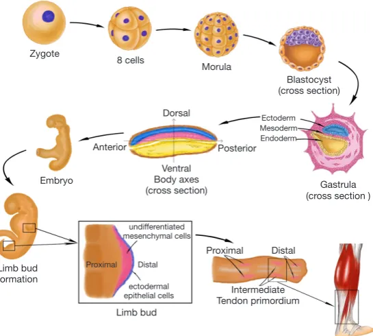
Human embryonic development begins with the cleavage of the fertilized egg. It then divides into many smaller cells leading to the formation of the blastula. Through the cell movements of gastrulation, the embryo is rearranged to form the three main germ layers: the ectoderm, mesoderm, and endoderm. The mesoderm then gives rise to chordamesoderm, paraxial mesoderm, intermediate mesoderm, and lateral plate mesoderm. With the establishment of body axes and formation of the neural tube, limb buds arise from lateral plate mesoderm at the appropriate levels along the ventral domain of the body (10). The early limb bud has two major components: an outer layer of ectodermal epithelial cells, and a core of loose undifferentiated mesenchymal cells derived from the lateral plate mesoderm (11,12). The skeletal elements and connective tissues (including tendon structure) of the
limb develop from these mesenchyme cells, but the limb muscles are derived from the somites which stem from the region of paraxial mesoderm and migrate into the limb bud. Therefore, the origin of tendon tissue is the same as that of bones, cartilage, and any other connective tissues, but not muscle. Analysis of the expression of scleraxis (Scx), a tendon specific transcription factor, revealed that the limb tendon progenitors originate from the subectodermal mesenchyme in proximomedial domains of the limb bud (13). Regulated by multiple morphogens and growth factors, three pairs of tendon primordium appear in a proximo-distal sequence in the developing limb (14,15). Proximal and intermediate tendon primordium appears the earliest on the dorsal side around limb junction. Following closely, intermediate tendon primordium appears on the ventral side around intermediate limb junction. The ventral proximal tendon primordium forms after the appearance of ventral intermediate tendon primordium. Subsequently, distal tendon primordium appears at both the dorsal and ventral sides of the distal limb. After the stimulation of growth factors and mechanical stress (16), the three pairs of tendon primordium will then differentiate into adult tendon structure in a proximal-to-distal sequence in the developing limb (Figure 1).
Various types of seed cells for tendon tissue engineering
Based on the process of limb tendon development, tenocytes originate from mesenchymal stromal cells (MSCs), which are also progenitor cells of skeletal elements and other connective tissues. Limb bud MSCs are derived from the lateral plate mesoderm of the embryo. These cells at different stages of tendon differentiation are candidate types of seed cells in tendon tissue engineering.
sources of MSCs (20). These MSC-derived stem cells can be differentiated into tenocytes under appropriate stimulation in vitro.
Another available cell type is pluripotent stem cells, which can differentiate into all types of cells constituting the human body. These include embryonic stem cells (ESCs) and induced pluripotent stem cells (iPSCs), which can be
derived from differentiated human dermal fibroblasts (21). In summary, the available seed cells for tendon tissue engineering include ESCs, iPSCs, MSCs (included BMSCs,
ADSCs, and TDSCs), dermal fibroblasts, and tenocytes, as
summarized in Figure 2. Next, we will discuss the respective advantages and disadvantages of these different cell types.
Comparison between different cell types
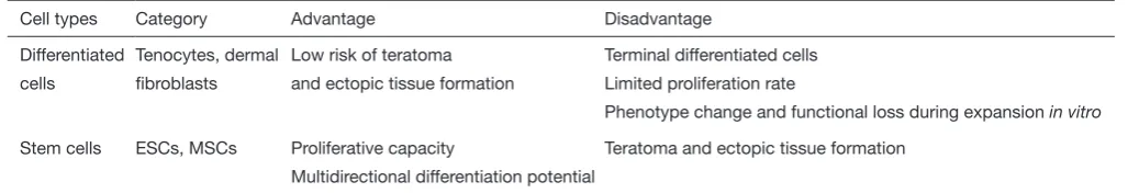
[image:3.595.160.433.85.330.2]The cells in the human body can be divided into two categories based on their differentiation status (Table 1). One is differentiated cells, such as tenocytes and dermal fibroblasts, which lack self-renewal capability in vivo. Differentiated tenocytes and dermal fibroblasts can be expanded in vitro in the presence of serum but have a limited expansion capacity. Culturing in vitro with the supplementation of growth factors may activate their ability of proliferation, but these cells still lack the capacity of differentiating into other cell types. Besides, their phenotype may change, which will cause a deficiency in their functions with increasing passaging (18). The Figure 1 The process of human limb tendon development.
Zygote
8 cells
Morula
Blastocyst (cross section)
Gastrula (cross section ) Posterior
Dorsal
Anterior
Ventral Body axes (cross section) Embryo
Limb bud formation
undifferentiated mesenchymal cells
ectodermal epithelial cells Proximal
Proximal Distal
Intermediate Tendon primordium
Adult tendon Limb bud
Distal
Ectoderm Mesoderm Endoderm
Figure 2 The derivation of tenocytes from ESCs and classification of cell types. Green solid arrows show the direction of differentiation, green dotted arrow shows that iPSCs can be obtained from dermal fibroblasts by dedifferentiation. The deepening of the font color means that the ability of cells to proliferare and differentiate is gradually reduced. ESCs, embryonic stem cells; iPSCs, induced pluripotent stem cells; MSCs, mesenchymal stromal cells; BMSCs, bone marrow-derived mesenchymal stromal cells; ADSCs, adipose-derived stem cells; TDSCs, tendon-derived stem cells.
Stem cells
iPSCs
ESCs MSCs
BMSCs ADSCs TDSCs Other sources
of mSCs
Mesenchymal
cells fibroblastsDermal
Tenocytes Differentiated cells
Pluripotent
[image:3.595.65.268.386.503.2]other is stem cells, which can replicate themselves as well as differentiate into specialized cells under appropriate conditions (22). At the same time, their ability to proliferate
and differentiate is difficult to control in vivo, which makes them susceptible to forming of tumor and undesired tissue cells during differentiation.
Differentiated cells
Tenocytes
Tenocytes are highly specialized mesenchyme-derived cells with important roles in synthesizing ECM and maintaining tendon structures in vivo (23). Cao et al. constructed tissue-engineered artificial tendons for the first time (24), but
they also indicated that tenocytes are relatively difficult to
grow and expand in vitro. Furthermore, some researchers indicated that with increased passaging, the gene expression of tendon-associated proteins such as collagen type I (col I), collagen type III (col III), tenascin, and tenomodulin (TNMD) exhibited a trend of decrease (25). In addition, there is also a change in the tenocyte cell phenotype, represented by more rounded cell morphologies instead of longer and thicker configurations (18). Thus, many researchers have explored the strategies to promote proliferation and at the same time, support a stable phenotype of tenocytes. At present, it has been reported that thyroid hormone T3, transforming growth factor (TGF)-b1, glycine, platelet-rich plasma, and insulin-like growth factor 1 could enhance the proliferation and differentiation capacity of tenocytes, as well as stimulate the secretion of ECM (23,26-29). Mechanical force can also regulate tenocyte differentiation (30).
Dermal fibroblasts
Dermal fibroblasts are terminally differentiated cells that
originate from mesoderm. In vitro, dermal fibroblasts exhibit similar cell morphology as tenocytes and mainly produce col I and col III. Compared with tenocytes, the harvest of dermal fibroblasts is less defective as only an easily accessible, small piece of skin is needed. Also, the cell expansion is less likely during in vitro culture (31). It has been revealed that there is no difference in their gross view between neo-tendon tissues engineered by human dermal
fibroblast or tenocytes. There was also no difference found
in the histologic structure, collagen superstructure, or mechanical property under the static strain in vitro (32-34). Therefore, researchers have used dermal fibroblast-engineered tendon to repair animal tendon defect, and the results are satisfactory in that the tensile stiffness and maximum load are expressly higher than those of
non-dermal fibroblast scaffolds (35-38).
When dermal fibroblasts and tenocytes are compared, both originate from mesoderm and have similar morphologies (36), and it was determined that dermal
fibroblasts were more advantageous compared to tenocytes.
First, dermal fibroblasts have good proliferative capacity and self-renewal potential (39). Second, dermal fibroblasts
have been shown to be easy to harvest with no major tissue defects at the donor site since the skin can regenerate in a short time (40). In contrast, tenocytes are more difficult
to collect because the density of tenocytes in a tendon is low, and there is an issue of donor site morbidity (41). However, dermal fibroblasts have a disadvantage in that they may produce fibrotic ECM which is involved in scar formation (42) (Table 2).
Stem cells
[image:4.595.42.556.96.184.2]According to the differentiation potential, stem cells can be divided into distinct kinds of cell types. Pluripotent stem cells can differentiate into all cell types of all organs, Table 1 Relative comparison between two human cell types using tissue engineerin
Cell types Category Advantage Disadvantage
Differentiated cells
Tenocytes, dermal fibroblasts
Low risk of teratoma and ectopic tissue formation
Terminal differentiated cells Limited proliferation rate
Phenotype change and functional loss during expansion in vitro
Stem cells ESCs, MSCs Proliferative capacity
Multidirectional differentiation potential
Teratoma and ectopic tissue formation
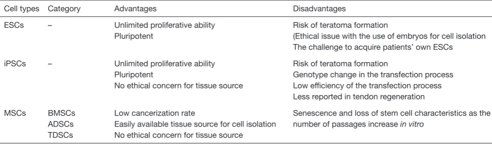
like ESCs and iPSCs (43). Multipotent stem cells can develop into many cell types within a particular lineage. For instance, the MSCs can give rise to bone cells, cartilage cells, adipose cells, and tendon cells. As ESCs have unlimited self-renewal potential and can differentiate into cells of all three embryonic germ layers (44), the risk of developing teratoma is expected to increase (45).
Meanwhile, considering that ESCs originate from the blastocyst, the harvest of human ESCs requires the destruction of the embryos, which may give rise to ethical concerns (46). iPSCs derived from differentiated cells may obviate these ethical issues. But, as they are pluripotent stem cells, the problem of forming teratoma during proliferation and differentiation remains unsolved. Furthermore, there are serious problems in the process of inducing iPSC formation (47).
By contrast, MSCs are more safe due to their limited proliferation capacity and the restricted multilineage differentiation potential of cells of the mesodermal lineage (48,49). The use of MSCs also bypasses certain ethical obstacles as they are adult stem cells that can be harvested from non-embryonic sources (50). However, it has been
accepted that MSC expansion in culture results in their accelerated aging, which will cause the deficiency of proliferation and multilineage differentiation potential (51,52) (Table 3).
ESCs
[image:5.595.41.557.96.179.2]ESCs can give rise to all tissues derived from the three germ layers (53). Because of this, ESCs hold great promise as seed cells for tendon tissue engineering (54). Thus, how to differentiate them into the tendon lineage has become a key point. Some researchers have investigated a stepwise differentiation approach by first inducing human ESCs to differentiate into MSCs, and subsequently allowing the MSCs to form tendon-like tissues. The results in vivo and in vitro showed that human ESC-derived MSCs exhibited tenocyte-like morphology and positively expressed tendon-related gene markers such as Scx, col I and col III, as well as other mechano-sensory structures and molecules (55,56). Moreover, the formation of teratomas could be avoided if ESCs are induced into MSCs before the transplantation (55). Table 2 Comparison between two differentiated cells types
Cell types Advantage Disadvantage
Tenocytes Major cell type in the tendon
Produce ECM for tendon function maintenance
Low cell number in the tendon Donor site morbidity
Dermal fibroblasts Easy to harvest from skin punch biopsy Production similar to the tenocyte
Not tenocyte Unstabled phenotype Scar formation ECM, extracellular matrix.
Table 3 Comparison between stem cells
Cell types Category Advantages Disadvantages
ESCs – Unlimited proliferative ability Pluripotent
Risk of teratoma formation
(Ethical issue with the use of embryos for cell isolation The challenge to acquire patients’ own ESCs
iPSCs – Unlimited proliferative ability Pluripotent
No ethical concern for tissue source
Risk of teratoma formation
Genotype change in the transfection process Low efficiency of the transfection process Less reported in tendon regeneration
MSCs BMSCs ADSCs TDSCs
Low cancerization rate
Easily available tissue source for cell isolation No ethical concern for tissue source
Senescence and loss of stem cell characteristics as the number of passages increase in vitro
[image:5.595.47.540.229.373.2]In addition, they demonstrated that the use of dynamic mechanical stress (1 HZ, 10% for 2 h/day) and bone morphogenetic protein (BMP)12 and BMP13 could promote differentiation of human ESCs into tenocytes (57-60).
iPSCs
The use of ESCs may be limited due to the need to sacrifice an embryo, which has aroused some ethical controversy. The discovery of iPSCs resolves the ethical problem of using ESCs, and recently, researchers were able to generate iPSCs from terminally differentiated cells (21,61). However, as their iPSCs were generated using retroviruses or lentiviruses (62), it might cause mutagenesis that would pose a risk for adverse effects in therapy (63). The efficiency of the transfection process also remains low. Thus, for the purpose of the safety of cell transplantation therapy, mRNA-delivered transcription factors have been applied to generate integration-free iPSCs (64,65). While these studies address some of the issues raised by the use of iPSCs in regenerative medicine, it has not been reported in tendon tissue engineering. For now, iPSCs are being used as a potential seed cell source for tendon regeneration research.
MSCs
MSCs are non-hematopoietic adult stem cells derived from the mesoderm germinal layer that can differentiate into mesenchymal-derived cell types and have the ability to self-renew (66). The membrane surface of MSCs expresses several antibodies, such as stromal cell antigen-1, CD271, stage-specific embryonic antigen-4, CD146, and so on, which can be considered as specific markers of MSCs (67,68). MSCs were initially isolated from bone marrow as precursors of stromal elements (69). From recent research, it is now clear that MSCs can be isolated from a wide range of adult and perinatal mesenchymal tissues, including those of bone, synovial membranes, periosteum, adipose tissue, tendons, skeletal muscles, and others (70). The use of MSCs for tendon repair has been extensively explored and may promote tendon regeneration.
BMSCs
The BMSCs have active self-proliferative and multi-differentiate capacity. The use of autologous BMSCs in the animal model could induce matrix production and
organization of injured tendon as well as restore histological structure and function (71). Because of the easy isolation from bone marrow and the enrichment of colony-forming units, BMSCs have become an attractive cell source for tissue engineering approaches. However, BMSCs may differentiate into osteoblasts instead of the expected tenocyte lineage when transplanted into the injured tendon, and form ectopic bone in vivo (72,73). To overcome such issue, a number of approaches are presently being developed to navigate the tenogenic differentiation of BMSCs (74). They have shown that cyclical uniaxial stretching and BMP14, TGF-β, connective tissue growth factor (CTGF), vascular endothelial growth factor (VEGF), and myostatin can initiate and maintain highly efficient growth and differentiation of BMSCs towards tenocytes in vitro (75-80). Furthermore, Zhang et al. (78) and Xie et al. (81) used a 3D culture environment combined with BMSCs sheets that have therapeutic effects on improving the healing quality of the tendon in vivo.
ADSCs
ADSCs are advantageous in tissue engineering due to their multipotency, high proliferation, easily isolated amounts of cells from the subcutaneous tissue, and low donor site morbidity (82,83). ADSCs have been regarded as one type of seed cell for tendon tissue engineering that can differentiate into tenocytes in vivo and increase the tensile strength of the repaired tendon (84,85). However, their innate tendency towards adipogenesis may hinder the application of ADSCs in tendon regeneration (1,86). For this purpose, intense research was conducted to overcome this obstacle. Yu et al. confirmed that hypoxia or activating the expression of hypoxia-inducible factor-1 could improve the tenogenesis of ADSCs (87). Additionally, several studies have indicated that the supplementation of growth factors could improve cell proliferation of ADSCs and promote tendon repair efficiency (88-90). Tendon ECM components (91) and TNMD (92) enhance the proliferation and tenogenesis of ADSCs. Physical stimulation such as uniaxial tensile cyclic loading (2% strain and 0.1 Hz frequency) (93) and extracorporeal shock waves in tenogenic medium (94) can also boost the differentiation of human ADSCs toward tendon-like cells.
TDSCs
regenerate tendon-like tissue structures in vivo
and avoid differentiating into other kinds of cell lines. Extensive progress and deep insights have emerged in the study of these cells for tendon tissue engineering and regeneration (19). ECM (97), biglycan (98), hepatocyte growth factor (99), and mechanical stimulation (100) that can promote TDSCs proliferation and tenogenic differentiation in vitro have been verified in research. CTGF and ascorbic acid can enhance the survival time, proliferation, and migration abilities of TDSCs in vivo (101). Recently, Zhang et al. demonstrated that genetic alterations of TDSCs following culture expansion could be prevented by pretreating TDSCs with histone deacetylase inhibitor, to retain their ability to accelerate tendon repair in vivo (102). Furthermore, Yin et al. identify a subpopulation of nestin+ TDSCs which exhibited a superior tenogenic capacity compared with nestin- counterparts (103). This study not
only redefines the subpopulation of tendon stem cells but
also provides new insights into a novel cell line for tendon tissue engineering.
Other sources of MSCs
Moshaverinia et al. tested the capacity of encapsulated dental MSCs to differentiate into tendon tissue in vitro and in vivo. Their findings indicate that periodontal ligament and
gingival tissue-derived MSCs can be considered as suitable stem cell sources for tendon engineering (104). Chen et al. explored dental pulp stem cells (DPSCs) for potential application in tendon tissue engineering and found that mature tendon-like tissue was formed after transplantation of DPSCs in a special fiber scaffold constructs under mechanical loading in a mouse model (105).
Comparison of the potential of various types of MSCs for tendon tissue engineering
MSCs can be harvested from various sources, such as bone marrow, adipose fat, and tendon tissue. They have paracrine effects, including immunomodulation, promoting
angiogenesis, or suppressing inflammation and immune cell
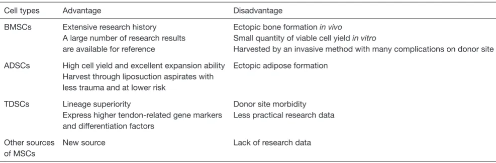
functions through secretory factors (106). BMSCs were the
first discovered MSCs and the most widely used seed cells in
tissue engineering. They are easy to harvest through bone marrow aspiration, but their limitations include relatively small quantities of viable cell yield through an invasive method, which cause many complications on the donor site and ectopic ossification after transplantation in vivo
(107,108). ADSCs can be obtained from liposuction aspirates, which is less invasive and involves lower risk. With the relatively large storage of adipose tissue in vivo, a mass of ADSCs can be isolated and expanded in vitro
(109,110). However, the main disadvantage of ADSCs is their tendency to undergo adipogenesis in vivo (111). TDSCs express higher tendon-related markers and differentiation factors than BMSCs and ADSCs (112,113), but the main drawback of TDSCs is that their isolation will lead to the injury of the donor site (114). Overall, there is not enough data from practical research on TDSCs to make
firm or extensive conclusions (Table 4).
Conclusions
[image:7.595.47.541.94.257.2]Tendons are an important component in the musculoskeletal system. Due to the limited regenerative ability of tendons, the healing process after an injury is slow, and the functions of the tendon are likely to be compromised. This results in Table 4 Comparison between MSCs
Cell types Advantage Disadvantage
BMSCs Extensive research history A large number of research results are available for reference
Ectopic bone formation in vivo
Small quantity of viable cell yield in vitro
Harvested by an invasive method with many complications on donor site
ADSCs High cell yield and excellent expansion ability Harvest through liposuction aspirates with less trauma and at lower risk
Ectopic adipose formation
TDSCs Lineage superiority
Express higher tendon-related gene markers and differentiation factors
Donor site morbidity Less practical research data
Other sources of MSCs
New source Lack of research data
T
able 5
Stimulation methods of different cell types for promoting tenogenesis and tendon healing
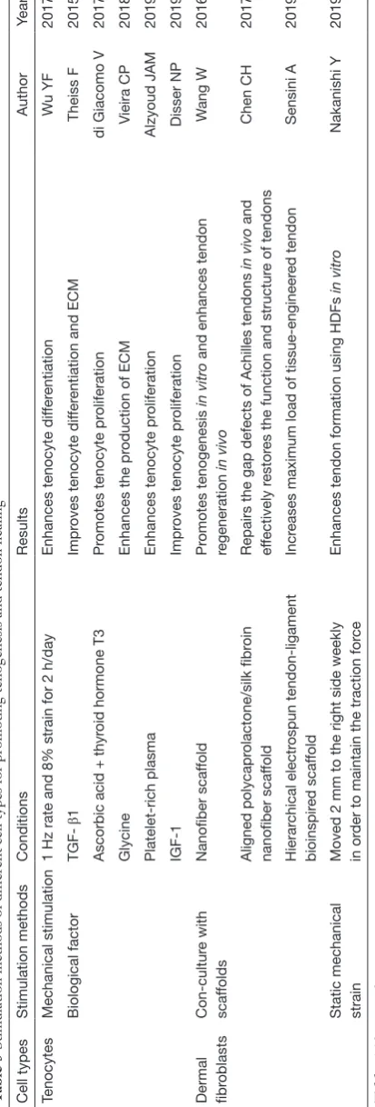
Cell types Stimulation methods Conditions Results Author Year Tenocytes Mechanical stimulation
1 Hz rate and 8% strain for 2 h/day
Enhances tenocyte dif
fer entiation W u YF 2017 Biological factor β 1 Impr
oves tenocyte dif
fer
entiation and ECM
Theiss F
2015
Ascorbic acid + thyr
oid hormone T3
Pr
omotes tenocyte pr
oliferation
di Giacomo V
2017
Glycine
Enhances the pr
oduction of ECM
Vieira CP
2018
Platelet-rich plasma
Enhances tenocyte pr
oliferation
Alzyoud JAM
2019
IGF-1
Impr
oves tenocyte pr
oliferation
Disser NP
2019
Dermal fibr
oblasts Con-cultur e with scaf folds Nanofiber scaf fold Pr omotes tenogenesis in vitro
and enhances tendon
regeneration in vivo W ang W 2016 Aligned polycapr olactone/silk fibr oin nanofiber scaf fold
Repairs the gap defects of Achilles tendons
in vivo
and
ef
fectively r
estor
es the function and structur
e of tendons
Chen CH 2017 Hierar chical electr ospun tendon-ligament bioinspir ed scaf fold Incr
eases maximum load of tissue-engineer
ed tendon
Sensini A
2019
Static mechanical strain Moved 2 mm to the right side weekly in or
der to maintain the traction for
ce
Enhances tendon formation using HDFs
in vitro Nakanishi Y 2019 T able 5 ( Continued )
a serious decline in the quality of life of patients. Cell-based tendon tissue engineering is a promising research area that aims to deliver adequate, regeneration-competent cells to the injured tendon and ultimately promote the restoration of its functions (20). This review provides an overview of tendon tissues from an embryonic stage and discusses the relative merits of each of the candidates that could be used as cell sources for tendon regeneration. Depending on the characteristics of each cell type, the researchers tested the most appropriate mechanical stimulation conditions and selected the different biological factors for improving the healing quality of the damaged tendon and reducing the adverse reactions during use (Table 5). Some major findings
are described below: Mechanical stimulation will enhance the tenocyte differentiation in different cell types. Growth factors can promote cell proliferation in differentiated cells. However, in stem cells, growth factors not only enhance tenogenic differentiation but also increase the rate of cell growth. Furthermore, cell ECM also can accelerate cell proliferation and the tenogenesis of MSCs. Therefore, the results of these studies may provide new concepts or methods in clinical treatment to improve the level of tendon repair in patients. Doctors can select different cells to treat tendon disease by measuring a patient’s expression level of growth factors, tendon transcription factors, the expression of collagen and cytoskeletal proteins in the extracellular matrix, and the microenvironment of the tendon. Clinical treatment of tendon injury can also provide an appropriate cell plan by avoiding the drawbacks of these seed cells. Massive ectopic ossification and fatty vesicles forming in the tendinopathy indicate that BMSCs and ADSCs are not the best choices in the treatment of tendinopathy (115).
In addition, scar formation and disorganization of fibers at
tendon damage or stump are factors to be considered when selecting candidate cells.
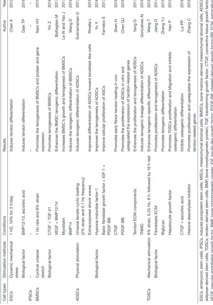
T able 5 ( Continued ) Cell types Stimulation methods Conditions Results Author Year ESCs
Dynamic mechanical stress
1 HZ, 10% for 2 h/day
Induces tendon dif
fer
entiation
Chen X
2012
Biological factor
BMP12/13, ascorbic acid
Induces tendon dif
fer entiation Dale TP 2018 iPSCs – – – – – BMSCs
Cyclical uniaxial stretch
1 Hz rate and 8% strain
Pr
omotes the tenogenesis of BMSCs and pr
otein and gene
expr
ession
Nam HY
2019
Biological factor
CTGF +
TGF-β1
Pr
omotes tenogenesis of BMSCs
Y
in Z
2016
VEGF + BMP12/14
Enhances the BMSC tenogenic dif
fer entiation Bottagisio M 2017 Myostatin Incr eases BMSC’ s gr
owth and tenogenesis of BMSCs
Le W and Y
ao J
2017
BMP14
Induces the tenogenic dif
fer
entiation of BMSCs
W
ang D
2018
ADSCs
Physical stimulation
Uniaxial tensile cyclic loading (2% strain and 0.1 Hz fr
equency)
Induces tenogenic dif
fer
entiation of ADSCs
Subramanian G
2017
Extracorpor
eal shock waves
Impr
oves dif
fer
entiation of ADSCs towar
d tenoblast-like cells
Rinella L 2018 Biological factor Hypoxia-inducible factor -1 Impr
oves the tenogenesis of ADSCs
Yu
Y
2016
Basic fibr
oblast gr
owth factor + IGF-1 +
PDGF-BB
Impr
ove cellular pr
oliferation of ASCs
Far
nebo S
2017
CTGF
Pr
omotes early flexor tendon healing
in vivo
Shen H
2018
PDGF-BB
Pr
omotes the pr
oliferation of ADSCs
in vitro
and
upr
egulated the expr
ession of tendon-r
elated genes
Chen QJ
2018
Tendon ECM components
Enhances the pr
oliferation and tenogenesis of ADSCs
Yang G
2017
TNMD
Induces tenogenic lineage dif
fer
entiation of ADSCs
Goncalves AI
2018
TDSCs
Mechanical stimulation
6% strain, 0.25 Hz, 8 h, followed by 16 h r
est
Enhances tenogenic-specific dif
fer entiation W ang T 2018 Biological factor Fibr oblasts ECM Pr omotes pr
oliferation and tenogenesis of ADSCs
Jiang D
2014
Biglycan
Pr
omotes tenogenic dif
fer entiation Zhang YJ 2019 Hepatocyte gr owth factor Pr
omotes TDSCs pr
oliferation and Migration and inhibits
osteogenic dif
fer
entiation
Han P
2019
CTGF + ascorbic acid
Inhibits osteogenic dif
fer
entiation
Lui PP
2016
Histone deacetylase inhibitor
Pr
omotes pr
oliferation and upr
egulates the expr
ession of
tendon-r
elated genes
Zhang C
2018
ESCs, embryonic stem cells; iPSCs, induced pluripotent stem cells; MSCs, mesenchymal str
omal cells; BMSCs, bone marr
ow-derived mesenchymal str
omal cells; ADSCs,
adipose-derived stem cells; TDSCs, tendon-derived stem cells; BMP
, bone morphogenetic pr
otein; TGF
, transforming gr
owth factor; CTGF
, connective tissue gr
should be customized designed on a case-by-case basis to achieve the best clinical outcomes.
Acknowledgments
Funding: This study was supported by the National Natural Science Foundation of China (81802214;81702191; 81371991), the National Natural Science Foundation of China Youth Science Foundation (81802222), the Major Program of Science and Technology of Guangdong (2015B020225007), the Natural Science Foundation of Guangdong Province (2018A030310694), the Fundamental Research Funds for the Central Universities, South China University of Technology (2018MS70), and the Guangdong Medical Science and Technology Research Foundation (2018114214430383). The Fundation of Traditional Chinese Medicine of Guangdong Province (20191004), The scientific Foundation of Guangdong Provincial People’s Hospital (2017bp01), The Outstanding Young Talents Foundation of Guangdong Provincial People’s Hospital (KJ012019091), Prrogam of Science and Technology of Guangzhou (201904010424).
Footnote
Conflicts of Interest: The authors have no conflicts of interest
to declare.
Ethical Statement: The authors are accountable for all aspects of the work in ensuring that questions related to the accuracy or integrity of any part of the work are appropriately investigated and resolved.
References
1. Docheva D, Muller SA, Majewski M, et al. Biologics for tendon repair. Adv Drug Deliv Rev 2015;84:222-39. 2. Nourissat G, Berenbaum F, Duprez D. Tendon injury:
from biology to tendon repair. Nat Rev Rheumatol 2015;11:223-33.
3. Wei Z, Reisdorf RL, Thoreson AR, et al. Comparison
of Autograft and Allograft with Surface Modification for
Flexor Tendon Reconstruction: A Canine in Vivo Model. J Bone Joint Surg Am 2018;100:e42.
4. Juneja SC. Cellular distribution and gene expression
profile during flexor tendon graft repair: A novel
tissue engineering approach(*). J Tissue Eng 2013;4:2041731413492741.
5. Andarawis-Puri N, Flatow EL, Soslowsky LJ. Tendon basic science: Development, repair, regeneration, and healing. J Orthop Res 2015;33:780-4.
6. Chang SK, Egami DK, Shaieb MD, et al. Anterior cruciate ligament reconstruction: allograft versus autograft. Arthroscopy 2003;19:453-62.
7. Langer R, Vacanti JP. Tissue engineering. Science 1993;260:920-6.
8. Glass ZA, Schiele NR, Kuo CK. Informing tendon tissue engineering with embryonic development. J Biomech 2014;47:1964-8.
9. Sharma P, Maffulli N. Biology of tendon injury: healing, modeling and remodeling. J Musculoskelet Neuronal Interact 2006;6:181-90.
10. Hwang SH, White KA, Somatilaka BN, et al. The G protein-coupled receptor Gpr161 regulates forelimb formation, limb patterning and skeletal morphogenesis in a primary cilium-dependent manner. Development 2018. doi: 10.1242/dev.154054.
11. Tickle C. How the embryo makes a limb: determination, polarity and identity. J Anat 2015;227:418-30.
12. Zhang YT, Alber MS, Newman SA. Mathematical modeling of vertebrate limb development. Math Biosci 2013;243:1-17.
13. Schweitzer R, Chyung JH, Murtaugh LC, et al. Analysis
of the tendon cell fate using Scleraxis, a specific marker for
tendons and ligaments. Development 2001;128:3855-66. 14. Kardon G. Muscle and tendon morphogenesis in the avian
hind limb. Development 1998;125:4019-32.
15. Guéro S. Developmental biology of the upper limb. Hand Surg Rehabil 2018;37:265-74.
16. Arvind V, Huang AH. Mechanobiology of limb musculoskeletal development. Ann N Y Acad Sci 2017;1409:18-32.
17. Di Giancamillo A, Deponti D, Raimondi MT, et al. Comparison between different cell sources and culture strategies for tendon tissue engineering. J Biol Regul Homeost Agents 2017;31:61-6.
18. Mazzocca AD, Chowaniec D, McCarthy MB, et al. In vitro changes in human tenocyte cultures obtained from proximal biceps tendon: multiple passages result in changes in routine cell markers. Knee Surg Sports Traumatol Arthrosc 2012;20:1666-72.
19. Ni M, Lui PP, Rui YF, et al. Tendon-derived stem cells (TDSCs) promote tendon repair in a rat patellar tendon window defect model. J Orthop Res 2012;30:613-9. 20. Costa-Almeida R, Calejo I, Gomes ME. Mesenchymal
Int J Mol Sci 2019. doi: 10.3390/ijms20123002. 21. Takahashi K, Tanabe K, Ohnuki M, et al. Induction of
pluripotent stem cells from adult human fibroblasts by defined factors. Cell 2007;131:861-72.
22. Gonzalez MA, Bernad A. Characteristics of Adult Stem Cells. Adv Exp Med Biol 2012;741:103-20.
23. Vieira CP, Viola M, Carneiro GD, et al. Glycine improves the remodeling process of tenocytes in vitro. Cell Biol Int 2018;42:804-14.
24. Cao Y, Liu YT, Liu W, et al. Bridging tendon defects using autologous tenocyte engineered tendon in a hen model. Plast Reconstr Surg 2002;110:1280-9.
25. Aparecida de Aro A, Vidal Bde C, Pimentel ER. Biochemical and anisotropical properties of tendons. Micron 2012;43:205-14.
26. Theiss F, Mirsaidi A, Mhanna R, et al. Use of biomimetic
microtissue spheroids and specific growth factor
supplementation to improve tenocyte differentiation and adaptation to a collagen-based scaffold in vitro. Biomaterials 2015;69:99-109.
27. di Giacomo V, Berardocco M, Gallorini M, et al. Combined supplementation of ascorbic acid and thyroid hormone T3 affects tenocyte proliferation. The effect of ascorbic acid in the production of nitric oxide. Muscles Ligaments Tendons J 2017;7:11-8.
28. Disser NP, Sugg KB, Talarek JR, et al. Insulin-like growth factor 1 signaling in tenocytes is required for adult tendon growth. FASEB J 2019;33:12680-95.
29. Alzyoud JAM, Al Najjar SA, Talat S, et al. Effect of light-emitting diodes, platelet-rich plasma, and their combination on the activity of sheep tenocytes. Lasers Med Sci 2019;34:759-66.
30. Wu YF, Huang YT, Wang HK, et al. Hyperglycemia Augments the Adipogenic Transdifferentiation Potential of Tenocytes and Is Alleviated by Cyclic Mechanical Stretch. Int J Mol Sci 2017. doi: 10.3390/ijms19010090.
31. Liu W, Chen B, Deng D, et al. Repair of tendon defect
with dermal fibroblast engineered tendon in a porcine
model. Tissue Eng 2006;12:775-88.
32. Deng D, Liu W, Xu F, et al. Engineering human
neo-tendon tissue in vitro with human dermal fibroblasts under
static mechanical strain. Biomaterials 2009;30:6724-30.
33. Wang W, He J, Feng B, et al. Aligned nanofibers direct human dermal fibroblasts to tenogenic phenotype in vitro
and enhance tendon regeneration in vivo. Nanomedicine (Lond) 2016;11:1055-72.
34. Busra FM, Lokanathan Y, Nadzir MM, et al. Attachment, Proliferation, and Morphological Properties of Human
Dermal Fibroblasts on Ovine Tendon Collagen Scaffolds: A Comparative Study. Malays J Med Sci 2017;24:33-43. 35. Chen CH, Chen SH, Kuo CY, et al. Response of Dermal
Fibroblasts to Biochemical and Physical Cues in Aligned
Polycaprolactone/Silk Fibroin Nanofiber Scaffolds for
Application in Tendon Tissue Engineering. Nanomaterials (Basel) 2017. doi: 10.3390/nano7080219.
36. Kwon J, Kim YH, Rhee SM, et al. Effects of Allogenic Dermal Fibroblasts on Rotator Cuff Healing in a Rabbit Model of Chronic Tear. Am J Sports Med 2018;46:1901-8. 37. Sensini A, Cristofolini L, Zucchelli A, et al. Hierarchical
electrospun tendon-ligament bioinspired scaffolds
induce changes in fibroblasts morphology under static
and dynamic conditions. J Microsc 2019. [Epub ahead of print].
38. Nakanishi Y, Okada T, Takeuchi N, et al. Histological evaluation of tendon formation using a scaffold-free three-dimensional-bioprinted construct of human dermal
fibroblasts under in vitro static tensile culture. Regen Ther
2019;11:47-55.
39. Vapniarsky N, Arzi B, Hu JC, et al. Concise Review: Human Dermis as an Autologous Source of Stem Cells for Tissue Engineering and Regenerative Medicine. Stem Cells Translational Medicine 2015;4:1187-98.
40. Chen B, Ding J, Zhang W, et al. Tissue Engineering of Tendons: A Comparison of Muscle-Derived Cells, Tenocytes, and Dermal Fibroblasts as Cell Sources. Plast Reconstr Surg 2016;137:536e-44e.
41. Qiu Y, Wang X, Zhang Y, et al. In vitro two-dimensional and three-dimensional tenocyte culture for tendon tissue engineering. J Tissue Eng Regen Med 2016;10:E216-26.
42. Thulabandu V, Chen DM, Atit RP. Dermal fibroblast in
cutaneous development and healing. Wiley Interdiscip Rev Dev Biol 2018. doi: 10.1002/wdev.307.
43. Dulak J, Szade K, Szade A, et al. Adult stem cells: hopes and hypes of regenerative medicine. Acta Biochim Pol 2015;62:329-37.
44. Yin Z, Chen X, Chen JL, et al. Stem cells for tendon tissue engineering and regeneration. Expert Opin Biol Ther 2010;10:689-700.
45. Li HL, Wei JF, Fan LY, et al. miR-302 regulates pluripotency, teratoma formation and differentiation in stem cells via an AKT1/OCT4-dependent manner. Cell Death Dis 2016;7:e2078.
46. Zheng YL. Some Ethical Concerns About Human Induced Pluripotent Stem Cells. Science and Engineering Ethics 2016;22:1277-84.
induced pluripotent stem cell-based therapeutics. J Biol Chem 2014;289:4585-93.
48. Dhinsa BS, Mahapatra AN, Khan WS. Sources of Adult Mesenchymal Stem Cells for Ligament and Tendon Tissue Engineering. Curr Stem Cell Res Ther 2015;10:26-30. 49. Lui PP. Stem cell technology for tendon regeneration:
current status, challenges, and future research directions. Stem Cells Cloning 2015;8:163-74.
50. Font Tellado S, Balmayor ER, Van Griensven M.
Strategies to engineer tendon/ligament-to-bone interface: Biomaterials, cells and growth factors. Adv Drug Deliv Rev 2015;94:126-40.
51. Bonab MM, Alimoghaddam K, Talebian F, et al. Aging of mesenchymal stem cell in vitro. BMC Cell Biol 2006;7:14. 52. Liu M, Lei H, Dong P, et al. Adipose-Derived
Mesenchymal Stem Cells from the Elderly Exhibit Decreased Migration and Differentiation Abilities with Senescent Properties. Cell Transplantation 2017;26:1505-19.
53. Thomson JA, Itskovitz-Eldor J, Shapiro SS, et al.
Embryonic stem cell lines derived from human blastocysts. Science 1998;282:1145-7.
54. Guillot PV, Cui W, Fisk NM, et al. Stem cell
differentiation and expansion for clinical applications of tissue engineering. J Cell Mol Med 2007;11:935-44. 55. Chen X, Song XH, Yin Z, et al. Stepwise differentiation
of human embryonic stem cells promotes tendon regeneration by secreting fetal tendon matrix and differentiation factors. Stem Cells 2009;27:1276-87.
56. Chen JL, Yin Z, Shen WL, et al. Efficacy of
hESC-MSCs in knitted silk-collagen scaffold for tendon tissue engineering and their roles. Biomaterials 2010;31:9438-51. 57. Schiele NR, Marturano JE, Kuo CK. Mechanical factors
in embryonic tendon development: potential cues for stem cell tenogenesis. Curr Opin Biotechnol 2013;24:834-40. 58. Chen X, Yin Z, Chen JL, et al. Force and scleraxis
synergistically promote the commitment of human ES cells derived MSCs to tenocytes. Sci Rep 2012;2:977. 59. Okech W, Kuo CK. Informing Stem Cell-Based Tendon
Tissue Engineering Approaches with Embryonic Tendon Development. Adv Exp Med Biol 2016;920:63-77. 60. Dale TP, Mazher S, Webb WR, et al. Tenogenic
Differentiation of Human Embryonic Stem Cells. Tissue Eng Part A 2018;24:361-8.
61. Takahashi K, Yamanaka S. Induction of pluripotent stem
cells from mouse embryonic and adult fibroblast cultures by defined factors. Cell 2006;126:663-76.
62. Nethercott HE, Brick DJ, Schwartz PH. Derivation of
induced pluripotent stem cells by lentiviral transduction. Methods Mol Biol 2011;767:67-85.
63. Lister R, Pelizzola M, Kida YS, et al. Hotspots of aberrant epigenomic reprogramming in human induced pluripotent stem cells. Nature 2011;471:68-73.
64. Engel M, Balez R, Munoz SS, et al. Viral-free generation and characterization of a human induced pluripotent
stem cell line from dermal fibroblasts. Stem Cell Res
2018;32:135-8.
65. McGrath PS, Diette N, Kogut I, et al. RNA-based Reprogramming of Human Primary Fibroblasts into Induced Pluripotent Stem Cells. J Vis Exp 2018. doi: 10.3791/58687.
66. Pittenger MF, Mackay AM, Beck SC, et al. Multilineage potential of adult human mesenchymal stem cells. Science 1999;284:143-7.
67. Samsonraj RM, Raghunath M, Nurcombe V, et al. Concise Review: Multifaceted Characterization of Human Mesenchymal Stem Cells for Use in Regenerative Medicine. Stem Cells Transl Med 2017;6:2173-85. 68. Rajpar I, Barrett JG. Optimizing growth factor induction of
tenogenesis in three-dimensional culture of mesenchymal stem cells. J Tissue Eng 2019;10:2041731419848776. 69. Friedenstein AJ, Chailakhjan RK, Lalykina KS. The
development of fibroblast colonies in monolayer cultures
of guinea-pig bone marrow and spleen cells. Cell Tissue Kinet 1970;3:393-403.
70. da Silva Meirelles L, Chagastelles PC, Nardi NB. Mesenchymal stem cells reside in virtually all post-natal organs and tissues. J Cell Sci 2006;119:2204-13. 71. Lu W, Xu J, Dong S, et al. Anterior Cruciate Ligament
Reconstruction in a Rabbit Model Using a Decellularized Allogenic Semitendinous Tendon Combined with Autologous Bone Marrow-Derived Mesenchymal Stem Cells. Stem Cells Transl Med 2019;8:971-82.
72. Awad HA, Boivin GP, Dressler MR, et al. Repair of patellar tendon injuries using a cell-collagen composite. Journal of Orthopaedic Research 2003;21:420-31. 73. Harris MT, Butler DL, Boivin GP, et al. Mesenchymal
stem cells used for rabbit tendon repair can form ectopic bone and express alkaline phosphatase activity in constructs. J Orthop Res 2004;22:998-1003
74. Tan SL, Ahmad RE, Ahmad TS, et al. Effect of growth differentiation factor 5 on the proliferation and tenogenic differentiation potential of human mesenchymal stem cells in vitro. Cells Tissues Organs 2012;196:325-38.
Formation and Defect Repair In Vivo. Stem Cells Transl Med 2016;5:1106-16.
76. Bottagisio M, Lopa S, Granata V, et al. Different combinations of growth factors for the tenogenic
differentiation of bone marrow mesenchymal stem cells in
monolayer culture and in fibrin-based three-dimensional
constructs. Differentiation 2017;95:44-53. 77. Le W, Yao J. The Effect of Myostatin (GDF-8) on
Proliferation and Tenocyte Differentiation of Rat Bone Marrow-Derived Mesenchymal Stem Cells. J Hand Surg Asian Pac Vol 2017;22:200-7.
78. Zhang B, Luo Q, Deng B, et al. Construction of tendon replacement tissue based on collagen sponge and mesenchymal stem cells by coupled mechano-chemical induction and evaluation of its tendon repair abilities. Acta Biomater 2018;74:247-59.
79. Nam HY, Pingguan-Murphy B, Abbas AA, et al. Uniaxial Cyclic Tensile Stretching at 8% Strain Exclusively Promotes Tenogenic Differentiation of Human Bone Marrow-Derived Mesenchymal Stromal Cells. Stem Cells Int 2019;2019:9723025.
80. Wang D, Jiang X, Lu A, et al. BMP14 induces tenogenic differentiation of bone marrow mesenchymal stem cells in vitro. Exp Ther Med 2018;16:1165-74.
81. Xie S, Zhou Y, Tang Y, et al. -Book-shaped decellularized tendon matrix scaffold combined with bone marrow mesenchymal stem cells-sheets for repair of achilles tendon defect in rabbit. J Orthop Res 2019;37:887-97.
82. Kryger GS, Chong AK, Costa M, et al. A comparison of tenocytes and mesenchymal stem cells for use in
flexor tendon tissue engineering. J Hand Surg Am
2007;32:597-605.
83. Uysal AC, Mizuno H. Tendon regeneration and repair with adipose derived stem cells. Curr Stem Cell Res Ther 2010;5:161-7.
84. Almeida H, Domingues RMA, Mithieux SM, et al. Tropoelastin-Coated Tendon Biomimetic Scaffolds Promote Stem Cell Tenogenic Commitment and Deposition of Elastin-Rich Matrix. ACS Appl Mater Interfaces 2019;11:19830-40.
85. de Lima Santos A, Silva CGD, de Sa Barretto LS, et al. Biomechanical evaluation of tendon regeneration with adipose-derived stem cell. J Orthop Res 2019;37:1281-6. 86. Baer PC, Geiger H. Adipose-derived mesenchymal
stromal/stem cells: tissue localization, characterization, and heterogeneity. Stem Cells Int 2012;2012:812693. 87. Yu Y, Zhou Y, Cheng T, et al. Hypoxia enhances tenocyte
differentiation of adipose-derived mesenchymal stem
cells by inducing hypoxia-inducible factor-1alpha in a co-culture system. Cell Prolif 2016;49:173-84.
88. Farnebo S, Farnebo L, Kim M, et al. Optimized
Repopulation of Tendon Hydrogel: Synergistic Effects of Growth Factor Combinations and Adipose-Derived Stem Cells. Hand (N Y) 2017;12:68-77.
89. Shen H, Jayaram R, Yoneda S, et al. The effect of
adipose-derived stem cell sheets and CTGF on early flexor tendon
healing in a canine model. Sci Rep 2018;8:11078. 90. Chen QJ, Chen L, Wu SK, et al. rhPDGF-BB combined
with ADSCs in the treatment of Achilles tendinitis via miR-363/PI3 K/Akt pathway. Mol Cell Biochem 2018;438:175-82.
91. Yang G, Rothrauff BB, Lin H, et al. Tendon-Derived Extracellular Matrix Enhances Transforming Growth Factor-beta3-Induced Tenogenic Differentiation of Human Adipose-Derived Stem Cells. Tissue Eng Part A 2017;23:166-76.
92. Gonçalves AI, Gershovich PM, Rodrigues MT, et al. Human adipose tissue-derived tenomodulin positive subpopulation of stem cells: A promising source of tendon progenitor cells. J Tissue Eng Regen Med 2018;12:762-74. 93. Subramanian G, Stasuk A, Elsaadany M, et al. Effect
of Uniaxial Tensile Cyclic Loading Regimes on Matrix Organization and Tenogenic Differentiation of Adipose-Derived Stem Cells Encapsulated within 3D Collagen Scaffolds. Stem Cells Int 2017;2017:6072406.
94. Rinella L, Marano F, Paletto L, et al. Extracorporeal shock waves trigger tenogenic differentiation of human adipose-derived stem cells. Connect Tissue Res 2018;59:561-73.
95. Bi Y, Ehirchiou D, Kilts TM, et al. Identification of tendon
stem/progenitor cells and the role of the extracellular matrix in their niche. Nature Medicine 2007;13:1219-27. 96. Tan Q, Lui PP, Rui YF. Effect of in vitro passaging on
the stem cell-related properties of tendon-derived stem cells-implications in tissue engineering. Stem Cells Dev 2012;21:790-800.
97. Jiang D, Xu B, Yang M, et al. Efficacy of tendon stem cells in fibroblast-derived matrix for tendon tissue engineering.
Cytotherapy 2014;16:662-73.
98. Zhang YJ, Qing Q, Zhang YJ, et al. Enhancement of tenogenic differentiation of rat tendon-derived stem cells by biglycan. J Cell Physiol 2019. [Epub ahead of print]. 99. Han P, Cui Q, Lu W, et al. Hepatocyte growth
factor plays a dual role in tendon-derived stem cell proliferation, migration, and differentiation. J Cell Physiol 2019;234:17382-91.
stimulation induces tenogenic differentiation of tendon-derived stem cells through a PI3K/AKT signaling pathway. FASEB J 2018;32:4804-14.
101. Lui PP, Wong OT, Lee YW. Transplantation of tendon-derived stem cells pre-treated with connective tissue growth factor and ascorbic acid in vitro promoted better tendon repair in a patellar tendon window injury rat model. Cytotherapy 2016;18:99-112.
102. Zhang C, Zhang E, Yang L, et al. Histone deacetylase inhibitor treated cell sheet from mouse tendon stem/ progenitor cells promotes tendon repair. Biomaterials 2018;172:66-82.
103. Yin Z, Hu JJ, Yang L, et al. Single-cell analysis reveals a nestin+ tendon stem/progenitor cell population with strong tenogenic potentiality. Sci Adv 2016;2:e1600874. 104. Moshaverinia A, Xu X, Chen C, et al. Application of stem
cells derived from the periodontal ligament or gingival tissue sources for tendon tissue regeneration. Biomaterials 2014;35:2642-50.
105. Chen YY, He ST, Yan FH, et al. Dental pulp stem cells express tendon markers under mechanical loading and are a potential cell source for tissue engineering of tendon-like tissue. Int J Oral Sci 2016;8:213-22.
106. Meirelles LS, Fontes AM, Covas DT, et al. Mechanisms involved in the therapeutic properties of mesenchymal stem cells. Cytokine Growth Factor Rev 2009;20:419-27. 107. Hsieh CF, Alberton P, Loffredo-Verde E, et al.
Scaffold-free Scleraxis-programmed tendon progenitors aid in
significantly enhanced repair of full-size Achilles tendon
rupture. Nanomedicine 2016;11:1153-67. 108. Behfar M, Javanmardi S, Sarrafzadeh-Rezaei
F. Comparative Study on Functional Effects of Allotransplantation of Bone Marrow Stromal Cells and Adipose Derived Stromal Vascular Fraction on Tendon Repair: A Biomechanical Study in Rabbits. Cell J 2014;16:263-70.
109. Mizuno H. Adipose-derived stem cells for tissue repair and regeneration: ten years of research and a literature review. J Nippon Med Sch 2009;76:56-66.
110. Hsiao ST, Asgari A, Lokmic Z, et al. Comparative analysis of paracrine factor expression in human adult mesenchymal stem cells derived from bone marrow, adipose, and dermal tissue. Stem Cells Dev 2012;21:2189-203.
111. Neo PY, See EYS, Toh SL, et al. Temporal profiling of the
growth and multi-lineage potentiality of adipose tissue-derived mesenchymal stem cells cell-sheets. J Tissue Eng Regen Med 2016;10:564-79.
112. Ni M, Rui YF, Tan Q, et al. Engineered scaffold-free tendon tissue produced by tendon-derived stem cells. Biomaterials 2013;34:2024-37.
113. Youngstrom DW, LaDow JE, Barrett JG. Tenogenesis of bone marrow-, adipose-, and tendon-derived stem cells in a dynamic bioreactor. Connect Tissue Res 2016;57:454-65. 114. Yan Z, Yin H, Nerlich M, et al. Boosting tendon repair:
interplay of cells, growth factors and scaffold-free and gel-based carriers. J Exp Orthop 2018;5:1.
115. Zhang X, Lin YC, Rui YF, et al. Therapeutic Roles of Tendon Stem/Progenitor Cells in Tendinopathy. Stem Cells Int 2016;2016:4076578.
116. Liu C, Luo JW, Zhang KK, et al. Tendon-Derived Stem Cell Differentiation in the Degenerative Tendon Microenvironment. Stem Cells Int 2018;2018:2613821.