Type: Full Length Article
Effect of A-PRF Application on Palatal Wound Healing after Free Gingival Graft Harvesting: A Prospective Randomized Study
Filipa Sousa1
Vanessa Machado1 0000-0003-2503-260X João Botelho1 0000-0002-1019-8263 Luís Proença2 0000-0002-8482-5936 José João Mendes2 0000-0003-0167-4077
Ricardo Alves1 0000-0001-7184-5965
1Periodontology Department, Clinical Research Unit (CRU), Centro de Investigação Interdisciplinar Egas Moniz (CiiEM), Instituto Universitário Egas Moniz
2Centro de Investigação Interdisciplinar Egas Moniz (CiiEM), Instituto Universitário Egas Moniz
Corresponding Author: Ricardo Alves
Periodontology Department
Clinical Research Unit (CRU), Centro de Investigação Interdisciplinar Egas Moniz (CiiEM) Instituto Universitário Egas Moniz, Egas Moniz Cooperativa de Ensino Superior
Campus Universitário, Quinta da Granja, 2829 - 511, Almada, Portugal Phone: +(351) 212 946 708
Fax: +(351) 212 946 733
E-mail: [email protected]
Abstract
This study aimed to investigate the healing effect of advanced platelet-rich fibrin (A-PRF) clot membranes in the reduction of palatal wounds resulting from free gingival graft (FGG) harvesting, in the re-epithelization rate and in the pain experience after surgery. Twenty-five patients requiring soft tissue augmentation (gingival recession coverage or keratinized gingiva augmentation) participated in this prospective randomized clinical study. After FGG harvesting, the test group (n=14) received A-PRF clot membranes at the palatal wound and the control group (n=11) a gelatin sponge. Epithelialization rate of the palatal wound, wound healing area, correspondent percentage of reduction and post-surgical pain experience were assessed. The follow-up period was 90 days. There was a significantly higher reduction of the palatal wound area in the A-PRF group vs. the control group, at 7 (p<0.001), 14 (p=0.009) and 30 days (p<0.001) follow-up. The maximum difference between groups was attained at 30 days (91.5% for A-PRF vs. 59.0% for the control group). At 14 days a significant difference in the proportion of patients showing total epithelization was found: 64.3% for A-PRF vs. 9.1% for the control group (p=0.012). At 90 days, both groups showed total recovery. Overall, the control group experienced a higher level of pain and discomfort until the 14th day, being significantly higher on the second day (p=0.013). The results suggest that A-PRF membranes haste the healing process by promoting a greater reduction along the recovery period and an apparent less painful postoperative period.
1 | Introduction
The hard palate is a usual source of soft-tissue grafts for both periodontal and peri-implant plastic surgery procedures (7,17). Despite the advantages against more conservative methods (1), free soft-tissue grafts involve a secondary intervention and demand an appropriate donor site, which, after the harvest, cures through secondary intention, and is more uncomfortable and painful requiring a more extended healing period (19).
Leukocyte- and platelet-rich fibrin (L-PRF) has a similar appearance to an autologous cicatricial matrix (6). L-PRF is a very dense fibrinogenic biomaterial with winning biomechanical and biological properties (Ehrenfest 2009) and broad application in several areas of Medicine (2,3,5,10,18). In this way, it serves as a biological healing matrix and acts as an immune regulation node with inflammation control abilities, supporting the cell migration and cytokines (6). Recently, it has emerged an advanced protocol with the decrease of rotations per minute (rpm) and increase of the centrifugation time, the so-called advanced platelet-rich fibrin (A-PRF), which results in the enhanced presence of neutrophilic granulocytes in the distal part of the clot (12). Also, literature points out that A-PRF is not only a scaffold per se but also a growth factors reservoir, with a continuous release action (11,15).
Thereupon, two randomized clinical trials have investigated the therapeutic potential of L-PRF on palatal wounds after free gingival graft harvesting (9,16). Whereas in (9) quadruple PRF membrane layers were placed over the wound and held with multiple sutures, in (16) the wound was spatially filled with the required and undefined amount of L-PRF and stabilized with cyanoacrylate adhesive on all borders and surfaces. Both trials have concluded that L-PRF stimulates palatal wound healing and improves patients’ postoperative morbidity. However, there is a lack of studies that have investigated the influence of A-PRF on the wound area reduction nor the re-epithelization.
Therefore, this study aimed to assess the healing effect of A-PRF clot membranes in the reduction of palatal wounds after free gingival graft harvesting and to compare the post-surgical pain experience and complications with a conventional procedure.
2 | Materials and Methods
Study design
This was a prospective case-control study, assessor-blinded, with flipping coin randomization. To participate in the study, the following inclusion and exclusion criteria needed to be satisfied.
2.1.1 | Inclusion criteria
Exclusion criteria
Exclusion criteria were (i) smokers, (ii) patients with removable upper denture, (iii) patients taking medication that could interpose the healing process, (iv) patients undergoing bisphosphonate therapy, (v) patients with history of radiation therapy of the jaws, and (vi) patients who dropped follow-up consults.
Patients who met these criteria were invited to participate in the study after being thoroughly informed about the purpose of the clinical research. All patients that agreed to participate in the study were invited to sign the informed consent form. The study was approved by a Portuguese state recognized Ethics Committee (Ethics Committee of Egas Moniz - Cooperativa de Ensino Superior, C.R.L. - Process Ref. nº 601) and a written consent was obtained from all participants. This study was conducted following the obligations of the Helsinki Declaration of 1975 and revised in 2013. After the beginning of the study, there were no changes in the protocol nor the inclusion/exclusion criteria.
This protocol followed the STrengthening the Reporting of OBservational studies in Epidemiology (STROBE) guidelines (8).
Participants and randomization
The study took place between March and June 2018, involving patients referred to the Periodontology Department of Egas Moniz Dental Clinic (Almada, Portugal).
At the beginning of the study, and according to the sample size calculation (section 2.6), 30 patients in need of soft tissue augmentation (gingival recession coverage or augmentation of keratinized gingiva), which met the inclusion criteria, were enrolled to participate. They were, a priori, randomly 1:1 allocated to control (n=15) or A-PRF (n=15) groups, via coin toss, carried out by an assistant not involved in the study. Five of the patients refused to participate, resulting in a final sample of 25 individuals (n=11 control, n=14 A-PRF). During the 90 days’ follow-up period, there were no subjects withdrawing the study.
Interventions
Prior to surgery, to ensure levels of plaque index below 15%, all participants received oral hygiene instructions and scaling, root planing and polishing. Alginate impressions were made for a palate protective splint production, to be used within two days after the surgery.
1. Preparation of A-PRF: It was performed a standard venipuncture (median basilica vein, median cubital vein, median cephalic vein). Ten mL of blood was drawn into a tube without anticoagulant (VACUETTE®; PRF Process™, Nice, France). A-PRF was prepared following (11). The tubes were immediately centrifuged according to the manufacturer instructions at 1,500 rpm for 8 minutes (DUO Quattro®; A-PRF Process™, Nice, France). After centrifugation, A-PRF clot from was removed from the tube and separated from the red element phase at the base with pliers. Then, A-PRF was delicately squeezed between a sterile metal plate and a metal box (gravity, no loading).
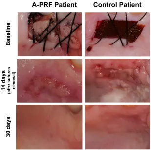
2. Palatal wound, as a result of free gingival graft harvesting, was occupied by two A-PRF clot membrane after careful positioning, and criss-cross sutures were done to hold it in position (Figure 1).
For the control group, the surgical wound as a result of the graft collecting was filled with lyophilized hydrolyzed collagen sponge (Technew™, Rio de Janeiro, Brazil) (Figure 1).
Figure 1. Surgical technique for application of advanced platelet-rich fibrin (A-PRF), and evaluation of postoperative epithelialization in the PRF and test groups at baseline, 14 days and 30 days after surgery.
Two days after surgery, all patients returned the protective splint to prevent continued usage. Palatal sutures were removed at seven days after surgery, and receiving zone sutures at 14 days. The control protocol consisted in the assessment of the palatal wound healing area and evaluation of postoperative pain and discomfort sensation.
Primary outcomes: epithelialization of the palatal wound, wound healing area and percentage of reduction
Follow-up control protocol included palatal wound measuring, using a CP-12 probe, and photography of the wound healing area. Additionally, post-surgery, postoperative complications (hemorrhage, suppuration, edema and necrosis) were searched and properly recorded. For the epithelialization clinical appraisal, it was used the wound closure visual criteria of (17).
Secondary outcomes: postoperative pain and discomfort sensation
Patient’s pain and discomfort perceptions were rated using the visual analogue scale (VAS) score (0-10), and registered during the follow-up period.
Sample size and statistical analysis
The sample size was calculated to provide a power 1-β = 90%, with α = 5%, to detect the difference in the proportion of patients who exhibited epithelialization after three weeks among patients whose palatal wounds were treated with L-PRF (test group) and with an absorbable gelatin sponge (control group), as reported in (9). Under this principle a minimum of 7 patients per group (14 in total) would have been required. To avoid a loss of statistical power, as a consequence of patient drop-out, a total of 30 patients, who met the inclusion criteria, were enrolled to participate.
Results
Demographic data
Twenty-five patients participated in this prospective randomized case-control study. The mean age was 36.4 ± 14.9 years (range 19-65 years) and the female/male ratio was 16/9.
Palatal surgical wound area decrease, percentage of reduction and epithelialization
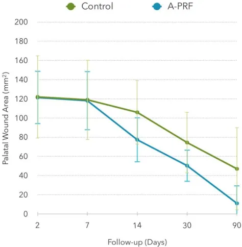
Results for the palatal wound area, as a function of the follow-up period, are displayed in Table 1 and represented in Figure 2. On the second day sutures where intact, and in the test group all A-PRF membranes where adherent to the palate.
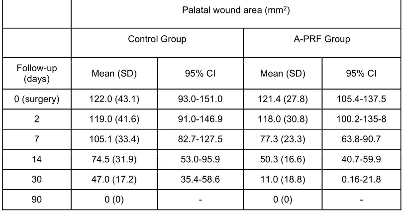
Table 1. Palatal wound area (mm2), presented as mean (standard deviation - SD) and estimated mean (95% CI), evaluated at surgery and along the follow-up visits, for the control (n=11) and A-PRF (n=14) groups.
Palatal wound area (mm2)
Control Group A-PRF Group
Follow-up
(days) Mean (SD) 95% CI Mean (SD) 95% CI
0 (surgery) 122.0 (43.1) 93.0-151.0 121.4 (27.8) 105.4-137.5
2 119.0 (41.6) 91.0-146.9 118.0 (30.8) 100.2-135-8
7 105.1 (33.4) 82.7-127.5 77.3 (23.3) 63.8-90.7
14 74.5 (31.9) 53.0-95.9 50.3 (16.6) 40.7-59.9
30 47.0 (17.2) 35.4-58.6 11.0 (18.8) 0.16-21.8
90 0 (0) - 0 (0) -
Table 2 presents the mean percentage reduction area of the palatal wound during the follow-up visits. A-PRF grofollow-up shows a significant larger decrease percentage than the control group, at 7 (p<0.001), 14 (p=0.009) and 30 days (p<0.001), conversely to the first two days after surgery, where no statistical significant difference was found (p=0.687). Maximum difference between groups was attained at 30 days (91.5% for A-PRF vs. 59.0% for the control group). At 90 days, both groups showed total recovery.
Table 2. Percentage decrease of the palatal wound area, presented as mean (standard deviation - SD), along the follow-up visits, for the control (n=11) and A-PRF (n=14) groups.
Palatal wound reduction area (%)
Follow-up
(days) Control Group Mean (SD) A-PRF Group Mean (SD) p *
2 2.0 (5.1) 2.9 (10.7) 0.687
7 12.9 (12.2) 36.4 (12.2) < 0.001
14 36.6 (20.4) 58.0 (14.2) 0.009
30 59.0 (14.3) 91.5 (14.6) < 0.001
90 100.0 (0.0) 100 (0.0) -
The baseline palatal depth ranged from 3 to 5 mm (control group) and 2 to 6 mm (A-PRF group), with an average of 3.8 (0.6) and 3.6 (1.1) mm, respectively. Moreover, when assessing the healing rate, via the the percentage reduction of wound area, as a function of the depth of the palate, a significant negative correlation occurred for the A-PRF group, at 30 days’ follow-up (Spearman’s correlation coefficient = -0.61, p = 0.021), indicating that in palates with greater baseline thickness, A-PRF accelerated the recovery. Additionally, correlation of palatal depth with the postoperative pain sensation was not found to be significant in either of the groups.
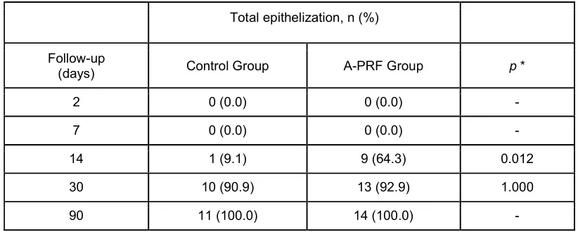
Table 3 displays the epithelization rate for both groups, along with the follow-up period (Figure 1). At 14 days a significant difference in the proportion of patients showing total epithelization was found: 64.3% for A-PRF vs. 9.1% for the control group (p=0.012). At 30 days, total epithelization was observed in more than 90% of the patients (92.9% for A-PRF vs. 90.9% for the control group), the difference being not statistically significant (p=1.000).
Table 3 Total epithelization, presented as n (%), along the follow-up visits, for the control (n=11) and A-PRF (n=14) groups
Total epithelization, n (%)
Follow-up
(days) Control Group A-PRF Group p *
2 0 (0.0) 0 (0.0) -
7 0 (0.0) 0 (0.0) -
14 1 (9.1) 9 (64.3) 0.012
30 10 (90.9) 13 (92.9) 1.000
90 11 (100.0) 14 (100.0) -
* Fisher’s exact test
Postoperative complications
Postoperative complications were identified at the second day (hemorrhage: one patient in the control group and two patients in the A-PRF group) and at the seventh day, in the control group, one patient exhibited necrosis of donor site margins.
Postoperative pain experience
Table 4. Postoperative pain experience, recorded through a VAS and presented as median (interquartile range - IQR), along the follow-up visits, for the control (n=11) and A-PRF (n=14) groups.
Postoperative pain experience (VAS)
Control Group A-PRF Group
Follow-up
(days) Median (IQR) Min.-Max. Median (IQR) Min.-Max. p *
2 2.0 (2) 0-9 0.0 (1) 0-7 0.013
7 1.0 (2) 0-9 0.0 (0) 0-0 -
14 0.0 (0) 0-5 0.0 (0) 0-0 -
30 0.0 (0) 0-0 0.0 (0) 0-0 -
90 0.0 (0) 0-0 0.0 (0) 0-0 -
* Mann-Whitney test
Discussion
In this prospective randomized clinical study, we used A-PRF to accelerate palatal wound healing, as a result of free gingival graft. The main results demonstrate that A-PRF benefits palatal tissue recovery up to 30 days after surgery, and, as expected, from there up to 90 days this difference vanishes. Thus, we can state that A-PRF as a palatal dressing enhances the patient's surgery experience by improving healing of the donor site. These outcomes are in accordance with previous studies that have investigated other types of PRF for the same purpose (9,13,16,20).
Nevertheless, although from the short-term view A-PRF significantly promotes the re-epithelialization of the wound (on day 14), from a long-term perspective it was not shown to be significant, as observed at 30 days after surgery. In fact, being the first time A-PRF is used in this palatal bandage procedure, there are no forms of comparison other than other types of PRF. Plausibly, this difference may be due to the smaller thickness / quantity of the PRF membranes used in our study, since similar results have been found using single membranes of PRF and T-PRF (13,20), and are different described in Femminella et al. (9), that used quadruple layer clots. Therefore, apparently the thicker the PRF the lower its degradation and the faster the re-epithelialization process. However, this should be confirmed with a more comprehensive and dedicated randomized clinical trial.
Noteworthy, deeper palates showed better healing results using A-PRF, which can be explained by the wealth of growth factors present in this type of platelet-concentrated biomaterial. Yet, according to Wyrębek et al. (22)pain had no association with the free gingival graft length and width, although they did not consider the initial thickness of the palate, and in our investigation the thickness of the graft was not measured, which may explain the powerlessness with post-surgery pain.
Since its introduction, A-PRF has been extensively studied in its composition, biocompatibility and performance in vitro (11,12,15,21). Clinically, A-PRF exhibited promising characteristics as biomaterial for ridge preservation (4) and regenerative periodontal therapy (14), although its limits are little explored. Regarding the limitations of this study, A-PRF maintains some major disadvantages of the PRF technique. PRFs require blood collection and careful handling, there is vague knowledge on the leukocytes, platelets and growth factors concentration on the clots, and the established protocols are highly variable. Additionally, while these results are encouraging, we can’t forget the fact that this study design lacks RCT methodology, such as random sequence generation and allocation concealment, since the remaining aspects were covered. Also, patient-centered outcome measures, like oral health-related quality of life, and thorough medication dosage should be pondered in future investigations, since these surgical adjunct healing procedures aim to improve patient experience periodontal surgeries.
Conclusion
Despite the limitations of this study, the results suggest that A-PRF membranes accelerate the healing process by promoting a higher reduction along the recovery period and an apparent less painful postoperative period.
Ethics statement/confirmation of patients’ permission
This study was approved by the Ethics Committee of Egas Moniz. All participants gave their signed informed consent.
Conflicts of interest
All authors declare no personal, commercial and financial relationships.
Funding information
Clinical Relevance
The use of A-PRF dressings may be a simple and effective method of accelerating the healing process and reducing post-operative discomfort associated with free gingival graft harvesting.
References
1. Bennani, V, Ibrahim H, Al-Harthi L LK. The periodontal restorative interface : esthetic considerations. Perio 2000 2017; 74: 74–101.
2. Castro AB, Meschi N, Temmerman A, et al. Regenerative potential of leucocyte- and platelet-rich fibrin. Part A: intra-bony defects, furcation defects and periodontal plastic surgery. A systematic review and meta-analysisJ Clin Periodontol 2017; 44: 67–82. 3. Castro AB, Meschi N, Temmerman A, et al. Regenerative potential of leucocyte- and
platelet-rich fibrin. Part B: sinus floor elevation, alveolar ridge preservation and implant therapy. A systematic review. J Clin Periodontol 2017; 44: 225–234.
4. Clark D, Rajendran Y, Paydar S, et al. Advanced platelet-rich fibrin and freeze-dried bone allograft for ridge preservation: A randomized controlled clinical trial. J
Periodontol 2018; 89: 379–387.
5. Ding H, Yuan JQ, Zhou JH, et al. Systematic review and meta-analysis of application of fibrin sealant after liver resection. Curr Med Res Opin 2013; 29: 387–394.
6. Dohan DM, Choukroun J, Diss A, et al. Platelet-rich fibrin (PRF): A second-generation platelet concentrate. Part III: Leucocyte activation: A new feature for platelet
concentrates? Oral Surgery, Oral Med Oral Pathol Oral Radiol Endodontology 2006; 101.
7. Dragan IF, Ms DDS, Paterno L, et al. Clinical Outcomes of Comparing Soft Tissue Alternatives to Free Gingival Graft: A Systematic Review and Meta-Analysis. J Evid
Based Dent Pract 2017; 17: 370–380.e3.
8. von Elm E, Altman DG, Egger M, Pocock SJ, Gøtzsche PC, Vandenbroucke JP. The strengthening the reporting of observational studies in epidemiology (STROBE) statement: Guidelines for reporting observational studies. Int J Surg 2014; 12: 1495– 1499.
9. Femminella B, Iaconi MC, Di Tullio M, et al. Clinical Comparison of Platelet-Rich Fibrin and a Gelatin Sponge in the Management of Palatal Wounds After Epithelialized Free Gingival Graft Harvest: A Randomized Clinical Trial. J Periodontol 2016; 87: 103–113. 10. Fredes F, Pinto J, Pinto N, et al. Potential Effect of Leukocyte-Platelet-Rich Fibrin in
Bone Healing of Skull Base: A Pilot Study. 2017 Epub.
11. Fujioka-Kobayashi M, Miron RJ, Hernandez M, Kandalam U, Zhang Y, Choukroun J. Optimized Platelet-Rich Fibrin With the Low-Speed Concept: Growth Factor Release, Biocompatibility, and Cellular Response. J Periodontol 2017; 88: 112–121.
12. Ghanaati S, Booms P, Orlowska A, et al. Advanced Platelet-Rich Fibrin: A New Concept for Cell-Based Tissue Engineering by Means of Inflammatory Cells. J Oral
13. Kulkarni MR, Thomas BS, Varghese JM, Bhat GS. Platelet-rich fibrin as an adjunct to palatal wound healing after harvesting a free gingival graft: A case series. J Indian Soc
Periodontol 2014; 18: 399.
14. Lei L, Yu Y, Ke T, Sun W, Chen L. The application of three-dimensional printing model and platelet-rich fibrin (PRF) technology in guided tissue regeneration surgery for severe bone defects.2018 Epub.
15. Masuki H, Okudera T, Watanebe T, et al. Growth factor and pro-inflammatory cytokine contents in platelet-rich plasma (PRP), plasma rich in growth factors (PRGF),
advanced platelet-rich fibrin (A-PRF), and concentrated growth factors (CGF). Int J
Implant Dent 2016; 2: 19.
16. Ozcan M, Ucak O, Alkaya B, Keceli S, Seydaoglu G, Haytac M. Effects of Platelet-Rich Fibrin on Palatal Wound Healing After Free Gingival Graft Harvesting: A
Comparative Randomized Controlled Clinical Trial. Int J Periodontics Restorative Dent 2017; 37: e270–e278.
17. Silva CO, Ribeiro ÉDP, Sallum AW, Tatakis DN. Free Gingival Grafts: Graft Shrinkage and Donor-Site Healing in Smokers and Non-Smokers. J Periodontol 2010; 81: 692– 701.
18. Theys T, Hoylandt A Van, Broeckx C, et al. Plasma-rich fibrin in neurosurgery : a feasibility study2018 Epub.
19. Tomar N, Singh R, Jain G, Kaushik M, Dureja D. Enhancement of healing of donor hard palate site using platelet-rich fibrin. J Curr Res Sci Med 2016; 2: 132.
20. Ustaoğlu G, Ercan E, Tunali M. The role of titanium-prepared platelet-rich fibrin in palatal mucosal wound healing and histoconduction. Acta Odontol Scand 2016; 74: 558–564.
21. Watanabe T, Isobe K, Suzuki T, et al. An Evaluation of the Accuracy of the Subtraction Method Used for Determining Platelet Counts in Advanced Platelet-Rich Fibrin and Concentrated Growth Factor Preparations. Dent J 2017; 5: 7.
22. Wyrebek B, Gorski B, Gorska R. Patient morbidity at the palatal donor site depending on gingival graft dimension.Dent Med Probl 2018; 55: 153–159.
23. Zuhr O, Bäumer D, Hürzeler M. The addition of soft tissue replacement grafts in plastic periodontal and implant surgery: Critical elements in design and execution. J Clin