THE EFFECT OF DONOR LIVER ORGAN PRESERVATION AND
REPERFUSION TECHNIQUES ON THE EXPRESSION OF
CELLULAR ADHESION MOLECULES
Mohamed Mohamed Zaki Mohamed El-Wahsh
A THESIS SUBMITTED FOR THE DEGREE OF
DOCTOR OF PHILOSOPHY
IN THE UNIVERSITY OF LONDON
2002
UNIVERSITY DEPARTEMENT OF SURGERY
ProQuest Number: U643754
All rights reserved
INFORMATION TO ALL USERS
The quality of this reproduction is dependent upon the quality of the copy submitted.
In the unlikely event that the author did not send a complete manuscript
and there are missing pages, these will be noted. Also, if material had to be removed, a note will indicate the deletion.
uest.
ProQuest U643754
Published by ProQuest LLC(2016). Copyright of the Dissertation is held by the Author.
All rights reserved.
This work is protected against unauthorized copying under Title 17, United States Code. Microform Edition © ProQuest LLC.
ProQuest LLC
789 East Eisenhower Parkway P.O. Box 1346
/
dedicate this thesis to my family, my supervisors Keith Rolles, Brian
Davidson and Barry Fuller in gratitude fo r all their support.
ACKNOWLEDGEMENTS
I would like to express my thanks to Professor Paul Dhillon from the
Histopathology Department at the Royal Free Hospital for teaching me the
rudiments of immunohistochemistry and pathology, and keeping an eye on
my histological interpretations. I owe a great debt of gratitude to many
members of the University Department of Surgery at the Royal Free
Hospital; to Alex Seifalian for introducing me to Laser Doppler Flowmetry
and for passionate debate about what it all meant; Karen Cheetham for
support and guidance in using laboratory facilities.
I would like also to thank Duncan Moore from Comparative Biology Unit at
the Royal Free Hospital for instructing me in the techniques of surgery,
ABSTRACT
Ischaemia/reperfusion (l/R) injury has a major influence on the outcome of liver
transplantation. The cell adhesion molecule ICAM-1 has a central role in the interaction
between polymorphonuclear neutrophils (PMNs) and sinusoidal endothelial cells (SEC's)
which is fundamental to the development of l/R injury. This thesis has investigated the effect
of liver preservation and reperfusion on ICAM-1 expression in both animal experimental
models and human liver transplantation. Factors which are likely to induce ICAM-1 were
analysed including endotoxic shock, warm and cold ischaemia and l/R injury using an animal
model of lobar l/R and an isolated perfused liver circuit.
In the rat model of endotoxaemia ICAM-1 expression was markedly induced. Warm
ischaemia for periods up to 45minutes or cold ischaemia up to 8 hours did not induce
ICAM-1 expression on the sinusoidal endothelium but it was markedly induced with longer periods.
ICAM-1 expression was found to correlate with biochemical evidence of hepatocyte damage
(ALT& AST levels) and histological changes in the liver. l/R injury had a more marked effect
on ICAM-1 expression than ischaemia alone. ICAM-1 expression was also induced in the non
ischaemic liver lobe. The influence of adhesion molecule induction on the liver
microcirculation was investigated in vivo using laser Doppler flowmetry. Increased SEC's
ICAM-1 expression was directly associated with impairment of the hepatic microcirculation.
Adhesion molecule induction is associated with activation of
inflammatory cytokines and the formation of oxygen free radicals. The effect of adding the free
radical scavenger reduced glutathione (GSH) to the liver perfusate on ICAM-1 expression
was investigated in an ex vivo perfused circuit. Isolated rat livers were perfused on bench with oxygenated buffer in a non-recirculating technique. GSH had no significant effect on the
histological grading of l/R injury. However, SEC's ICAM-1 expression was significantly reduced
in GSH perfused livers with cold ischaemia up to 16 hours but not with 24 hours.
The timing and significance of ICAM-1 induction was then investigated in 83 patients
undergoing orthotopic liver transplantation (OLT). Liver biopsies were assessed for ICAM-1
and compared with histological evidence of l/R injury, biochemical indicators of early graft
function and the incidence of acute cellular rejection. ICAM-1 induction following graft
TABLE OF CONTENTS
DEDICATION AND ACKNOWLEDGMENTS... 2
ABSTRACT... 3
LIST OF FIGURES... 9
LIST OF PLATES... 10
LIST OF TABLES... 11
LIST OF ABBREVIATIONS...
14CHAPTER 1: INTR O D U CTIO N AND REVIEW OF LITER A TU R E 16 1.1 Liver transplantation... 16
1.1.1 Experimental Background...16
1.1.2 Liver transplantation in Human... 16
1.2 Liver graft preservation...19
1.3 University of Wisconsin (UW) solution... 24
1.4 The Clinical problem...26
1.4.1 Preservation injury (ischaemia injury)... 26
1.4.2 Reperfusion injury...30
1.5 Role of Kupffer cells in organ preservation and ischaemia/ reperfusion injury...32
1.6 Leukocytes and ischaemia/reperfusion injury...34
1.7 Free Oxygen Radicals (FRO)... 37
1.8 Reduced Glutathione... 38
1.9 Cell Surface Receptor... 40
1.9.1 Immunoglobulin Superfamily... 41
1.9.2 Molecular characterisation of Intercellular Adhesion Molecule-1 42 1.9.3 ICAM-1 Ligand...42
1.9.4 Tissue distribution of ICAM-1... 44
1.9.5 ICAM-1 expression in general... 44
1.9.6 ICAM-1 expression in normal and transplanted liver... 45
1.9.7 Effect of ischaemia/reperfusion on ICAM-1 expression... 47
CHAPTER 2: MATERIALS AND METHODS... 50
2.1 Experimental study... 50
2.1.1 Animals... 50
2.1 .2 Surgical procedures... 50
2.1.3 Standard liver function tests...53
2.1.4 Liver biopsies...53
2.1.5 Tissue preparation... 53
2.1.6 Immunohistochemistry staining... 54
2.1.7 Assessment of ischaemia and ischaemia/reperfusion injury.... 55
2.1.8 Measurement of flow of the hepatic microcirculation by Laser Doppler Flowmetry (LDP)... 56
2 .2 Clinical study... 58
2.2.1 Recipients... 58
2.2 .2 Donors... 58
2.2.3 Surgical procedures...59
2.2.4 GSH addition to the wash out solution...61
2.2.5 Liver allograft biopsies... 61
2.2.6 Immunohistochemistry staining... 62
2.2.7 Grading of graft acute cellular rejection...64
2.2.8 Graft function tests... 64
2.3 Statistical analysis... 65
CHAPTER 3: A STUDY ON LIVER ICAM-1 INDUCTION INCLUDING SEPSIS, WARM ISCHAEMIA, COLD ISCHAEMIA AND REPERFUSION INJURY...66
3.1 Introduction and objective...66
3.2 Methodoiogy... 68
3.2.1 Experimental groups... 68
3.2.2 Surgical procedures... 69
3.2.3 Investigation of ICAM-1 expression...71
3.3 Results...71
3.3.1 Effect of intraperitoneal injection of LPS toxin on ICAM-1 expression... 71
3.3.2 Effect ischaemia on ICAM-1 expression...72
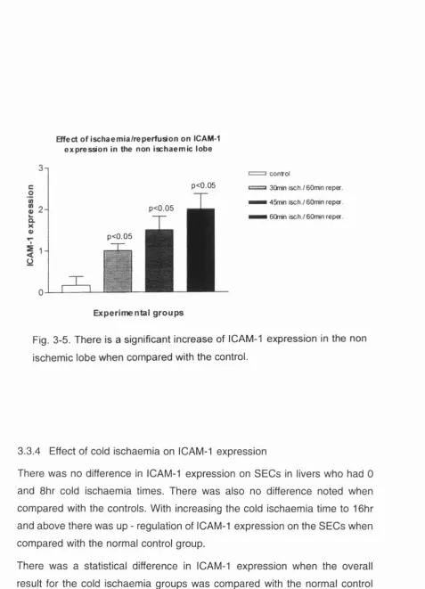
3.3.3 Effect of warm ischaemia/reperfusion on ICAM-1 expression., 76 3.3.4 Effect of cold ischaemia on ICAM-1 expression... 79
3.3.5 Effect of on bench reperfusion on ICAM-1 expression... 81
3.3.6 Effect of GSH addition to the liver perfusate on ICAM-1 expression... 81
CHAPTER 4: A STUDY ON THE EFFECT OF ISCHAEMIA AND ISCHAEMIA/REPERFUSION ON LIVER HISTOLOGY
AND ITS CORRELATION WITH ICAM-1 EXPRESSION 88
4.1 Introduction and objective... 88
4.2 Methodology... 89
4.2.1 Experimental groups...89
4.2.2 Surgical procedures... 90
4.2.3 Assessment of ischaemia and ischaemia/reperfusion injury... 91
4.2.4 Investigation of ICAM-1 expression... 91
4.3 Results... 92
4.3.1 Effect of warm ischaemia on liver histology and ICAM-1 expression... 92
4.3.2 Effect of warm ischaemia/reperfusion on liver histology and ICAM-1 expression... 94
4.3.3 Effect of cold ischaemia on liver histology and ICAM-1 expression...97
4.3.4 Effect of on bench reperfusion {in vitro) on liver histology and ICAM-1 expression... 101
4.3.5 Effect of GSH addition to the liver perfusate on histology and ICAM-1 expression... 107
4.4 Discussion...109
CHAPTER 5: CORRELATION OF HEPATOCELLULAR INJURY AND ADHESION MOLECULE ACTIVATION WITH LIVER ISCHAEMIA AND ISCHAEMIA/REPERFUSION INJURY... m 5.1 Introduction and objective...m 5.2 Methodology... 112
5.2.1 Experimental groups...112
5.2.2 Surgical procedure... 112
5.2.3 Blood samples...113
5.2.4 Liver biopsies...113
5.2.5 Investigation of ICAM-1 expression...113
5.3 Results...114
5.3.1 Control group... 114
5.3..2 Effect of warm ischaemia on ALT & AST and ICAM-1 expression 114
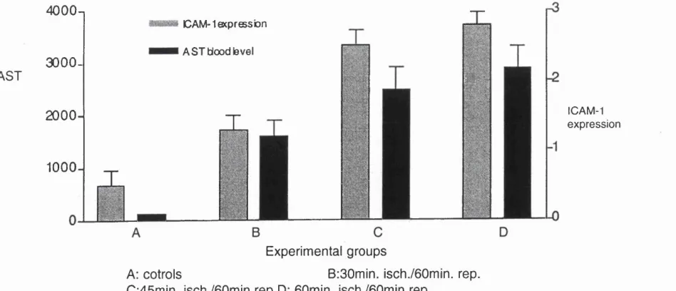
5.3.3 Effect of warm ischaemia/reperfusion on ALT & AST and ICAM-1 expression...116
CHAPTER 6 : THE CORRELATION BETWEEN FLOW OF HEPATIC MICROCIRCULATION AND ADHESION MOLECULE ACTIVATION FOLLOWING ISCHAEMIA AND
ISCHAEMIA/REPERFUSION... 123
6.1 Introduction and objective... 123
6 .2 Methodology... 124
6.2.1 Experimental groups... 124
6.2.2 Surgical procedure...125
6.2.3 Liver biopsies... 125
6.2.4 Investigation of ICAM-1 expression... 125
6.2.5 Measurement of hepatic microcirculation by Laser Doppler Flowmetry... 126
6.3 Results... 126
6.3.1 Control group... 126
6.3.2 Effect of warm ischemia on hepatic microcirculation and ICAM-1 expression... 126
6.3.3 Effect of warm ischaemia/reperfusion on hepatic microcirculation and ICAM-1 expression... 128
6.4 Discussion...130
CHAPTER 7: EFFECT OF WARM AND COLD ISCHAEMIA AND FREE RADICAL SCAVENGER (GLUTATHIONE) ON MARKERS OF l/R INJURY AND ACUTE CELLULAR REJECTION IN CLINICA HEPATIC TRANSPLANTATION...133
7.1 Introduction and objectives...133
7.2 M ethodology...134
7.2.1 Patients... 134
7.2.2 Surgical procedures... 135
7.2.3 Liver allograft biopsies... 135
7.2.4 Blood samples... 135
7.2.5 Assessment of Ischaemia/reperfusion injury (l/R)... 135
7.3 Results... 136
7.3.1 Effect of l/R on liver functions during the first 5 post-operative days... 136
7.3.2 Effect of l/R on acute cellular rejection (ACR)... 135
7.3.3 Effect of cold ischaemia time (CIT) on l/R and ACR...133
7.3.4 Effect of warm ischaemia time (WIT) on l/R and ACR...142
7.3.5 Effect of GSH addition to the wash out solution on l/R and ACR... 142
CHAPTER 8: A STUDY ON HUMAN LIVER ICAM-1 INDUCTION AND ITS CORRELATION WITH REPERFUSION INJURY AND ACUTE
CELLULAR REJECTION... 150
8.1 Introduction and objectives...150
8.2 Methodology... 151
8.2.1 Surgical procedures... 151
8.2.2 Reduced glutathione addition...151
8.2.3 Liver allograft biopsies... 152
8.2.4 Immunohistochemistry staining...152
8.3 Results... 153
8.3.1 ICAM-1 expression in controls... 153
8.3.2 ICAM-1 expression in liver allograft biopsies... 153
8.3.3 Effect of cold ischaemia time (CIT) on ICAM-1 expression 155
8.3.4 Effect of warm ischaemia time (WIT) on ICAM-1 expression... 153
8.3.5 Effect of reduced glutathione addition to the wash out solution on ICAM-1 expression...159
8.3.6 Correlation between ICAM-1 expression and ischaemia/ reperfusion injury...159
8.3.7 Correlation between ICAM-1 expression and acute cellular rejection... 160
8.4 Discussion... 162
CHAPTER 9: Thesis Discussion...166
9.1 Analysis of methodoiogy... 166
9.1.1 Measurement of ICAM-1 expression by Immunohistochemistry 166 9.1.2 Experimental animal models... 167
9.1.3 Clinical evaluation of ICAM-1 expression... 167
9.1.4 Experimental measurement of the hepatic microcirculation by Laser Doppler Flowmetry... 168
9.1.5 Assessment of ischaemia/reperfusion injury in animal model by histological analysis... 168
9.1.6 Assessment of ischaemia/reperfusion injury in human OLT by biochemical analysis...170
9.2 Hypothesis and Aims...170
9.3 Conclusions, Reflections and Future perspectives... 176
List of publications;... 182
References... 184
LIST OF FIGURES
CHAPTER 1
Figure 1-1
Figure 1-2
Figure 1-3
Figure 1-4
Effect of storage and reperfusion on hepatocytes, sinusoidal endothelial cells (SEC's) and Kupffer cells (Kc).
A scheme representing mechanism of leukocytes migration at an inflammatory site.
ICAM-1 structure.
A scheme representing factors involved in the mechanism of ICAM-1 up-regulation.
CHAPTER 3
Figure 3-1
Figure 3-2
Figure 3-3
Figure 3-4
Figure 3-5
Figure 3-6
Figure 3-7
Effect of warm ischaemia on ICAM-1 expression.
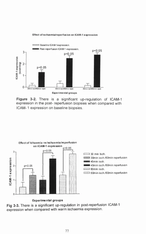
Effect of ischaemia/reperfusion on ICAM-1 expression.
Effect of ischaemia vs ischaemia/reperfusion on ICAM-1 expression.
Effect of ischaemia vs ischaemia/reperfusion on ICAM-1 expression.
Effect of ischaemia/reperfusion on ICAM-1 in non ischaemic liver lobe.
Comparison between the effect of ischaemia/reperfusion on ICAM-1 expression in non ischaemia lobe and
ischaemic lobe.
Effect of cold ischaemia on ICAM-1 expression.
CHAPTER 4
Figure 4-1
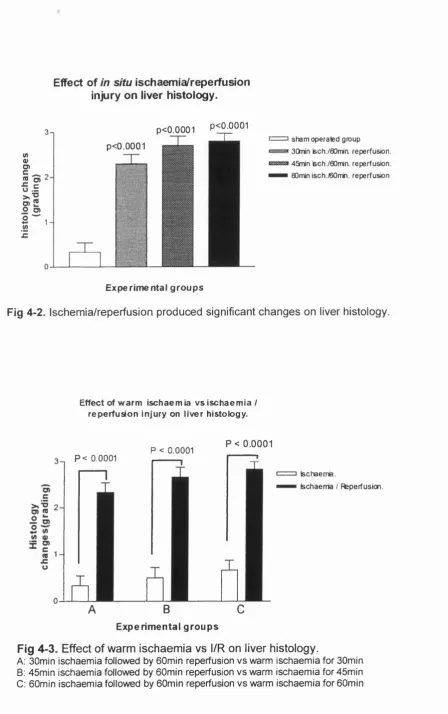
Figure 4-2
Figure 4-3
Figure 4-4
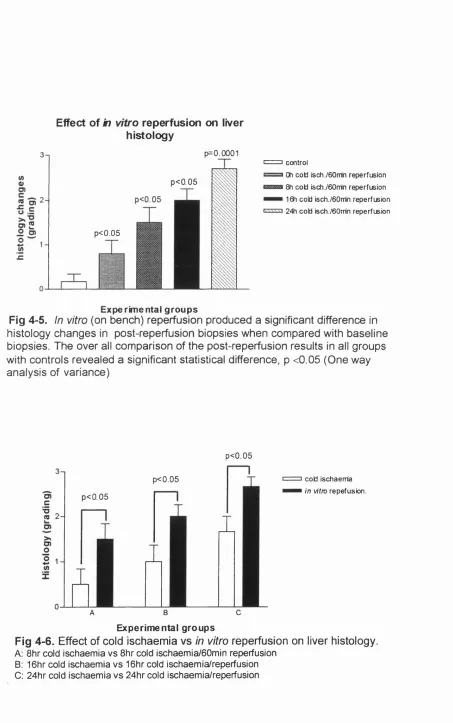
Figure 4-5
Figure 4-6
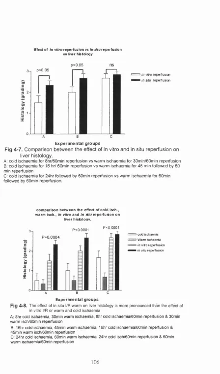
Figure 4-7
Figure 4-8
Effect of warm ischaemia on liver histology.
Effect of in situ ischaemia/reperfusion injury on liver histology.
Comparison of the effect of warm ischaemia vs ischaemia/reperfusion on liver histology.
Effect of cold ischaemia on liver histology.
Effect of cold ischaemia/reperfusion on liver histology.
Effect of cold ischaemia vs cold ischaemia/reperfusion on liver histology.
Effect of cold ischaemia/reperfusion vs warm ischaemia/reperfusion on liver histology.
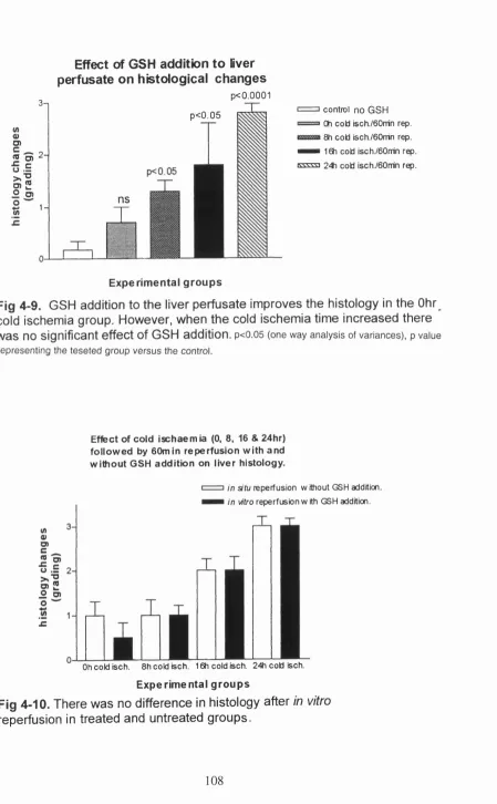
Figure 4-9
Figure 4-10
CHAPTER 5
ischaemia, cold ischaemia/reperfusion and warm ischaemia/reperfusion on liver histology.
Effects of GSH addition to liver perfusate on histological changes.
Effect of cold ischaemia followed by reperfusion with and without GSH addition on liver histology.
Figure 5-1
Figure 5-2
CHAPTER 6
Correlation between ICAM-1 and ALT blood level, post reperfusion.
Correlation between ICAM-1 and AST blood level, post reperfusion.
Figure 6-1 Effect of warm ischaemia on blood flow in the hepatic microcirculation.
Figure 6-2 Effect of ischaemia/reperfusion on blood flow in the hepatic microcirculation.
CHAPTER 8
Figure 8-1 ICAM-1 expression in human liver allograft.
LIST OF PLATES
CHAPTER 2
Plate 2-1 Needle biopsy from liver allograft after overnight storage.
CHAPTER 3
Plate 3-1
Plate 3-2
Plate 3-3
Plate 3-4
Faint ICAM-1 staining on SEC's in normal rat liver.
Mild ICAM-1 expression on SEC's.
Moderate ICAM-1 expression on SEC's.
Intense ICAM-1 expression on SEC's and hepatocytes.
CHAPTER 4
Plate 4-1
Plate 4-2
Histology of normal rat liver.
Plate 4-3
Plate 4-4
Plate 4-5
Plate 4-6
Effect of warm iscfiaemia (up to 90 minutes) on rat liver fiistology.
Effect of warm iscfiaemia (more tfian 90 minutes) on rat liver histology.
Effect of warm ischaemia/reperfusion on rat liver histology.
Effect of warm ischaemia/reperfusion on rat liver histology.
CHAPTER 7
Plate 7-1
Plate 7-2
Plate 7-3
Plate 7-4
Histology of accepted liver allograft.
Mild acute cellular rejection.
Moderate acute cellular rejection.
Severe acute cellular rejection.
CHAPTER 8
Plate 8-1
Plate 8-2
Plate 8-3
Plate 8-4
CHAPTER 1
Table 1-1
Table 1-2
Table 1-3
No ICAM-1 staining in normal human liver.
Mild ICAM-1 expression on SEC's.
Moderate ICAM-1 expression on SEC's.
Intense ICAM-1 expression on SEC's and hepatocytes.
LIST OF TABLES
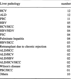
Indications for liver transplantation.
Balanced electrolytic fluid used to perfuse canine kidneys.
Composition of University of Wisconsin solution.
CHAPTER 2
Table 2-1
Table 2-2
Table 2-3
Table 2-4
Krebs-Henseleit bicarbonate buffer solution.
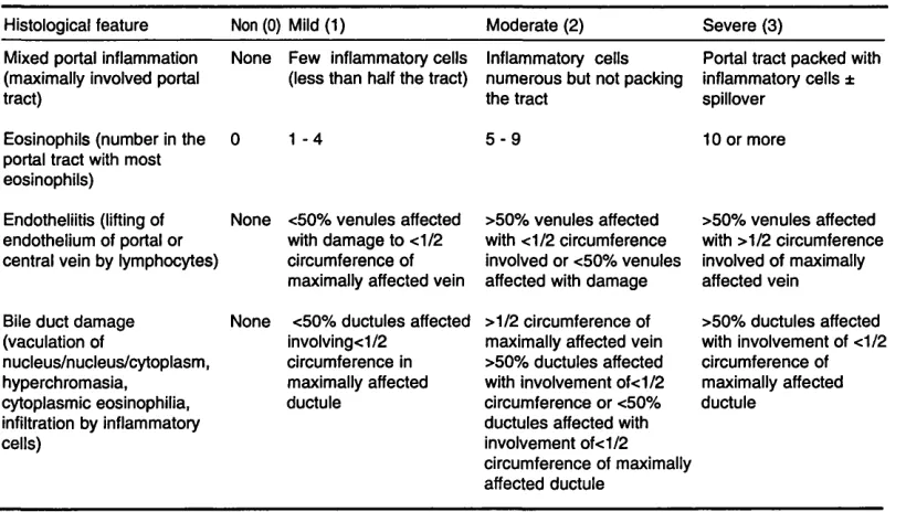
scorring of ICAM-1 expression.
Underlying liver pathology of the transplanted patients.
The Royal Free Hospital scoring system of the features of cellular rejection.
CHAPTER 3
Table 3-2
Table 3-3
Table 3-4
Table 3-5
Table 3-6
Table 3-7
CHAPTER 4
Table 4-1
Table 4-2
Table 4-3
Table 4-4
Table 4-5
Table 4-6
CHAPTER 5
administration.
Baseline ICAM-1 expression and following warm ischaemia
ICAM-1 expression at baseline and post-reperfusion.
Effect of cold ischaemia on ICAM-1 expression.
ICAM-1 expression at baseline and after ischaemia/ reperfusion.
ICAM-1 expression in the GSH treated group.
Post-reperfusion ICAM-1 expression in the GSH treated and untreated group.
Histological score of warm ischaemia.
Comparison between the effect of cold ischaemia vs warm ischaemia on liver histology.
Effect of warm ischaemia/reperfusion on liver histology.
Histological score of cold ischaemic liver injury.
Effect of cold ischaemia/warm reperfusion on liver histology.
Effect of reduced glutathione addition to the liver perfusate on liver histology.
Table 5-1
Table 5-2
Table 5-3
Table 5-4
Table 5-5
Table 5-6
Table 5-7
CHAPTER 6
Table 6-1
Table 6-2
Table 6-3
Table 6-4
Effect of warm ischaemia on ALT level.
Effect of warm ischaemia on AST level.
ICAM-1 expression at baseline biopsies and the end of warm ischaemia time.
Effect of ischaemia/reperfusion on ALT level.
Effect of ischaemia/reperfusion on AST leve.l
Effect of warm ischaemia on ALT and AST levels vs the effect of ischaemia/reperfusion.
ICAM-1 expression on baseline and post-reperfusion liver biopsies.
Hepatic microcirculation (HM) at baseline and end of ischaemia.
ICAM-1 expression at baseline biopsies and the end of warm ischaemia.
HM at baseline and after ischaemia/reperfusion.
CHAPTER 7
Table 7-1
Table 7-2a
Table 7-2b
Table 7-3a
Table 7-3b
Table 7-4
Table 7-5
Table 7-6
Table 7-7
Table 7-8
Table 7-9
Table 7-10
Table 7-11
Table 7-12
Table 7-13
Table 7-14
Table 7-15
CHAPTER a
Table 8-1
Table 8-2
Table 8-3
Table 8-4
Table 8-5
Table 8-6
Table 8-7
Scoring parameters of ischaemia/reperfusion injury.
AST level in GSH and saline groups.
ALT level in GSH and saline groups.
Bilirubin level in GSH and saline groups.
FT level in GSH and saline groups.
Comparison of ischaemia/reperfusion injury with grade of early rejection.
Comparison of ischaemia/reperfusion injury with grade of late rejection.
Minimum and maximum cold ischaemia time.
Cold ischaemia time in accepted allografts and in grafts which developed early post-transplant mild, moderate or severe acute cellular rejection.
Cold ischaemia time in accepted allografts and in grafts which developed late post-transplant mild, moderate or severe acute cellular rejection.
Warm ischaemia time in allografts which had mild, moderate or severe ischaemia/reperfusion injury.
Warm ischaemia time and early post-transplant rejection.
Warm ischaemia time and late post-transplant rejection.
Warm ischaemia time in GSH and saline groups.
Cold ischaemia time in GSH and saline groups.
Ischaemia/reperfusion injury in GSH and saline groups.
Acute cellular rejection in GSH and saline groups.
Analysis of ICAM-1 expression in post-storage and post-reperfusion biopsies
Effect of cold ischaemia time on ICAM-1 expression on post-storage biopsies
Effect of cold ischaemia time on ICAM-1 expression on post-reperfusion biopsies
Effect of warm ischaemia time on ICAM-1 expression in post-reperfusion biopsies
ICAM-1 expression in GHS and saline groups in post storage biopsies
ICAM-1 expression in GSH and saline groups in post reperfusion biopsies
Table 8-8 Correlation between the degree of ischaemia/reperfusion injury and ICAM-1 expression in post-reperfusion allografts biopsies.
Table 8-9 Correlation between the degree of early acute cellular rejection and ICAM-1 expression in post-reperfusion biopsies.
Table 8-10 Correlation between the degree of late acute cellular rejection and ICAM-1 expression in post-reperfusion biopsies.
LIST OF ABBREVIATIONS
ACR acute cellular rejection
ADP adenosine diphosphate
ATP adenosine triphosphate
ALT alanine transaminase
ALD alcoholic liver disease
AST aspartate transaminase
Bp blood pressure
BW body weight
BT body temperature
BSA bovine serum albumin
°C degree centigrade
Ca2+ calcium ion
CIT cold ischaemia time
cr chloride ion
DAB 3,3/diaminobenzidine tetrahydrochloride dihydrate
dl decilitre
DNA deoxyribonucleic acid
EM electron microscopy
FOR free oxygen radicals
g gram
GSH reduced glutathione
GSSG glutathione disulphide
Mb haemoglobin
HR, bpm heart rate beat/minute
HBV hepatitis B virus
HCV hepatitis C virus
HCC hepatocellular carcinoma
HDV hepatitis D virus
Hz Hertz
hr hours
H202 hydrogen peroxide
IVC inferior vena cava
IL-1 interleukin-1
IL-8 interleukin-8
IP intraperitoneal
iv intravenous
l/R ischaemia reperfusion
KHz Kiiohertz
KD kilo Dalton
Kc Kupffer cells
K+ potassium ion
LDF laser Doppler Flowmetry
LPS lipopolysaccharides toxin
1 iitre
LFA-1 iymphocyte function associated antigen-1 LFA-3 iymphocyte function associated antigen-3
MgCL2 magnesium dichloride
MHC major histocompatibility complex
ml millilitre mmol millimole mW milliwatt mg miiligram min minutes mol moies
MAb monoclonal antibody
MAG myelin associated giycoproteins
n number in a single group
nm nanomoles
NCAM neyral cell adhesion molecuie
ns non significant
Na+ sodium ion
NaCL sodium chioride
NaOH sodium hydroxide
NaHC03 sodium hydrogen bicarbonate OLT orthotopic liver transplantation
02 oxygen
02 sat. oxygen saturation
0-2 superoxide radical
PMNs polymorphonuclear neutrophils pH potential of hydrogen (acidity)
PT prothrombin time
PBC primary biliary cirrhosis PSC primary sclerosing choiangitis
PMA phorbole-12-myristate acetate
PNF primary non function
RBCVF Red Blood Cells Volume Fraction
RFH Royai Free Hospitai
sec seconds
SEC's sinusoidal endothelial cells
SD Sprague Dawley rat
SE standard error
s.c. subcutaneous
IN F tumour necrosis factor
TBS tris buffered saiine
um micrometer
ul microlitre
UW University of Wisconsin
VCAM-1 vascular adhesion molecule-1
vs versus
V volt
WIT warm ischaemia time
CHAPTER 1
INTRODUCTION AND REVIEW OF LITERATURE
1.1 Liver transplantation
1.1.1 Experimental Background
The first report of experimental liver transplantation was by Welch in 1955
(Rolles et al., 1987). An auxiliary liver was accommodated in the lower
abdomen with the portal vein being supplied with systemic blood from the
inferior vena cava of the recipient and the hepatic artery anastomosed to an
iliac artery. Venous drainage was via the inferior vena cava and bile was
drained via the gall bladder into the duodenum (Rolles at a!, 1987). In 1959
and 1960 Moore and colleagues of Boston and Starzl and colleagues of
Chicago published the first report on the technique of one stage total
hepatectomy and orthotopic liver transplantation (OLT) in the dog (Caine etal,
1987). It was clear that the operation was unlikely to be successful unless a
technique was developed to prevent the damming up of blood in the
splanchnic and inferior vena cava drainage areas. Moore was able to
decompress these two systems by means of external shunts. Starzl
performed a sutured side-to-side portocaval anastomosis and then shunted
the two drainage systems through an external shunt from the femoral vein to
the external jugular vein. (Caine at a!., 1987) . Biliary drainage in the early
canine experiments was usually by means of a choledocho-duodenostomy,
the common bile duct having been ligated (Caine at al, 1987).
1.1.2 Liver Transplantation in Human
The first human OLT program performed in Denver between May and July
1963. Out the four OLT performed, one patient survived for 23 days (Starzl at
al, 1963). The next seven transplants performed in Denver, Boston and Paris
heterotopic liver transplant in a child with biliary atresia who died from biliary
leakage 13 days after transplantation (Abouna et al., 1982). Caine and
Williams initiated the procedure at Cambridge and King’s College in London
and Roy Caine performed the first successful OLT in the UK in 1968 (Caine
e ta l, 1977).
The first long-term survival after liver transplantation was in 1967. The
patient, an 18 month old child with a hepatoma, survived for 13 months before
dying from widespread tumour metastasis (Starzl etal., 1968). The late 1960s
and 1970s saw very slow progress in this field, with an overall 1 year patient
survival of only 35% (Starzl et al., 1974). Disasters were recorded using the
cystic duct for drainage, not only because it is a narrow passage for bile but
also because the cystic duct in 15% of cases enters the common hepatic duct
very low down behind the duodenum and has been inadvertently caught in the
ligature with consequent prevention of drainage (Caine et al., 1987; Collins
et al., 1982)
Several major advances in the early 1980s - the introduction of cyclosporine,
the progress in donor surgery and organ preservation, and the refinement of
the surgical technique led to improve of the results. As from February 1st 1981
through May 1982, 32 OLT were performed. The in hospital post-operative
stay period averaged 57.1 ±7.9 day. In this series 11 recipient died (3
intra-operatively and 8 post-operative).
Currently OLT has proven to be clinically useful and often life saving in a
variety of irreversible acute and chronic liver diseases for which no
satisfactory medical therapy is available (table 1-1), with more than 4000
Table 1-1. indications for iiver transplantation
I Unresectable primary malignancy of the liver - hepatocellular carcinoma
- cholangiocarcinoma
- rare nonhepatocellular or bile ductular tumors that arise within the hepatic parenchyma
- isolated hepatic metastatic disease carcinoid
pancreatic islet cell tumor
II Various types of end-stage cirrhosis - predominantly cholestatic disease
primary biliary cirrhosis primary sclerosing cholangitis biliary atresia
familial cholestatic syndromes - predominantly hepatocellular disease
chronic viral-induced liver disease chronic drug-induced liver disease alcoholic liver disease
idiopathic autoimmune liver disease - predominantly vascular disease
Budd- Chiari syndrome Veno-occlusive disease - polycystic disease
III Acute or rapidly progressive liver failure from hepatic necrosis - viral hepatitis
- drug induced liver disease (Halothane, Gold, Disulfiram, Acetaminophen) - metabolic liver disease (Wilson’s disease, Reye’s syndrome. Organic
aciduria)
IV Inborn errors of metabolism - alpha-1- antitrypsin deficiency - Wilson’s disease
- Homozygous type II hyperlipoproteinemia - Crigler-Najjar syndrom type I
- Erythropoietic protoporphyria - Urea cycle deficiencies - Glycogen storage disease - Tyrosinemia
1.2 Liver graft preservation
The viability of the organ and its maintenance is very important during any
transplant procedure and there is usually a period between the stoppage of
the circulation in the donor and re-vascularisation of the organ again in the
recipient. During this period the harvested organ may have an irreversible
harmful series of events rendering it not suitable for transplantation. This
non-reversible damage which occurs during the ischaemic period depends partly
on the metabolic requirements and pathways in the specific tissues
(Jamieson et al., 1991). In the field of kidney transplantation these problems
have been overcome with live related kidney transplantation in which the
ischaemic time is kept to a minimum by performing donor and recipient
operations in adjacent operating theatres.
During the early days of Cambridge and Kings College Hospital’s experiences
in liver transplantation, they transferred the recipient to the donor’s hospital
with all the surgical team to keep the ischaemic time to a minimum. This was
not a practical procedure (Wall e ta l., 1977). The need for organ preservation
was increased to make transplantation procedures semi-elective as seen now
in living related kidney transplantation. This semi-elective transplantation
yields substantial benefits. An unnecessary recipient laparotomy may be
prevented if the donor organ appears unsuitable for transplantation, or if the
laparotomy findings indicate that the recipient is unsuitable for transplantation.
Proper organ preservation gives time for another patient to be prepared for
transplantation. Preservation will also allow for preparation time and well
rested support in anaesthesia, nursing operating room, blood bank and
Intensive Care Unit.
In 1960 Starzl et al developed the earliest device to protect the donor liver
from ischaemia and this was based on hypothermia. Whole body cooling of
the donor was achieved by immersing in an ice bath and the body
temperature reduced to 25°C - 30°C. The liver was then perfused with cold
Ringer’s lactate solution. In this way the hepatic core temperature fell to
approximately 10°C - 20°C. Some of these livers sustained life up to 20 days in the
time in Starzl's studies resulted in acute liver failure. The liver became tense
and dark in colour, with histologic evidence of intense congestion. Death
followed few hours post- operatively due to haemorrhagic gastroenteritis as a
result of haemorrhage from small capsular tears in the distended liver, or from
acute hepatic failure due to outflow block. In the case of outflow block the
transplanted liver was unable to transmit the required venous flow due to
sequestration of the blood inside the liver. Histologic sections of the liver
revealed acute congestion and early disruption of hepatic parenchymal
architectural. The outflow block was likely to be caused by spasm of the small
intra-parenchymal hepatic veins as these vessels have extra ordinarily well
developed muscular coats in the dog.
Starzl’s technique of infusion with an electrolyte solution was the first effective
method in liver preservation. He perfused the liver in situ through the portal
vein. One liter of cooled (5 -10°C ) ringer's lactate was used for gravity
perfusion. As soon as perfusion was begun, the animal was bled to death
through a catheter inserted into the aorta. During the perfusion the interior of
the liver cooled to 10 -20°C.
In 1961 Kestens and McDermott reported a technique for isolated canine liver
perfusion in which both the portal vein and hepatic artery were ex vivo
perfused with blood and that physiological conditions of pressure and oxygen
saturation. When they tried to re-implant the liver to the hepatectomised
animal, they required an additional shunt from the splenic vein to the left
jugular vein as well as from the infra-hepatic vena cava to the right jugular
vein to prevent splanchenic pooling. This added to the complexities of the
apparatus and presented additional metabolic and coagulation problems in
the donor animals. Few years later Brettschneider and co-workers
(Brettschneider et ai., 1968) described a combined preservation technique for
extended storage of orthotopic liver homograft. They used total body cooling
of the donor animal to 30°C in an ice bath. During donor hepatectomy the
liver was further cooled by an intra-portal infusion with a balanced electrolyte
solution, which has been buffered to pH 7.45 and chilled to 2°C. These
homografts could be effectively preserved for 8 hours, they were put into cold
blood through the portal vein and hepatic artery. They succeeded to extend
the liver preservation period to 8 hours. However the technique was too
complex to apply clinically.
In the history of organ preservation experiences in kidney and liver
preservation frequently supplemented one another. Caine (Caine et al .,1963)
reported preservation of canine kidneys for periods up to 12 hours using
surface cooling with ice. Caine concluded the report on renal preservation by
ice cooling as follows "the cooling and rewarming period can be reduced by
perfusion techniques, which may be especially pertinent to the clinical
application of cadaveric transplants since there is an inevitable delay in
removing the kidney after death and the relative large mass of the human
kidney takes longer to cool by surface methods".
Colin and co-workers (Caine ef a/.,1969) explored the combination of surface
cooling with hypothermic perfusion with a variety of solutions. They found that
simple surface cooling of the canine kidney provided good storage for up to
16 hours. With extended storage times to 24 hours they noticed irreversible
damage to the kidney tissue. So, they combined the surface cooling with cold
solutions (0 - 4°C) perfused through the renal artery. The perfusate was an
attempt to simulate the electrolyte constitution of the intracellular fluid and
was modified through a series of steps (Table 1-2).
This technique extended the storage time up to 30 hours by using the
perfusate C4. The performance of the stored kidney was assessed by its
capacity to maintain normal blood urea and creatinine level in the
transplanted animal.
Table 1-2. Balanced electrolytic fluid (solutions Cl - 04) used to perfuse canine kidneys (pH 7.00 , temperature 25°C). X, the component is included in the solution.
components g. per litre C l C2 C3 C4
KH2PO4 2.05 X X X X
K2HPO4.3H2O 9.7 X X X X
KCL 1.12 X X X X
NaHCOa 0.84 X X X X
Procaine HCL 0.10 X X
Heparine(5000 u/1) X X X X
Phenoxybenzamine 0.025 X
Glucose 25.0 X X X
The Cambridge group continued to use capsular hypothermia with perfusions
of modified plasma protein solution. Clinically the limitation of up to 4 hours of
hypothermic storage in many cases resulted in transplantation operations
outside their own institutions with transferal of medical and nursing personnel
and the recipient to the hospital where the donor had died. This caused many
logistic problems and it was felt if the advantages of the lower temperature
used in simple storage could be combined with a continuous perfusion a
useful system of organ preservation might be developed. Caine and
colleagues (Caine et a/.,1972) introduced a new technique of continuous
perfusion, the perfusate was administered intermittently at higher pressures
as a squirt of a bolus of perfusate. In their experiments they perfused the
donor portal vein with 500ml - 1500ml of chilled electrolyte solution containing
1000 units of Heparin. Hartmann’s solution was used in most of the
experiments. In two experiments the initial cooling was with Collin’s solution.
The hepatic artery was perfused with 100ml of chilled solution. The perfusate
consisted of plasma protein fraction. The interval between each bolus was
five minutes. The bolus consisted of 20ml, which was introduced in 15
seconds. The pressure recorded was between 40 and 60 mmHg. This
technique allowed preservation of porcine liver for up to 17hr. However, the
disadvantage of this technique was that it needed a continuous supply of
perfusate fluid and 4 litres of perfusate was required to perfuse the liver for 17hr.
Clarins (Clarins e ta l., 1973) reported successful 24hr preservation of canine
livers using pulsatile hypothermic perfusion with membrane oxygenation and
continuous monitoring and titration of pH to physiological levels. In all
experiments all livers were flushed through the portal vein with 3 liters of cold
solution consisting of lactate Ringer’s solution.
In the same year 1973 Sung ef a/reported 48 hour preservation of the canine
liver using a similar technique used by Clarins. However, the perfusion circuit
used by Sung allowed the hepatic artery to receive a pulsatile flow and the
portal vein received a continuous flow. The temperature of the perfusate was
maintained between 5°C and 7°C over the 48hr period of perfusion.
A comparison of the organ preservation methods is difficult as there are
solutions, the chemical composition of the perfusate and whether the storage
technique used continuous or intermittent perfusion. All this led to variations
in the results. Added to that, continuous perfusion techniques have the
disadvantages of being expensive, technically complex, bulky and not easily
portable.
Wall and associates (Wall et al., 1977) reported successful preservation of
human liver allograft by using a simple hypothermic preservation. Gravity
infusion of heparinised chilled Hartmann's solution was used for initial rapid
cooling of the liver. The flushing solution was then changed to ice cold plasma
protein fraction (PPF). The liver was stored in a plastic bag containing
physiological saline at 4°C. They used this technique in 12 livers, 5 out of 12
were preserved for approximately 4hr before transplantation. Then Benishou
and colleagues (Benishou at a!.,^ 977) reported successful preservation of a
canine liver for 9 and 18hr using 3 different solutions (lactate Ringer’s, plasma
solution and Collin’s solution). The results of preservation for 9hr of all 3
solutions were the same, as there were no differences in the criteria of post
operative behavior and mortality. At 18hr both the plasma solution and Collin’s
solution gave better results than lactate Ringer’s. In the same reports they
used Collin’s solution for preservation of 7 human allografts and they reported
acceptable liver function in 6 allografts after lOhr of preservation using this
technique. Subsequently most centres started to use Collin’s solution or its
more recent derivative Euro Collins for human liver preservation and
continued to do so until 1988. In that year Belzer and Southard (Belzer &
Southard 1988) at the University of Wisconsin succeeded in producing a
remarkably effective solution for liver preservation, the so called University of
Wisconsin solution (UW). This cold storage solution has provided for the first
time consistent 72hr preservation of the pancreas (Wahlberg eta l., 1987) and
1.3 University of W isconsin (U W ) Solution:
One of the most important advances in liver transplantation in recent years
has been the development of newer preservation solutions which have
extended the safe storage times. Belzer and Southard (Belzer & Southard.,
1988) at the University of Wisconsin, succeeded in producing a remarkably
effective solution for liver preservation the so called UW solution. It was
conceived on the basis of a thorough understanding of organ damage and
protection mechanisms. This knowledge was gathered by extensive analysis
of the basic principles of anaerobic hypothermic ischaemia and organ specific
metabolism.
The rational for the special composition of UW is as follows:
1- Minimization of cell swelling by two impermeants with large relative
molecular masses, lactobionate and raffinose.
2- Prevention of extracellular space expansion by using the non toxic
colloid, hydroxyethyl starch.
3- Prevention of intracellular acidosis by omitting glucose.
4- Prevention of injury from oxygen free radicals by allopurinol and glutathione.
5- Facilitation of ATP resynthesis by the precursor adenosine.
Table 1 -3 showed composition of and rationale for ingredients of UW solution
The UW solution is based on lactobionate and raffinose as impermeants to
suppress hypothermic induced tissue swelling, replacing glucose and
mannitol in Collins solution and hypertonic citrate respectively. The latter
sugars are effective as impermeants in the kidney, but the liver shows free
permeability to these small carbohydrates rendering them unsuitable as
impermeants. The major effect of UW solution seems to be minimizing
hepatocyte swelling rather than direct protection of sinusoidal cells (Belzer &
Southard., 1988).
The superiority of UW solution has been demonstrated in experimental
conditions. In vivo, it successfully preserved six out of eight dog livers for 48hr,
and six out of 11 transplanted dogs survived for as long as 6-9 days (Moen et
Euro-Table 1-3. Composition of University of Wisconsin (UW) solution. This solution is brought to ph 7.4 at room temperature with NaOH. The final concentration are Na: 30±5 mmol, K+ : 120±5 mmol, mOsm/ liter:320±5.
Substance Amount in one litre Rational
K^- Lactobionate 100 mmol Barrier
KH2PO4 25 mmol Buffer
MgS04 5 mmol Membrane stabilization
Raffmose 30 mmol Oncotic support
Adenosine 5 mmol ATP precursor
Glutathione 3 mmol Scavenger
Allopurinol Im M Xanthin Oxide inhibitor
Hydroxyethyl starch 50 g Colloid
Insulin 100 U
Penicillin 40 U
Dexamethasone 8 mg
Collins (EC) solution versus UW solution. The median duration of
preservation for the grafts in the EC group was 6.3hr (range 3.6-11.7hr),
compared with the duration of 10.2hr (range 5.3 - 16.4hr) in the UW group.
The prolonged liver preservation (up to 16hr) with UW solution had no
adverse effect on peri-operative blood loss or results of post-operative serum
biochemistry studies. After use of UW solution, biliary and vascular
complications as well as evidence of histologic damage were less compared
to EC solution.
Jamieson and Colleagues (Jamieson et al.,1998) confirmed in a clinical report
of 179 human liver transplants using UW solution or a simplified liver
preservation solution, that the preservation time of livers for transplantation
can be safely prolonged using UW solution to allow the operation to be
1.4 The clinical problem
1.4.1 Preservation injury (ischaemic injury)
Preservation is the first phase of transplantation and the quality of it affects
the outcome (Manner et al., 1990; Howard et al., 1998). The degree of
damage due to ischaemia depends on the duration of the ischaemia,
preservation temperature and the tissue sensitivity, which varies between
organs and species. The relationship between reduced temperature and
decreased rates of many biological reactions became evident in studies of
isolated tissues. The finding that deliberate hypothermia could ameliorate
ischaemic damage was an important advance in transplantation surgery
(Caine and Williams., 1968). However, the pathological changes resulting
from ischaemia could only be delayed and not prevented by hypothermia and
might introduce new damaging factors, such as molecular reorganisation of
cell membranes or cytoskeletal elements (Stansby et al., 1993; Churchill et
al,. 1995). For normothermic animals they showed a 1.5 - 2.0 fold decrease
in enzymatic activity for every 10°C decrease in temperature. Most organs
can tolerate warm ischaemia ranging between 30 to 60 minutes without
complete loss of viability and function. Therefore cooling an organ from 37°C
degrees to 0°C would extend the preservation time to 12 - 1 3 hours (Belzer &
Southard., 1988).
Pathophysiology of Hypothermia
Suppressed sodium pump activity during the anaerobic hypothermic period
leads to a decrease in the membrane potential of the plasma membrane.
Sodium and chloride will enter the cell down a concentration gradient, and the
cell swells because it accumulates water (Belzer and Southard., 1988).
The mitochondrial coupling of oxidation and phosphorilation is a very
important process for ATP synthesis. Kim (Kim etal., 1991 ) conducted a study
to demonstrate the effect of hypothermic preservation of rat livers (up to 96hr)
and mitochondrial function. After only 20hr of preservation, there was a loss
of mitochondrial function that increased up to 96hr. Mitochondrial dysfunction
in cold stored liver may be due to free fatty acid accumulation during cold
functions. However, the origin of the free fatty acids could not identified.
Cooling of the liver graft may results in rapid reductions in ATP because
residual energy requirements exceed the capacity of the cell to generate ATP
by anaerobic glycolysis (Kemiike ef a/., 1988).
During ischaemia and anoxia, glyconeogenesis and anaerobic glycolysis are
stimulated causing increased production of lactate and hydrogen ions. This
will lead to a decrease in the intracellular pH (Boudjema et al., 1990). This
may explain why the GSH in UW solution is unstable and rapidly oxidised in
the plastic storgae bags (Boudgema e t a l1990)
In an experimental animal study on rabbit livers Boudgjema and co-workers
(Boudjema et al., 1990) showed that hepatic preservation injury is more severe in
rabbits following depletion of hepatic glycogen stores by fasting. This finding was
confirmed by Anundi (Anundi et al., 1987) who reported that in livers from fed rats,
damage as assessed by trypan blue uptake, enzyme release and ultrastructural
alterations was not observed, possibly due to the high capacity for ATP production
from endogenous glycogen. It was found that the cold preservation time affected the
ATP content of rabbit livers. The ATP levels decreased from 1640 to 270 nMol/g
tissues after 6hr in fed animals and 90 nmol/g tissues after 24hr of cold storage. In
fasted animals it decreased to 66 and 33 nmol/g tissue after 6 and 24hr preservation
respectively (Boudgjema et al., 1990). Sorrentino and co-workers (Sorrentino etal.,
1991) showed that extended hypothermic preservation up to 48hr has no major effect
on the hepatocytes (parenchymal cells) and that they can regenerate their ATP
content on rewarming after preservation in UW solution. This data suggested that
cold ischaemia may damage non-parenchymal rather than parenchymal cells. Fratte
and colleagues (Fratte et al., 1991) studied the effects of hypothermic preservation
on the sinusoidal endothelial cells of rat livers and showed that cold preservation
causes SECs to become rounded and eventually to slough into the sinusoidal lumen.
Most SECs remain alive for long periods after detachment, as assessed by trypan
blue staining (Holloway et al., 1990). At the beginning of cold ischaemia the highly
specialised SECs lie adjacent to the hepatocyte microvilli, the long fenestrated cell
processes forming the sinusoidal lining. Soon after initiation of cold storage the
fenestrations enlarge and the large cell processes appear mesh-like. After a
detached cell bodies. Eventually the sinusoidal cells become almost
completely detached and hepatocyte microvilli are exposed to the sinusoidal
lumen. The cells are still alive at this time, but if preservation is continued, cell
death will be detectable on reperfusion. In a study reported by Pober and
Cotran (Pober & Cotran., 1998) they showed that during the inflammatory
processes, endothelial cells contract and undergo cytoskeletal and junctional
changes that ultimately lead to cell injury, lysis and denudation. The process
of endothelial cell retraction leads to extravasation of fluid and plasma
proteins through the vessel wall and subsequently contributes to blood stasis.
Manner (Manner et al., 1990) assessed preservation injuries in orthotopic
liver transplants in a pig model. After 9hr of preservation of the liver at 4°C,
the microcirculation was reduced by 15% 30 minutes after transplantation and
when the cold preservation time was extended to 18 hr, the microcirculation
was reduced by 50%. The longer preservation period was associated with
poor survival post-transplant. Manner also demonstrated that AST levels
correlate with the preservation period, levels being significantly higher for
18hr compared to 9hr preservation. In a clinical study conducted by Howard
and associates (Howard et a!., 1998) 215 liver grafts were graded by AST
levels. Those producing an initial AST of 2000 u/l or greater when they arrived
in the Intensive Care Unit, were considered as having severe preservation
injuries and those with an AST level of between 600 - 2000 u/l as having a
moderate preservation injury. Patients with an initial AST of 600 u/l or less as
having minimal preservation injury. They analysed the outcome of these
patients and showed that the incidence of graft cellular rejection was much
higher in patients who have severe graft preservation injury, 71% compared
to 33% in the group with minimal preservation injuries. Grafts which sustained
severe preservation injury also required significantly longer to return the
bilirubin to less than 2.0 mg/dl. Patients with severe preservation injuries
stayed in the Intensive Care Unit longer than patients with minimal
preservation injuries. When comparing the clinical outcome at 6 months, it
was found that survival of patients in the severe preservation injury group
was worse than in the minimal preservation injury group. In a study of 151
liver grafts by Katz and Colleagues (Katz et a/.,1993) graft survival at 3
However, in this study the investigators failed to demonstrate a relationship
between the incidence and severity of early rejection and the degree of
preservation injury. They suggested that there could be other factors other
than cold ischaemic time, which could predominate in the pathophysiology of
preservation injury
Effect of hypothermia on hepatocytes and sinusoidal cells
Fratte and co-workers (Fratte et al., 1991) conducted an experimental study
to compare hypothermic changes in hepatocytes and sinusoidal cells. In this
study rat livers were harvested after combined arterial and portal perfusion
with cold storage solution. They were then preserved for 0 (controls), 2, 4, 6,
10, 16, 24 and 48hr in the same solution at 4°C before being perfusion-fixed
and processed for light and electron microscopy (EM). In the control group the
sinusoidal cells were intact without any alteration and the sinusoidal
endothelial cells forming the sinusoidal wall had numerous fenestrations. The
thin endothelial processes came into close contact with hepatocyte microvilli
through the space of Diss. They reported that the first preservation damage
occured in endothelial cells. After 2hr of storage in UW solution, there was
enlargement of fenestrations in sinusoidal endothelial cells with the microvilli
swelling at their tips. With regards to hepatocytes, the glycogen content
remained abundant. After 4 to 6hr of preservation the enlargement of the
fenestration was more pronounced and the gaps were more numerous. Then
after 10 to 16hr, the sinusoidal endothelial cells became string like. After 24
hr of preservation in UW solution, the sinusoidal wall was composed of highly
swollen cytoplasmic portions of sinusoidal endothelial cells. After 48hr, there
was a slight disjunction of rough endoplasmic reticulum which was apparent
in the hepatocyte, but glycogen was still present. Cellular cohesion between
parenchymal cells was never compromised, bile canaliculi kept their normal
appearance and neither Kupffer cells nor perisinusoidal cells were
significantly modified by 48hr storage. Their findings suggested that
endothelial cells were highly susceptible to preservation damage, whereas
perisinusoidal cells and Kupffer cells are well preserved even at 48hr. There
suggesting that these changes occur only during reperfusion.
Adam (Adam e ta l., 1992) studied the effect of extended cold ischaemia using
UW solution and graft function after analysing the data of 306 consecutive
elective human liver transplantations. Grafts were divided into 2 groups
according to cold ischaemic times, less than, or more than, 12hr. He found a
direct correlation between the longer cold ischaemic time and elevated liver
function tests, increased primary non-function, greater frequency of
histological lesions, and decreased prothrombin time and bile output. Long
term graft survival was also poorer in the group which had extended cold
ischaemic time and he drew a conclusion from this analysis that there should
be a limit of cold ischaemic time, as increasing this time could be a great risk
factor and might affect the post-operative outcome of the liver grafts.
Lukewarm ischaemia
This kind of ischaemia occurs between the time that the liver graft is removed
from the cold storage box to start implantation and revascularisation. During
this period the temperature is higher than 4°C, substantial organ damage may
occur during this rewarming period because temperature increases, enzyme
systems become more active and metabolic activity increases putting stress
on energy availability in the cells (Cywes e ta l., 1991).
1.4.2 Reperfusion injury
Reperfusion is a prerequisite for recovery from ischaemic injury and has 2
beneficial consequences for ischaemic tissue; restoring the energy supply
and removing the toxic metabolites. However, these toxic metabolites return
to the systemic circulation and may produce serious metabolic effects beyond
local tissue injury (Nagano e ta l., 1990).
Pathophsyology of the reperfusion injury
The pathophysiology of reperfusion injury is complex and involves free radical
mediated lipid peroxidation, arachidonic acid metabolites, complement,
cytokines and tissue accumulation of neutrophils (Blanot et al., 1993). Of