Benchmarks
1
Preactivation Crosslinking – An Efficient Method for
2
the Oriented Immobilization of Antibodies
3
Barbara Schroeder1,2, Hoa Le Xuan1, Jule L. Völzke1, Michael G. Weller1*
4
1 Federal Institute for Materials Research and Testing (BAM), Division 1.5 Protein Analysis,
Richard-5
Willstätter-Strasse 11, 12489 Berlin, Germany
6
2 Institute of Pharmacy, Medicinal Chemistry, Freie Universität Berlin, Königin-Luise-Strasse 2+4, 14195
7
Berlin, Germany
8
* Correspondence: [email protected]; Tel.: +49-30-8104-1150
9
Received: date; Accepted: date; Published: date
10
Abstract: Crosslinking of proteins for their irreversible immobilization on surfaces is a proven and
11
popular method. However, many protocols lead to random orientation and the formation of
12
undefined or even inactive by-products. Most concepts to obtain a more targeted conjugation or
13
immobilization requires the recombinant modification of at least one binding partner, which is often
14
impractical or prohibitively expensive. Here a novel method is presented, which is based on the
15
chemical preactivation of Protein A or G with selected conventional crosslinkers. In a second step,
16
the antibody is added, which is subsequently crosslinked in the Fc part. This leads to an oriented
17
and covalent immobilization of the immunoglobulin with a very high yield. Protocols for Protein A
18
and Protein G with murine and human IgG are presented. This method may be useful for the
19
preparation of columns for affinity chromatography, immunoprecipitation, antibodies conjugated
20
to magnetic particles, permanent and oriented immobilization of antibodies in biosensor systems,
21
microarrays, microtitration plates or any other system, where the loss of antibodies needs to be
22
avoided, and maximum binding capacity is desired. This method is directly applicable even to
23
antibodies in crude cell culture supernatants, raw sera or protein-stabilized antibody preparations
24
without any purification nor enrichment of the IgG. This new method delivered much higher signals
25
as a traditional method and, hence, seems to be preferable in many applications.
26
Keywords: Antibody coating, proximity-enhanced reaction, immunoglobulins, IgG, Protein A,
27
Protein G, bio-interaction, immunoprecipitation, pull-down assay, immunocapture, stabilization,
28
yield, regeneration, nanoparticles, microparticles, biochips, immunosensor, photochemical
29
crosslinker, click chemistry, Herceptin, Trastuzumab.
30
31
1. Introduction
32
Antibodies are one of the most important biochemical reagents. They can be used in
33
immunoassays [1,2], biosensors [3-7], microarrays [8,9], atomic force microscopy [10], surface
34
plasmon resonance [11,12], affinity chromatography [13,14], affinity purification-mass spectrometry
35
[15], mass spectrometric immunoassay [16], immunoprecipitation [17], and magnetic particle
36
separation [18] for the application in diagnostics, food and environmental analysis, medical and
37
biochemical research. Many of these techniques require the immobilization of the respective antibody
38
to a surface. Although the random attachment of the immunoreagent is common due to its simplicity,
39
oriented immobilization is usually considered to be preferable [12,19-23]. A multitude of techniques
40
has been proposed for the oriented immobilization of antibodies. However, only the use of secondary
41
antibodies, (strept)avidin, Protein A [24] or G [25] and the periodate method [26] have been used
42
more frequently. In some cases, the reversibility of such complexes is seen as an advantage since the
43
surface can be regenerated by the release of the primary binding reagent. However, for preparative
44
applications or sample preparation for mass spectrometry (e.g., immunocapture LC-MS/MS), the
45
elution of the immunoreagent leads to unwanted contamination of the sample or product. Besides,
46
the expensive antibody may be lost during the elution step. In these cases, either non-oriented
47
covalent techniques are used, or the oriented protein A/G/antibody complex needs to be stabilized
48
with crosslinking reagents. Unfortunately, with conventional crosslinkers, a targeted approach is
49
challenging, which leads to the random derivatization of many antibody side chains and
amino-50
termini. Since crosslinkers have been used heavily for the examination of protein-protein interactions
51
in general, these reactions have been studied in some detail. However, up to now, the
random-52
derivatization characteristics was accepted an inevitable consequence of this approach. It must be
53
noted that the N-termini of antibodies are quite near to their binding sites, which makes a potentially
54
negative influence of amino-reactive reagents quite likely. Since the variable region of antibodies
55
shows individual structures and properties, the prediction of such problems, e.g., the loss of binding
56
capacity, is nearly impossible today.
57
To overcome these limitations, we developed a novel two-step crosslinking method. In these
58
protocols, the antibody capturing molecule is pre-activated with “slow” crosslinkers, and
59
subsequently, any residual reagent is washed away to avoid any contact of the free crosslinking
60
reagent with the antibody. “Slow” in this context means the property that in a bifunctional
61
crosslinker, the first reaction does not lead to the hydrolysis or otherwise deactivation of the second
62
function. This concept shows some similarity with photochemical crosslinking [27], which has been
63
used in the exploration of nearly all types of bio-interactions. However, photochemical linkers have
64
some significant disadvantages, which may have limited their more widespread application. The
65
most obvious drawback is their light sensitivity, which requires appropriate countermeasures during
66
synthesis, purification, and use. Accidental exposure to light might reduce the conjugation yield in
67
an irreproducible way. Furthermore, the reaction yields of photochemical reactions often are low [27].
68
Also, the required setup for UV irradiation adds complexity to the experiments, the progression of
69
the reaction is difficult to monitor, and unwanted photochemical byproducts may be formed. Some
70
short wavelength lamps also need additional safety measures to avoid unwanted exposure of the
71
laboratory workers. Finally, the possibility of the direct introduction of a photo-inducible group in a
72
recombinant protein [28], leads to a complicated and expensive production, which might preclude
73
commercial availability even in the future.
74
One of the most popular applications for which chemical crosslinking plays an important role is
75
the immobilization of antibodies on magnetic or other beads. Particles pre-coated with Protein A or
76
G are readily available from many commercial suppliers. Most of the protocols delivered by the
77
manufacturer suggest crosslinking the protein G/IgG complex by use of chemical crosslinkers, such
78
as bis(sulfosuccinimidyl)suberate (BS3), to avoid co-elution of the antibody. However, the formation
79
of many byproducts and the potential inactivation of the antibodies is rarely considered at all.
80
In recent years, some quite smart concepts have been presented, to achieve
“proximity-81
enhanced” or “proximity-enabled” crosslinking reactions in biochemical complexes. Xiang et al.
[29-82
32] showed the introduction of haloalkane-modified tyrosine residues for this purpose. Very recently,
83
a similar concept was published based on haloalkane-modified lysines [33]. Furthermore, lysines
84
modified with a fluoroacetamide group were used in combination with a cysteine to introduce
85
defined crosslinks in proteins or protein complexes [34]. In addition, Furman et al. [35] and Xuan et
86
al. [36] presented other reactive groups for the same purpose. All of them require the site-specific
87
introduction of artificial amino acids [37,38], e.g., by tRNA-synthetases. This limits the applicability
88
to genetically modified proteins [39] and may be the reason for their lack of practical use. In contrast,
89
our approach can be used for any protein or peptide, irrespective of their source, if a favorable (bio-)
90
interaction can be formed.
93
94
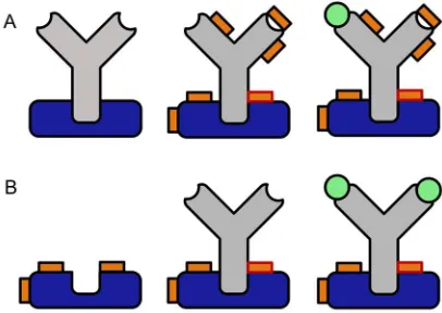
Fig 1: Comparison of conventional crosslinking (A) to the proposed preactivation crosslinking
95
method (B). Please note the potentially higher binding capacity of the immobilized antibody and the
96
complete lack of chemical modification in the Fab region (blue: Protein A or G, grey: antibody, orange:
97
crosslinker, orange with red rim: protein-protein crosslink, orange with dark rim: intramolecular or
98
half crosslink, green: antigen).
99
100
2. Materials and Methods
101
2.1 Chemicals and reagents
102
Laboratory water was obtained from a Milli-Q water purification system (Millipore, Bedford,
103
MA, USA). Dimethylsulfoxide (DMSO), was from AppliChem (Darmstadt, Germany), bovine serum
104
albumin (BSA), disodium hydrogen phosphate dihydrate, sodium dihydrogen phosphate dihydrate,
105
sodium chloride, citric acid monohydrate, and trisodium citrate dihydrate, sodium
106
cyanoborohydride, sulfuric acid, Tween 20, sodium tetraborate decahydrate were obtained from
107
Sigma-Aldrich (Steinheim, Germany). The reagents for crosslinking were obtained from the
108
following sources: Succinimidyl iodoacetate (SIA), bis(sulfosuccinimidyl) suberate,
1-ethyl-3-(3-109
dimethylaminopropyl) carbodiimide hydrochloride (EDC), N-hydroxysuccinimide (NHS) were
110
obtained from Thermo Scientific, succinimidyl(4-iodoacetyl) aminobenzoate) (SIAB) and
111
sulfosuccinimidyl (4-iodoacetyl) aminobenzoate) (Sulfo-SIAB) were obtained from Apollo Scientific
112
(Bredbury, UK). Glutaraldehyde, 1,3-butadiendiepoxide and formaldehyde were obtained from
113
Sigma-Aldrich (Steinheim, Germany), disuccinimidyl tartrate was from CovaChem, glyoxal was
114
from Merck (Darmstadt, Germany) and tris(hydroxymethyl)phosphine was bought from abcr
115
(Karlsruhe, Germany). Protein G (pro-402-c) and recombinant Protein A (pro-774) were purchased
116
from ProSpec. The microtitration plates were from Greiner bio-one (Germany). Mouse Monoclonal
117
antibodies to horseradish peroxidase (HRP), clone HP-03 (IgG1), product No. 11-262-C100 were
118
obtained from EXBIO (Praha, Czech Republic). The humanized antibody Herceptin (Trastuzumab)
119
was kindly supplied by Roche. In this article, Herceptin is referred to as “human IgG1”, due to its
120
human Fc domain. It was purified from any additives by Protein A chromatography. TMB substrate
121
was obtained from Seramun GmbH, Germany.
122
123
2.2 Buffers
124
Phosphate-buffered saline (PBS), pH 7.4: 2.3 mM KH2PO4, 10 mM Na2HPO4*2 H2O, 136.9 mM NaCl
125
Phosphate buffer, pH 6: 12.3 mM Na2HPO4*2H20, 61.0 mM NaH2PO4*2 H2O, Phosphate buffer, pH 7:
126
87.7 mM Na2HPO4*2H20, 39.0 mM NaH2PO4*2 H2O, Sodium borate buffer (SB), pH 8 and 9: 10.0 mM
127
Na2B4O7*10 H2O, Citrate buffer, pH 5: 35.0 mM Citric Acid*H2O, 65.0 mM Trisodium citrate*H2O
128
Washing buffer (PBS-Tween), pH 7.4: pH 7.4: 1.3 mM KH2PO4, 6.6 mM Na2HPO4*2 H2O, 0.5 mM
129
Tween 20, Elution buffer (Glycine/HCl), pH 2.3: 0.1 M Glycine, titrated to pH 2.3 with 0.1 N HCl.
2.3 Equipment
134
ELISA washer: 405 Select BioTek, ELISA reader: EPOCH 2 BioTek, multichannel pipettes: Eppendorf
135
Xplorer plus, Brand Transferpipette S, balance Mettler Toledo XS105 Dual Range, centrifuge Hettich
136
Mikro 220R, UV VIS spectrophotometer ThermoScientific Evolution 220
137
138
2.4 Crosslinking protocol with SIAB or sulfo-SIAB
139
Crosslinking assays (Fig. 2) were performed in 96-well polystyrene microtitration plates (MTP).
140
Protein G was diluted in phosphate-buffered saline (PBS, pH 7,4) to a final concentration of 10 mg/L.
141
100 µL of this solution was pipetted into each well of the MTP, which was shaken for at least 90 min
142
at 750 rpm. The plate was washed three times with PBS-Tween. In the next step, 300 µL of a 1%
143
solution of bovine serum albumin (BSA) was added to each well. This blocking step was performed
144
for at least one hour at a shaking frequency of 750 rpm. The plate was subsequently washed three
145
times. Then 100 µL of the crosslinker solution was added to the wells. Suitable concentrations have
146
been determined as follows: 0.25 mM for SIAB and 1 mM for sulfo-SIAB. For poorly soluble
147
crosslinkers, such as SIAB, DMSO can be used as solubilizer with a subsequent dilution in a buffer to
148
a maximum final concentration of 40% of solvent. After a reaction time of 15 min, residual crosslinker
149
was removed by three washing steps. The monoclonal anti-peroxidase antibody was diluted
150
1:100,000 (10 ng/L) in phosphate buffer (pH 6) and added to the wells (100 µL per cavity). This
151
solution was incubated at 750 rpm for sixteen hours and removed by three washing steps with
PBS-152
Tween. A solution of 10 mg/L of horseradish peroxidase was prepared in PBS-Tween-BSA as
153
described above. 100 µL was added to each well and shaken at 750 rpm for 15 min. Subsequently, the
154
plate was washed three times with PBS-Tween. Finally, 100 µL per well of TMB substrate was added
155
and incubated for 1 to 30 minutes, as required to reach a sufficient absorbance. After this development
156
time, the reaction was stopped with 0.25 M sulfuric acid. The absorbances were recorded with a
157
microplate reader at 450 nm (650 nm reference wavelength).
158
159
2.5 Crosslinking protocol with glutaraldehyde
160
First, the protocol was performed as described in section 2.4 with protein G. Instead of SIAB
161
solution, 100 µL of glutaraldehyde (2 mM) dissolved in sodium borate buffer (pH 8) was added to
162
the wells. After a reaction time of 15 min, residual crosslinker was removed by three washing steps.
163
The monoclonal anti-peroxidase antibody was diluted 1:100,000 (10 ng/L) in phosphate-buffer (pH 6)
164
with 0.1% of Tween 20 and 1% of bovine serum albumin (BSA) and added to the wells (100 µL per
165
cavity). This solution was incubated at 750 rpm for one hour and removed by three washing steps
166
with PBS-Tween. In the following step, 100 µl of NaCNBH3 (200 µg/ml) in PBS was added to the wells
167
to reduce imines to stable secondary amines. A one-hour incubation was required for the reduction
168
(750 rpm), and subsequently, the solution was removed by three washing steps with PBS-Tween. The
169
rest of the protocol followed the steps described in section 2.4.
170
171
2.6 Comparison of crosslinking protocols
172
The experiment was performed in analogy to the protocol 2.4. 100 µL of a Protein G solution (10
173
mg/L, PBS pH 7.4) was pipetted to a microtitration plate and incubated for 90 min at room
174
temperature under shaking. After a washing step (PBS-Tween, pH 7.4), blocking was performed with
175
300 µL of BSA solution (PBS pH 7.4) for one hour. After another washing step, 100 µL of sulfo-SIAB
176
(100 µL, 1 mM, PBS, pH 7.4) was incubated for 30 min. The wells for the BS3 crosslinker were supplied
177
with 100 µL of PBS. After a subsequent washing step, the whole plate (except controls) was incubated
178
with a murine monoclonal antibody (HP-03, IgG1, 100 µL, 1:100,000, phosphate buffer, pH 6.0) for 16
179
hours. After the next washing step, the sulfo-SIAB wells were supplied with 100 µL of PBS, and the
180
BS3 wells were incubated with 100 µL of BS3 in PBS (pH 7.4) for 30 min under shaking. After the next
181
washing step, any non-crosslinked antibody was removed with elution buffer (pH 2.3, glycine/HCl,
182
30 min) under shaking. After a further washing step, the plate was supplied with 100 µL of
183
horseradish peroxidase (HRP, 10 mg/L, PBS-Tween + 1% BSA) and incubated for 15 minutes. After a
184
final washing step, the 100 µL of TMB substrate was added, incubated for 10 minutes and stopped
with diluted sulfuric acid. The signal was recorded at 450 nm. A blank value (without crosslinker)
186
was subtracted.
187
188
3. Results
189
190
3.1. Crosslinking assay based on Protein G–Mouse IgG Interaction
191
Well-known protein interaction pairs were chosen for this study. Recombinant Protein G, a
192
protein from Streptococcus, (or Protein A), and a murine monoclonal antibody (IgG1) against
193
horseradish peroxidase (HRP) were used. The advantage of the latter is its antigen, which can be
194
easily determined in a microtitration plate (MTP) format and hence is ideally suited for screening
195
purposes. Protein G (or A) were adsorbed to the MTP, washed and subsequently activated with the
196
respective crosslinker. Any excess of the reagents was easily removed by washing steps; this is a big
197
plus of any heterogeneous format. Furthermore, this setup simplifies any pH variation by complete
198
buffer exchange. After the activation step, the antibody was added in a suitable conjugation buffer.
199
After a suitable conjugation time, the conjugation yield was determined by elution of the antibody
200
by acidic buffer (glycine/HCl). Any non-conjugated antibody will be released; the conjugated fraction
201
will stay immobilized on the plate surface. In the next step, horseradish peroxidase was added and
202
incubated. After the next washing step, a chromogenic substrate based on tetramethylbenzidine and
203
hydrogen peroxide were added. After a suitable development time, the color reaction was stopped
204
by acid and absorbance was measured with an MTP-reader. This assay (Fig. 2) was designed for the
205
convenient examination of the preactivation crosslinking procedure.
206
207
208
209
Fig 2: Crosslinking assay for the screening of potential crosslinkers (blue: Protein A or G, grey:
210
anti-horseradish peroxidase antibody, orange: crosslinker, orange with red rim: protein-protein
211
crosslink, orange with black rim: intramolecular or half crosslink, green: horseradish peroxidase
212
(antigen), yellow: chromogenic product. A: Coating with Protein G, B: Preactivation with the
213
crosslinker, C: Antibody binding to Protein G, D: Formation of crosslink, E: Binding of antigen
214
(enzyme), F: Enzymatic formation of chromogenic product (washing steps are not shown).
215
216
217
3.2 Crosslinker Screening
218
A considerable number of crosslinkers have been proposed, and quite a few of them are
219
commercially available. The most frequently used ones seem to be based on N-hydroxysuccinimide
220
(NHS) chemistry or their sulfo-derivatives [40-42], targeted against the ε-amino group of the lysine
221
side chain and the N-terminus of a peptide or protein. We have chosen a series of crosslinkers based
222
on different chemistries for a prescreening. There are several criteria, which are relevant for the
223
selection of a suitable crosslinker, for example, the chemical reactivity, the linker length and
224
flexibility, the hydrophobicity, the solubility and stability of the reactive groups in aqueous buffers,
225
their pH preference and many more. In Table 1, a list of crosslinkers is shown, which have been used
226
for the screening.
Table 1: Compounds used for the preactivation crosslinker screening with Protein G at pH 7.4.
228
229
Crosslinker Abbr. Efficiency
Formaldehyde [43] FA -
Disuccinimidyl tartrate [44] DST -
Tris(hydroxymethyl) phosphine THP -
1-Ethyl-3-(3-dimethylaminopropyl) carbodiimide [45] / N-Hydroxysuccinimide
EDC/NHS -
Bis(sulfosuccinimidyl)suberate [44] BS3 +
1,3-Butadiendiepoxide BDDE +
Glutaraldehyde [46-48] GA ++
Succinimidyl iodoacetate [49] SIA ++
Succinimidyl (4-iodoacetyl)aminobenzoate [44] SIAB +++ Sulfosuccinimidyl (4-iodoacetyl)aminobenzoate [44,50] Sulfo-SIAB +++
230
In Table 1, the results are summarized as – to +++, where – stands for no crosslinking and +++ denotes
231
a very high signal caused by crosslinked Protein G/antibody complex. It has to be noted that many
232
crosslinkers, which are perfectly suitable for normal crosslinking protocols (reaction with the
233
preformed complex), such as BS3, are not or only weakly active in the new format. In the novel
234
preactivation format only a few crosslinkers have proven to be suitable: Particularly glutaraldehyde
235
(GA) [46,51,52], succinimidyl iodoacetate (SIA) [49], sulfosuccinimidyl iodoacetate (sulfo-SIA),
236
succinimidyl (4-iodoacetyl)aminobenzoate (SIAB) [44] and sulfosuccinimidyl
(4-237
iodoacetyl)aminobenzoate (sulfo-SIAB) [50] can be recommended. All further experiments have been
238
focused on these pre-selected reagents (Fig. 3).
239
240
241
Fig. 3: Chemical structures of crosslinkers, which have been found particularly suitable for
242
preactivation protocols: Glutaraldehyde (1), Succinimidyl iodoacetate (2), Succinimidyl(4-iodoacetyl)
243
aminobenzoate (3), Sulfosuccinimidyl(4-iodoacetyl)aminobenzoate (4).
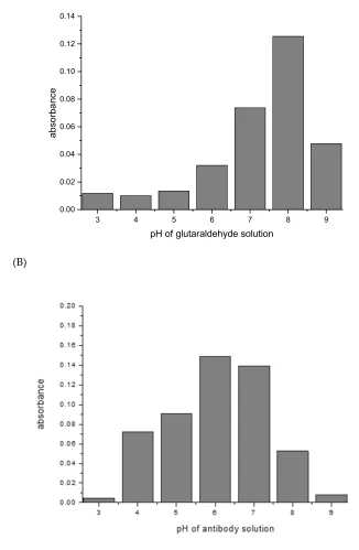
3.3 Influence of pH on the crosslinking of Protein G with murine IgG1
246
247
Most crosslinking reactions are highly pH dependent. This was explored with the examples
248
glutaraldehyde/mouse IgG1/Protein G, and SIAB/mouse IgG1/Protein G. For glutaraldehyde, it could
249
be shown that a pH of 8 seems to be optimal for preactivation (Fig. 4). This means that a standard
250
buffer, such as PBS of a pH 7.4 is a suitable option. In the second step, the addition of the mouse IgG1,
251
a pH of 6 seems to be preferable (Fig. 5). This might be dominated by the binding optimum of the
252
murine IgG1/Protein G pair, which has been determined as pH 4-6 [53,54]. In addition, it was
253
observed that increasing the incubation time of the glutaraldehyde leads to increasing immobilization
254
yields (data not shown).
255
(A)
256
(B)
257
258
Fig. 4 (A): Protein G: pH influence of glutaraldehyde solution on the preactivation immobilization of
259
mouse IgG1. (B): pH influence of antibody solution in the same experiment.
260
3 4 5 6 7 8 9
0.00 0.02 0.04 0.06 0.08 0.10 0.12 0.14
absorba
nce
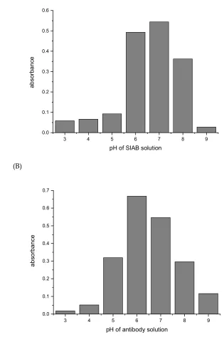
For SIAB a pH around 7 was found to be optimal for preactivation (Fig. 6). This also means that a
261
standard buffer, such as PBS pH 7.4 might be a good option. Similar to the situation with
262
glutaraldehyde, a pH of 6 was found to be preferable (Fig. 7) for the antibody (mouse IgG1). In
263
experiments with Protein A, a preferable pH of 7.4 was found (not shown), in accordance with the
264
reported IgG/Protein A optimum. This also supports the notion that not the linker, but the
protein-265
protein interaction governs the second step. This has to be taken into consideration when new
266
crosslinking pairs should be explored.
267
(A)
268
269
(B)
270
271
Fig. 5(A): Protein G: pH influence of SIAB solution on the preactivation immobilization of mouse
272
IgG1. (B) pH influence of antibody solution in the same experiment.
273
274
In the next step, the sequential crosslinking with the systems Protein A and Protein G in combination
275
with murine IgG1 and human IgG was examined. In the case of IgG1 from mouse, a monoclonal
276
antibody against horseradish peroxidase was used as a model system. In Fig. 8A it could be shown
277
that Protein A/IgG1 leads to a much lower signal than Protein G/IgG1. In the case of human IgG,
278
Protein A and Protein G lead to very similar immobilization results (Fig. 8B).
279
3 4 5 6 7 8 9
0.0 0.1 0.2 0.3 0.4 0.5 0.6
a
b
s
o
rb
a
n
c
e
pH of SIAB solution
3 4 5 6 7 8 9
0.0 0.1 0.2 0.3 0.4 0.5 0.6 0.7
a
b
s
o
rb
a
n
c
e
280
281
Fig. 8A: Preactivation immobilization of mouse IgG1 with Protein A (dark grey) or Protein G (light
282
grey),8B: Preactivation immobilization of human IgG with Protein A (dark grey) or Protein G (light
283
grey).
284
285
The species specificity of Protein A, G, and other IgG binding molecules had been explored in detail
286
[55]. Hence, it is well-known that mouse IgG1 binds only weakly to Protein A, in contrast to human
287
IgG, which is a strong binder. These properties could be confirmed in our system. This is also clear
288
support of the selectivity of this crosslinking procedure. If the crosslinker alone would be responsible
289
for the immobilization, no such behavior would be expected. Furthermore, the addition of BSA to the
290
antibodies did not influence the immobilization significantly (data not shown). This also
291
substantiates the highly selective mechanism and contradicts any simple protein crosslinking
292
hypothesis. In this case, any presence of any irrelevant protein should heavily compete with the
293
desired immobilization process, which is a frequent problem in conventional immobilization
294
procedures.
295
296
297
without SIAB with SIAB
0.0 0.2 0.4 0.6 0.8 1.0 1.2
re
la
ti
v
e
a
b
s
o
rb
a
n
c
e
A
without SIAB with SIAB
0.0 0.2 0.4 0.6 0.8 1.0 1.2
re
la
ti
v
e
a
b
s
o
rb
a
n
c
e
3.4 Incubation time of SIAB-activated Protein G with mouse IgG1
298
299
In the next experiments, the time-dependency of the crosslinking process with SIAB was explored. It
300
could be shown that some of the crosslinkers seem to bind relatively fast, in contrast to others, which
301
need several hours to reach a maximum signal. After one hour of antibody incubation, about 50% of
302
the maximum was achieved already. After 16 hours, the signal doubled. Further extension of the
303
incubation time did not increase the response anymore. The non-linear increase indicates that at least
304
two different rate constants, and hence two different crosslinking sites may be involved. In general,
305
24 hours should be more than sufficient to reach a maximal signal (Fig. 9).
306
307
Fig. 9: Influence of the incubation time on the SIAB-based preactivation immobilization of mouse
308
IgG1 on Protein G.
309
310
3.5 Influence of solvents on the crosslinking process
311
The water solubility of different crosslinkers varies widely. Particularly, SIAB is poorly water soluble.
312
Hence, concentrated SIAB solutions cannot be prepared in the usual buffers. Hence, SIAB should be
313
pre-dissolved in organic solvents such as methanol or DMSO. Interestingly, we found that DMSO
314
leads to more efficient activation of Protein G than methanol. With 40% of DMSO, a concentration of
315
only 0.25 mM of SIAB is sufficient to obtain a maximum signal in the model system. In contrast, with
316
40% of methanol, more than 2.5 mM of SIAB is necessary (data not shown). This leads to the
317
conclusion that the activation of Protein G with SIAB should be preferentially performed in PBS pH
318
7.4 with 40% of DMSO. In the case of SIA, the exceptionally high reactivity even with methanol has
319
to be taken into consideration [49]. For SIA, a stock solution in acetonitrile seems to be preferable.
320
321
3.6 Crosslinking yield of the Protein A/G IgG system
322
The crosslinking yield of the method was determined by an additional dissociative elution step with
323
acidic glycine/HCl buffer, which is a proven approach to elute IgG from Protein A or G columns. Any
324
non-crosslinked IgG should be lost during this step, leading to a loss of binding capacity. Fig. 10
325
shows a schematic representation of this test.
326
1 h 2 h 5 h 16 h 24 h 40 h
0.0 0.2 0.4 0.6 0.8 1.0
re
la
ti
v
e
a
b
s
o
rb
a
n
c
327
Fig. 10: Elution test for the determination of the crosslinking yield. (A): Bound antibodies before the
328
elution step (B): Bound antibodies after the elution step. Any non-crosslinked antibody is lost in a
329
subsequent washing step.
330
In contrast to most photochemical crosslinking protocols, the crosslinking yield with SIAB seems to
331
be quantitative (Fig. 11), at least in systems of sufficient binding strength of the protein-protein
332
complex. However, even in the case of incomplete crosslinking, a simple pre-elution step easily gets
333
rid of any traces of non-crosslinked antibody. This avoids leakage of antibodies into the
affinity-334
purified sample.
335
336
Fig. 11: Examination of the crosslinking yield of SIAB-activated Protein G with the recombinant
337
antibody Herceptin. Residual non-crosslinked human IgG was eluted with a glycine/HCl buffer at
338
pH 2.2 (black: before elution, red: after elution). The crosslinking yield of the protein G/ human IgG
339
system was apparently quantitative.
340
341
3.7 Comparison of the efficiency of the traditional and the novel immobilization method
342
A traditional crosslinking protocol with the reagent bis(sulfosuccinimidyl)suberate (BS3) was
343
compared to the proposed 2-step (preactivation) method based on sulfo-SIAB (Fig. 12). It is evident
344
that the novel method leads to a much higher signal in this model assay. Besides, the experiment
345
shows that a higher concentration of BS3 leads to lower signals, which is most likely caused by the
346
unwanted chemical modification of the antibody binding site. The used concentration range of BS3
347
0.0 0.2 0.4 0.6 0.8 1.0 1.2 1.4 1.6 1.8 2.0 2.2 2.4
0.00 0.05 0.10 0.15 0.20 0.25 0.30 0.35
A
b
s
o
rb
a
n
c
e
is based on the manufacturer's recommendation. The concentration of sulfo-SIAB in the preactivation
348
step was derived from our optimization experiments.
349
Fig. 12: Comparison of the traditional with the novel immobilization method. BS3: Post-crosslinking
350
of Protein G/antibody complex according to the manufacturer’s recommendation [56], sulfo-SIAB:
351
Preactivation of Protein G with the subsequent addition of the antibody. Any non-crosslinked
352
antibody was removed by elution buffer (glycine/HCl pH 2.2).
353
354
4. Discussion
355
It could be shown that some known crosslinkers can be used in a novel, 2-step protocol for
356
oriented antibody immobilization. Up to now, protein G/IgG or protein A/IgG complexes have been
357
treated with crosslinkers after the protein-complex had been formed, which inevitably leads to
358
chemical changes in and near the variable region of the antibody, which is critical for selective
359
binding and preservation of binding capacity. In our approach, Protein A or G is chemically
pre-360
activated by an excess of homo- or heterobifunctional reagents. This did not eliminate the bioselective
361
interaction between Protein A or G and the immunoglobulin. It can be assumed that this 2-step
362
reaction is generally applicable for most immunoglobulins, which have some affinity to Protein A or
363
G. A further advantage of this approach is the flexibility of the conjugation conditions, such as pH,
364
salt concentration, additives and so on. The resulting conjugate should show no loss of binding
365
capacity by the chemical crosslinking step since any covalent bonds are restricted to the Fc part of the
366
antibody far away from the antigen binding site. Also, it can be assumed that no optimization of the
367
conjugation should be required for known IgG subclasses since the regions involved in binding to
368
Protein A or G are highly conserved. We noticed that the presence of other proteins (such as albumin)
369
did not significantly influence the conjugation efficiency and hence, neither a pre-purification nor
370
preconcentration of the antibody or serum is necessary. Even very raw or diluted antibody
371
preparations might be used directly for the conjugation, which is in strong contrast to common
372
products with pre-activated surfaces.
373
This selective and covalent immobilization protocol should be useful in many fields: The
374
preparation of immunoaffinity columns, magnetic beads, the coating of nanoparticles, such as
375
quantum dots or gold particles, the activation of glass or other slides for microarray technology, the
376
BS3 0.25 mM BS3 0.5 mM BS3 5 mM sulfo-SIAB 1mM
0.0 0.5 1.0 1.5
abs
or
banc
robust coating of immunosensor surfaces, the oriented and irreversible immobilization of antibodies
377
on microtitration plates and even homogeneous variants, such as the labeling of antibodies might be
378
feasible. It should be stressed that in contrast to most other recent concepts, neither the production of
379
genetically modified proteins [57] nor the introduction of synthetic amino acids is required. Most
380
buffers, preservatives or protein additives do not limit the applicability of this approach. However,
381
the transfer of this protocol to other biochemical binding pairs has still to be explored.
382
This approach might be even useful for crosslinking experiments in solution, which are highly
383
popular in proteomics [58] and structural biology [59,60]. All experiments, which are performed with
384
traditional thermal or photochemical crosslinkers today, could be alternatively performed with
385
protocols analogous to those presented here. The crosslinking site might be more restricted and hence
386
better to control.
387
Regarding the reaction mechanism, it seems to be evident that crosslinkers suitable for this
388
approach need at least one active group, which does not hydrolyze or otherwise deactivate too fast.
389
Therefore, bifunctional NHS esters [41] seem to be suboptimal. In contrast, haloacetyl-residues, such
390
as SIA or SIAB possess a good balance between stability and reactivity towards nucleophiles. We
391
assume that at neutral pH values, primarily histidine residues are involved in the crosslinking with
392
haloacetyl groups, in contrast to the mechanism with glutaraldehyde, which should be dominated
393
by lysines [48]. Due to the slow reaction, even complexes with a relatively low affinity at low protein
394
concentrations may be accessible, in contrary to photochemical groups, which have very short
395
reactive lifetimes and hence often low reaction yields [27] with various side reactions. Finally, we
396
want to stress that all required reagents necessary for this novel approach are commercially available
397
from standard suppliers. Considering the much lower signals obtained with the old method, in most
398
applications the proposed approach should be preferred.
399
400
Acknowledgments: All costs were covered by internal funding of the Federal Institute for Materials Research
401
and Testing (BAM), Berlin, Germany. We want to thank Frank Osl, Roche Diagnostics GmbH, for the donation
402
of the Herceptin product sample and Marco Wilke, BAM, for the purification of this antibody.
403
Author Contributions: All authors conceived and designed the experiments; B.S., H.L.X. and J.L.V. performed
404
the experiments and analyzed the data; M.G.W. wrote the paper.
405
Conflicts of Interest: The authors declare no conflict of interest.
406
© 2018 by the authors. Submitted for possible open access publication under the terms and
407
conditions of the Creative Commons Attribution (CC BY) license
408
(http://creativecommons.org/licenses/by/4.0/).
409
410
References


![Fig. 12: Comparison of the traditional with the novel immobilization method. BS3: Post-crosslinking of Protein G/antibody complex according to the manufacturer’s recommendation [56], sulfo-SIAB: Preactivation of Protein G with the subsequent addition of th](https://thumb-us.123doks.com/thumbv2/123dok_us/7913220.1313904/12.595.114.402.131.378/comparison-traditional-immobilization-crosslinking-manufacturer-recommendation-preactivation-subsequent.webp)
