Integrating single cell RNA-sequencing and functional assays to decipher mammary cell states and 1
lineage hierarchies 2
3
Joseph L. Regan1* 4
Matthew J. Smalley2 5
6
1Charité - Universitätsmedizin Berlin, Charitéplatz 1, 10117 Berlin, Germany
7
2European Cancer Stem Cell Research Institute, School of Biosciences, Hadyn Ellis Building, Cardiff
8
University, CF24 4HQ, UK 9
10
*Correspondence: [email protected] 11
12
The identification and molecular characterization of cellular hierarchies in complex tissues is key to 13
understanding both normal cellular homoeostasis and tumorigenesis. The mammary epithelium is a 14
heterogeneous tissue consisting of two main cellular compartments, an outer bas al layer containing 15
myoepithelial cells and an inner luminal layer consisting of estrogen receptor negative (ER-) ductal cells 16
and secretory alveolar cells (in the fully functional differentiated tissue) and hormone responsive estrogen 17
receptor positive (ER+) cells. Recent publications have used single cell RNA-sequencing (scRNA-seq) 18
analysis to decipher epithelial cell differentiation hierarchies in human1 and murine2–4 mammary glands and
19
reported the identification of new cell types and states based on the expression of the luminal progenitor cell 20
marker KIT (c-Kit)1,2. These studies allow for comprehensive and unbiased analysis of the different cell 21
types that constitute a heterogeneous tissue. Here we discuss scRNA-seq studies in the context of previous 22
research in which mammary epithelial cell populations were molecularly and functionally characterized, 23
and identified c-Kit+ progenitors and cell states5 analogous to those reported in the recent scRNA-seq studies.
24
Previous studies to elucidate the cellular identities of mammary epithelial subpopulations have involved 26
functional and molecular characterization by flow cytometric and functional (down to single cell) 27
transplantation assays6,7,16–19,8–15 as well as, more recently, lineage tracing studies20,21,30,31,22–29. 28
Transplantation experiments have generally supported a model in which facultative MaSCs, cells capable 29
of regenerating the epithelium when injected into a cleared mammary fat pad (one free of endogenous 30
epithelium)6,32, are localized to the basal cell layer5,10,18,33–35. Progenitor cells, which are functionally defined
31
by high colony forming and proliferative potential in vitro and limited repopulating ability when transplanted 32
into cleared fat pads, are localized to the luminal layer5,11,19,34. Differentiated cells do not transplant or
33
generate colonies in vitro. The molecular profiling of mammary epithelial subpopulations functionally 34
defined by their transplantation potential has been extensive10,30,44,45,36–43. 35
36
Supporting this model, in situ evidence, including lineage tracing studies from early mammary 37
development, puberty and alveolargenesis during pregnancy have shown that basal cells can contribute 38
to the luminal layer22,46–48. We previously proposed, based on in situ analysis, that basal MaSCs located 39
in the cap cell layer of terminal end buds (TEBS), the outermost cell layer of the specialized growth 40
structure that drives ductal growth during puberty, are bipotent and produce daughter cells that contribute 41
to both the basal and luminal cell lineages48. Lineage tracing experiments from Rios et al. (2014) and 42
Wang et al. (2014) were in agreement with transplantation data and our in situ analysis suggesting that 43
MaSCs in the developing postnatal gland are bipotent20,21,48. However, more recently it has been shown that, 44
rather than a transcriptionally defined bipotent TEB MaSC, a group of transcriptionally heterogeneous 45
lineage committed MaSCs mediate development of the pubertal mammary gland and contribute transiently 46
to ductal expansion26, mirroring the organization and neutral drift of adult stem cells observed in the 47
intestine49,50. This model of postnatal mammary gland development is in agreement with saturation,
single-48
cell genetic and neutral lineage-tracing studies demonstrating that bipotent fetal MaSCs (fMaSCs), first 49
molecular heterogeneity) by Spike et al. (2012)40, exist in the embryo but that in the postnatal gland,
51
basal and luminal lineages are maintained by separate lineage committed stem/progenitor 52
populations22,23,53,24–27,31,47,51,52. During oncogenic transformation basal and luminal cell populations may 53
lose this restricted lineage potential and acquire multipotency23,27,54,55. 54
55
Recent studies have used scRNA-seq, which unlike functional and population based sequencing studies, 56
allows for unbiased analysis of individual cells in a heterogeneous tissue, to decipher lineage hierarchies 57
and cell states in the mammary epithelium1–4. To investigate cellular heterogeneity and lineage relationships
58
in the human breast, Nguyen et al. (2018) performed scRNA-seq analysis on fluorescence-activated cell 59
sorted (FACS) breast epithelial cells and reported the identification of additional cell types within the 60
three main mammary epithelial cell populations, previously identified as basal (B: CD49fHigh EPCAM+, 61
K14+), luminal progenitors (L1: CD49f+ EPCAM+, ER-, K8/18+), and mature luminal (L2: CD49f− 62
EPCAM+, ER+, K8/18+) cells1,9,11. Significantly, the authors detected replicating KIT+ cells in all three
63
main populations (Basal, L1, and L2), suggesting that each cluster may be maintained by its own KIT+
64
progenitor cell population and proposed a continuous lineage hierarchy connecting the basal lineage to 65
the two luminal branches via a bipotent MaSC. Furthermore, the authors highlight adult luminal cells 66
that co-express both luminal (KRT8/18) and basal (KRT14) markers in situ. 67
68
The receptor tyrosine kinase KIT (c-Kit) has previously been identified as a defining marker of mammary 69
epithelial progenitor cells (summarized in Table 1) and of the cells of origin of BRCA1-mutation breast 70
cancer, luminal ER- cells5,30,37,43,55,56. Similar to Nguyen et al. (2018), in Regan et al. (2012) we identified 71
in the mouse, and also functionally tested via in vitro colony forming assays and cleared mammary fat pad 72
transplantation, c-Kit- and c-Kit+ cell states within each of the mammary epithelial basal (CD24+/Low Sca-1
-73
CD49f+/High c-Kit- and c-Kit+), myoepithelial (CD24+/Low Sca-1- CD49f+/Low c-Kit- and c-kit+), luminal ER -74
c-kit- and c-kit+) cellular compartments5. The expression of KIT, as well as the luminal markers KRT8/18
76
and ESR1 and basal marker KRT14, in each of Nguyen et al.’s human breast populations of B, Myo, L1.1, 77
L1.2 and L2 are consistent with the expression levels reported in Regan et al. (2012) in the corresponding 78
murine basal, myoepithelial, luminal ER- c-Kit+/High, luminal ER- c-Kit+/Low, and luminal ER+ cells, 79
respectively (Figure 1). The KIT+ cells identified by Nguyen et al. (2018) are therefore likely equivalent 80
to the c-Kit+ progenitor cells previously reported in Regan et al. (2012), which was the first study to
81
functionally characterize c-Kit as a progenitor marker in the mammary gland (Table 1). When discussing 82
KIT as a progenitor cell marker, Nguyen et al. incorrectly cites Stingl et al. (2001)57 and Shehata et al.
83
(2012)11. These papers, respectively, did not investigate or functionally test c-Kit as a progenitor marker 84
in the mammary gland. 85
87
Study (year) Method(s) Cells / Progenitor cell markers Results
Nataliet al. (1992) Matsudaet al. (1993) Hineset al. (1995) Uliviet al. (2004) Tsudaet al. (2005) Westburyet al. (2009)
Immunohistochemistry Normal human breast tissue High levels of c-Kit protein detected in the
luminal alveolar/ductal epithelium but not in the basal/myoepithelial layer.
Shackleton et al. (2006) Stingl et al. (2006) Sleemanet al. (2006) Sleemanet al. (2007) Asselin-Labatet al. (2007)
FACS
Colony-forming assays Gland reconstitution Immunostaining Gene expression analysis
Mouse mammary cell populations
Basal CD24+/Low Sca1-CD49f/CD29+/High Luminal ER-CD24+/High Sca1-/CD61+
Luminal ER-cells are in vitroprogenitors and possess limited mammary gland repopulation potential.
Basal cells contain facultative MaSCs.
Kendricket al. (2008) Transcriptome analysis Mouse mammary cell populations
Basal CD24+/LowSca-1 -Luminal ER−CD24+/HighSca-1
-Luminal ER−CD24+/HighSca-1-progenitor cells are enriched for c-Kit expression.
Limet al. (2009) Limet al. (2010)
FACS
Colony-forming assays Gland reconstitution Immunostaining Transcriptome analysis
Mouse mammary cell populations
Basal CD29hiCD24loCD61+ Luminal ER-CD29loCD24+CD61+
Human mammary cell populations
Basal CD49f+/hiEpCAM+/lo Luminal ER-CD49f+EpCAM+/hi
c-Kit is highly expressed in mouse and human luminal progenitor cells. Functional testing of isolated c-Kit+cells was not carried out in these studies.
Reganet al. (2012) [Epub 18 July 2011]
FACS
Colony-forming assays Gland reconstitution Immunostaining Gene expression analysis
Mouse mammary cell subpopulations
Basal CD24+/LowSca-1-CD49f+/Highc-Kit -Basal CD24+/LowSca-1-CD49f+/Highc-Kit+ Luminal ER-CD24+/HighSca-1-c-Kit+/Low Luminal ER-CD24+/HighSca-1-c-Kit+/High Luminal ER+CD24+/HighSca-1+c-kit+
c-Kit is anin vitroandin vivofunctional marker of mammary progenitors and lineage primed cell states in basal, luminal ER-and luminal ER+cell populations.
Facultative MaSCs are CD24+/Low Sca-1 -CD49f+/Highc-Kit-.
Asselin-Labatet al. (2011) [Epub 19 Sept. 2011]
FACS
Colony-forming assays Gland reconstitution Immunostaining Gene expression analysis
Mouse mammary cell subpopulations
Luminal ER-CD29loCD24+CD14+c-kit−/lo Luminal ER-CD29loCD24+CD14+c-kit+
c-Kit+ luminal cells expand during early pregnancy and are in vitro colony forming progenitors.In vivofunctional testing of isolated c-Kit+cells was not carried out.
Shehataet al. (2012) FACS
Colony-forming assays Gland reconstitution Immunostaining Gene expression analysis
Mouse mammary cell subpopulations
Luminal ER-EpCAM+Sca-1-CD49b+CD14+ Luminal ER+EpCAM+Sca-1+CD49b+CD14+
Human mammary cell subpopulations
Luminal CD49f+EpCAM+/hiALDH+ERBB3+ Luminal CD49f+EpCAM+/hiALDH-ERBB3+ Luminal CD49f+EpCAM+/hiALDH-ERBB3
-Identified luminal ER- and luminal ER+ progenitor cells in mouse and human.
Detected c-Kit+cells in the luminal populations of FVB/N mice but not in C57Bl6/J mice. Functional testing of isolated c-Kit+cells was not carried out in this study.
Palet al. (2017) scRNA-Seq Mouse mammary cell populations
Basal CD29hiCD24+ Luminal CD29loCD24+
Hierarchical clustering revealed luminal progenitors are enriched for c-Kit. Transcriptome mapping identified rare c-Kit+ lineage primed basal cells.
Bachet al. (2017) scRNA-seq Nulliparous, embryonic, lactating and post-involution mouse mammary cells
EpCAM+
Identified c-Kit+ luminal progenitor cells that give rise to intermediate, alveolar and hormone-sensitive progenitors.
Kim & Villadsen. (2018) Immunohistochemistry Normal human breast tissue
EpCAM+Ki-67+KIT+
KIT+ cells constitute a proliferating (Ki-67+) luminal progenitor compartment during homeostasis of the resting gland.
Nguyenet al. (2018) scRNA-seq Human mammary cell populations
Basal (B) CD49fHighEPCAM+ Luminal (L1) ER-CD49f+EPCAM+ Luminal (L2) ER+CD49f−EPCAM+
Identified KIT+ progenitor cells in each mammary population, including L1.1 luminal (ER-KIT+/High) and L1.2 luminal (ER-KIT+/Low) progenitors.
Giraddiet al. (2018) Chunget al. (2019)
scRNA-seq snATAC-seq
Embryonic and post-natal mouse mammary cells
EpCAM+
Table 1: Studies demonstrating that luminal ER- cells are enriched for c-Kit and that c-Kit identifies 88
progenitor cells in the mammary epithelium1,2,34,37,43,56,58–63,3,64–66,4,5,10,11,18,19,30. 89
90
91
92
93
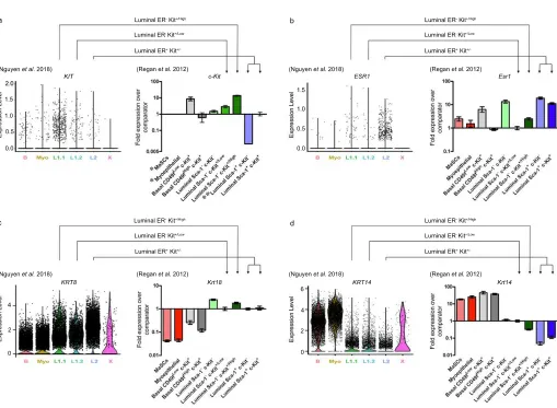
Figure 1: Comparison of gene expression in cell populations identified by Nguyen et al. (2018) and
94
Regan et al. (2012). Nguyen et al. (2018) violin plots showing the expression pattern of progenitor 95
marker KIT (a; LHS), luminal genes ESR1 and KRT8 (b – c; LHS) and basal gene KRT14 (d; LHS) 96
grouped by final cluster determination in human mammary epithelium. B = Basal (cont aining facultative 97
MaSCs), Myo = Myoepithelial. Regan et al. (2012) gene expression in the different cellular 98
subpopulations as determined by qPCR for progenitor gene c-Kit (a RHS) relative to comparator luminal 99
comparator luminal Sca-1− c-Kit+/Low cells, in murine mammary epithelium. Data are presented as fold 101
expression levels ±95% confidence intervals (n=three independently harvested isolates of each ce ll 102
population). *Gene expression was undetectable in these populations in all three independent isolates. 103
**Gene expression was only detected (at very low levels) in two of three isolates of the luminal Sca -1+ 104
c-Kit− population. Therefore, no error bars are shown for this sample. Images used with permission under 105
a CC-BY 4.0 license from Nguyen et al. (2018)1 and Regan et al. (2012)5.
106
107
Nguyen et al. (2018) observed fractions of cells that co-express both luminal K8 and basal K14 markers 108
and report that such K8+ K14+ cells had previously been observed in mouse fMASCs by Spike et al. 109
(2012)40 (such fetal cells were also previously described by Sun et al. (2010)67), but not in adult human 110
tissue in homeostasis. However, while the canonical view amongst mouse mammary developmental 111
biologists is that the K5/14 pair is a basal marker and the K8/18 pair is a luminal marker68–70, breast 112
pathologists have known for many years that keratins 5 and 14 (and indeed another ‘basal’ keratin, 17) 113
are in fact expressed in basal cells of human breast ducts and in the luminal cells of the terminal ductal 114
lobuloalveolar units (TDLUs)68,71–74. Indeed, K5/K18 and K14/K18 double positive cells are not 115
uncommon in human TDLUs71. More recently, Boecker et al. (2018), identified K5+ K18/19- and K5+ 116
K18/K19+ populations in the luminal layer of ductal and TDLU breast tissue in situ75, while in human 117
breast epithelial populations isolated by flow cytometry, the progen itor populations (Lin− CD49f+
118
EpCAMhi) include cells double positive for K5/6 and K14 – and notably are also c-KIT+43. To add to the 119
complexity of these marker patterns, K19 has been described both as a marker of progenitors76–78 and 120
highly expressed in differentiated luminal ER+ cells19,79. 121
122
Boecker et al. (2018) termed the populations they identified as progenitors and intermediary cells, 123
respectively, but it is difficult to definitively assign such functions purely on the basis of marker 124
transgene activation as one can in the mouse but use of cytochrome C oxidase (CCO) mutations in the 126
mitochondrial genome has proven feasible as an approach. Cereser et al. (2018) report the presence of 127
CCO-deficient clonal expansions in both ducts and TDLUs of normal breast80. Notably, the expansions
128
were limited to the luminal layers and they found no evidence of luminal CCO-deficient clones 129
contributing to the basal layer. Therefore, if the K5/K14/c-KIT+ luminal cells of the human breast are 130
indeed progenitors, they are lineage restricted. 131
132
Keratin expression patterns in the mouse mammary epithelium are somewhat easier to define, but also 133
not as straightforward as often suggested. Unlike in the human, when analyzed in situ, K14 and K8/18 in 134
the mouse appear to be restricted to the basal and luminal cell layers, respectively. Indeed, we have rarely 135
(if ever) observed a luminal cell in the normal resting adult mammary gland we could confidently say is 136
K14 positive, or a basal cell which is K8/18 positive, by immunofluorescence in situ, and this is in 137
agreement with most studies. However, immunohistochemical analysis of the mouse mammary gland by 138
Mikaelian et al. (2006) has detected rare weak K14 staining of luminal cells from birth to puberty and 139
weak K8/18 labelling of basal cells during mammary morphogenesis, which were most easily visualized 140
during lactation69. As an added complication, it should be noted that in the mammary alveoli, the 141
basal/myoepithelial cells form a classic ‘basket-like network’ around the secretory cells, and in that 142
location the ‘luminal’ cells are in fact touching the basement membrane through the gaps between the 143
myoepithelial cells. Interestingly, therefore, in agreement with Mikaelian et al. (2006), when basal and 144
luminal sub-populations were isolated by flow cytometry and stained by immunofluorescence, we found 145
that c-Kit+ luminal cells (which were approx. 50% of the total mammary epithelium) were all strongly 146
K18+ but also weakly K14+ and that c-Kit+ basal cells were strongly K14+ and weakly K18+ (Figure 2b)5. 147
c-Kit negative single luminal and basal cells prepared and stained at the same time were respectively 148
K18+ K14- and K14+ K18-, suggesting we were not seeing background staining in the c-Kit positive cells. 149
enhancing the K14 staining to a level where it can be detected in luminal cells would result in a huge 151
excess of staining from the basal cells as well as background signal from other cell types in the mammary 152
gland (and likewise for K18 detection in basal cells), which is notorious for background fluorescence 153
coming from adipocytes. Thus, only approaches based on single cell separation will accurately detect 154
mouse cells expressing the ‘luminal’ keratin 18 and the ‘basal’ keratin 14, and as we report using such 155
approaches, such cells express the c-Kit marker5. Note that the scRNA-seq analysis of mouse mammary
156
epithelium by Bach et al. (2017) shows that a subset of luminal cells have Krt14 expression levels 157
equivalent to the mean expression level of Krt14 in basal cells. Their differentiation trajectory maps show 158
that the Krt14 expressing luminal cells are enriched in a progenitor population which is also c-Kit-159
positive3 160
161
In contrast, we find cells double positive for ‘basal’ keratin 5 and ‘luminal’ keratin 19 are readily 162
detectable in the mouse luminal epithelium in situ (Figure 2 c-d). Interestingly, K19 has been proposed 163
to be a neutral switch keratin that permits the changeover of one type of cytoskeleton to the other78,81. 164
We have particularly noted K5 positive cells in the body cell region of terminal end buds in situ (Figure 165
2c). The origin of these cells is unclear. Rios et al. (2014) reported that using a Krt5-promoter driven cell 166
labelling approach, labelled cells were only observed in the basal compartment but generated both 167
luminal and basal daughter clones, and hence proposed the existence of bipotent basal stem cells arising 168
from the basal layer of the TEBs21. However, the work of Scheele et al. (2018)26 and others22–26,31,51,53 169
suggests that cap cells (the basal cell layer of the TEBs) do not contribute to the luminal layer of the 170
subtending duct, therefore K5 positive body cells, if they are cap cell derived, are unlikely to contribute 171
to outgrowth of the ducts. In contrast, if these cells are derived from the body cells, they are switching 172
on high levels of K5 expression, but whether this is only transient – perhaps a temporary failure of lineage 173
specification in a newly established daughter cell which is later corrected – is unclear. 174
Therefore, while use of keratins as basal/luminal lineage markers is more robust in the mouse mammary 176
epithelium than in the human, single cell analysis approaches have demonstrated that even the mouse has 177
a more promiscuous pattern of keratin expression than previously suspected, and that this promiscuous 178
expression of keratins is seen in c-KIT+ stem/progenitor cells. Plasticity in the expression of keratins and 179
other genes within c-Kit+ luminal progenitors may relate to their potential to contribute to multiple cell 180
lineages during epithelial remodeling, e.g. at involution of the mammary gland after weaning82. In
181
addition, the phenotypic plasticity and multilineage differentiation potential of these luminal progenitors 182
is consistent with their ability to give rise to tumors with basal features43,55 as well as lineage switching
183
in response to injury and oncogene activation23,27,54. It is clear, therefore, that a great deal of caution must 184
be used when keratin promoters are being used for lineage tracing studies in the mouse or for assigning 185
luminal/basal identity in human cells. Indeed, in a dissociated human breast epithelial cell population, 186
keratin expression levels alone cannot be used to assign basal/luminal identity to a cell with any 187
confidence. 188
189
To address the debate as to whether homeostasis and development in the postnatal mammary gland is 190
maintained by bipotent MaSCs20,21,48 or lineage-restricted basal and luminal cells4,22–25, Nguyen et al. 191
(2018) performed pseudotemporal reconstruction-based lineage hierarchy analysis. This analysis 192
identified a continuous lineage connecting the basal lineage, via a bipotent MaSC, to the two luminal 193
branches. These results agree with previous models of mammary differentiation wherein a bipotent basal 194
MaSC generates daughter cells that differentiate into myoepithelial and luminal cell lineages20,21,48. 195
However, Nguyen et al. propose that their results differ from previous studies in that L1.2 cells (luminal 196
ER- c-kit+/Low cells) are progenitors to L1.1 cells (luminal ER- c-Kit+/High cells) and that c-Kit+/High L1.1 197
cells are another type of mature differentiated luminal cell rather than a luminal proge nitor upstream of 198
luminal ER+ L2 cells. Based on this pseudotemporal analysis the authors suggest that KIT is not a marker 199
express KIT (Figure 1), which as well as being a defining marker of mouse and human progenitor cell gene 201
expression signatures2–4,30,37,43,65, has been functionally demonstrated as a progenitor cell marker5 (Table 1). 202
Figure 2. Basal and luminal marker expression suggests potential for differentiative plasticity in
205
the mouse mammary gland in situ. (a)Immunofluorescence of sections though the mammary fat pads 206
of adult virgin female FVB mice stained with antibodies against the luminal markers K18 and c -Kit and 207
the basal marker K14. c-Kit staining is located predominantly in the K18+ K14- luminal layer, although 208
occasional K14+ c-Kit+ basal cells are detected (arrowhead). Bar = 40 µm. (b) K18 and K14 staining of 209
freshly isolated single c-Kit+ luminal and c-Kit+ basal cells from adult virgin mice sorted directly onto
210
slides. Insets show c-Kit- luminal and basal cells negative for K14 (LHS) and K18 (RHS), respectively. 211
(Bar = 3 µm). The numbers of cells examined and overall staining patterns are given in Table 1 of Regan 212
et al. 20125. (c) Basal K5 staining in the terminal end buds (TEBs) and subtending duct of four -week-213
old pubertal mouse mammary epithelium. K5 staining is located predominantly in the basal layer. 214
Occasional K5+ cells are detected in the luminal layer (arrowheads). Bar = 40 μm. (d) Section through a 215
cleared fat pad outgrowth double stained for basal K5 and luminal K19. A K5+ K19+ double positive cell 216
is observed in the basal layer (arrowhead). Bar = 40µm. All cells were counterstained with DAPI (blue). 217
218
Similar to Nguyen et al. (2018), Pal et al. (2017) used scRNA-seq to identify lineage relationships in the 219
mouse mammary gland and also suggested that bipotent basal MaSCs give rise to basal and luminal 220
lineages2. Supporting our previous assessment of intermediate cells in the luminal lineage5, the authors 221
also described the identification of intermediate luminal cells. Significantly, Pal et al. report the 222
identification of rare mixed-lineage or “lineage-primed” c-Kit-expressing basal cells in the adult 223
mammary gland and state, “It is presumed that these cells represent a transient population that is poised 224
for commitment to the luminal lineage, reminiscent of ‘lineage-primed’ stem and progenitor cells initially 225
reported in the hematopoietic system.” These lineage-primed c-Kit+ basal cells comprised approximately
226
5% of the basal compartment and expressed luminal genes such as Esr1, Prlr, Csn2 and Areg in addition 227
to basal genes. Pal et al. state, “these data suggest that the basal state may precede commitment to a 228
230
In Regan et al. (2012) we also identified cells that we described as lineage-primed basal cells (CD24+/Low 231
Sca-1- CD49f+/High c-Kit+) in the adult mammary gland that expressed luminal genes, including those 232
described by Pal et al. (Esr1, Prlr, Csn2, Areg), but that clustered with the basal facultative MaSCs5. 233
Significantly, we functionally tested these cells by single cell cleared mammary fat pad transplantation 234
and demonstrated that they can reconstitute an entire ductal tree, although at a lower frequency (1 in 8 ± 235
95% CI 1 in 3 – 1 in 21.3) than facultative c-Kit- MaSCs (1 in 3 ± 95% CI 1 in 1.69 – 1 in 6.27), the 236
highest enrichment of facultative MaSCs reported to date and potentially a pure facultative MaSC 237
population. Based on these data we came to the same conclusion as Pal et al. (2017) and described these 238
c-Kit+ basal cells as intermediate MaSCs that were undergoing “lineage priming”, in which stem cells 239
express genes associated with their differentiated daughter populations83,84. This was the first time that 240
lineage-primed basal cells in the adult mammary gland had been reported and functionally tested. 241
242
In contrast to Nguyen et al. (2018) and Pal et al. (2017), scRNA-Seq by Bach et al. (2017)3 on mouse 243
mammary epithelial cells at nulliparous, mid gestation, lactation and post involution concluded that, 244
rather than clearly defined clusters maintained by their own stem/progenitor population, a continuous 245
spectrum of differentiation exists. In this model, a common luminal progenitor cell, which notably 246
expressed c-Kit at high levels, gives rise to intermediate, restricted alveolar, and hormone-sensitive 247
progenitors. 248
249
More recently, Giraddi et al. (2018) used scRNA-seq and transposase-accessible chromatin sequencing
250
(ATAC-seq), which examines global chromatin accessibility85, of embryonic, postnatal and adult mouse 251
mammary epithelia, to elucidate the lineage hierarchies and biological programs that generate mature 252
cell types from their embryonic precursors4. This work was more consistent with the conclusions of Bach 253
that while embryonic mammary cells are bipotent, in the adult gland, basal and luminal cell lineages are 255
derived from and maintained by separate lineage committed progenitor populations22,23,53,24–27,31,47,51,52. 256
257
Similar to Pal et al. (2017), Giraddi et al. (2018) also identified rare c-Kit+ basal cells, although they did 258
not occur at a frequency greater than the expected doublet frequency (1%) of the 10X Genomics 259
Chromium System sequencing platform4; a frequency similar to the c-Kit+ basal cells that Pal et al. (2017) 260
also detected using the 10X platform. In contrast, the lineage primed c-Kit+ basal cells that we identified 261
in our 2012 study were visually confirmed to be single cells prior to performing the single cell trans plants, 262
in which they displayed a transplantation frequency intermediary to facultative c-Kit- MaSCs and c-Kit+
263
luminal progenitor cells. In addition, immunofluorescence staining of single c -Kit+ basal cells 264
demonstrated that they expressed both K14 and K18 (Figure 2B)5. 265
266
Transcriptional profiling by Giraddi et al. (2018) did not detect any distinct adult basal stem cell 267
subpopulation. However, ATAC-seq revealed that adult basal cells display an embryonic MaSC-type 268
chromatin accessibility at luminal gene loci, which the authors speculate allows for lineage plasticity4,65,86. 269
Such plasticity may account for acquisition of multilineage potential upon perturbation of a homeostatic 270
niche environment, such as during cell isolation and ex vivo culture, transplantation assays, wounding 271
and cancer4,54,86–89. The performance of a particular cell type during functional assays may therefore be 272
a product of both their transcriptional heterogeneity and the context in which they are challenged54.
273
Similar functional stem cell capacities have also been described in embryonic tissue, intestine, bone 274
marrow, skin and lung90–92. These observations challenge the concept of fixed cell identities in complex
275
tissues and suggest a more fluid concept of cell state (for a more detailed discussion of this concept see 276
Wahl & Spike (2017))54. With this in mind, a potential mammary epithelial cell hierarchy based on lineage 277
279
Figure 3: Proposed model (adapted with permission from Giraddi et al., 2018)4 of the mammary
280
epithelial cell state lineage hierarchy in the postnatal gland based on lineage tracing, functional assays,
281
scRNA-seq and snATAC-seq. Bipotent fetal mammary stem cells (fMaSCs) are present in the embryo and 282
become lineage restricted after birth. In the adult gland each lineage is maintained by its own c-Kit+ 283
progenitor. Loss of homeostasis (e.g. injury, cell isolation, ex vivo culture, transplantation) or tumorigenesis 284
may trigger a wound response that leads to acquisition of multi-lineage potential by facultative inducible 285
MaSCs (iMaSCs), c-Kit+ lineage-primed and progenitor cell states. Lineage-primed c-Kit+ basal cells that 286
express intermediate levels of luminal genes may represent a transient or intermediate population that 287
precedes commitment to the luminal lineage2,5. Gene expression analysis suggests that an alternative route 288
for generating ER+ cells from intermediate luminal cell states may also exist. 289 290 Acquisition of multi-lineage potential Differentiated Cells Myoepithelial Progenitor States c-Kit+ K14+K18+
/-c-Kit
-K14+K18- c-Kit
-K14+K18 -ER+c-Kit
-K14-K18+
ER-c-Kit+/Low
K14+/-K18+ ER +c-Kit+ K14+/-K18+ ER-c-Kit+/High
K14+/-K18+
ER+c-Kit -K14+/-K18+
fMaSCs
c-Kit+ K14+K18+
Birth Lineage Restriction L U M I N A L B A S A L Facultative MaSCs Lineage Primed Ductal Alveolar Intermediate Alveolar Ductal Stemness
Mammary Development & Homeostasis
Heterogeneity in single cell expression profiles
•Mixed-lineage gene expression •Multi-lineage chromatin access •Cell state plasticity
Wound Response / Tumorigenesis iMaSCs P L A S T I C I T Y
Future studies that aim to map fluid cell state dynamics and their regulatory mechanisms will require the 291
use of single-cell and single-molecule epigenomic technologies that reveal a cells regulatory potential, 292
rather than its current state, as indicated by its transcriptome93,94. Indeed, Chung et al. (2019) recently 293
demonstrated that single-cell chromatin accessibility mapping of mammary gland development using 294
single-nucleus ATAC-seq (snATAC-seq) enables greater resolution of cell state heterogeneity and to be 295
a better indicator of cell state during development than scRNA-seq65. The lineage relationships delineated
296
in this study were consistent with those of Bach et al. (2017) and Giraddi et al. (2018) and also found c-297
Kit to be most highly expressed and chromatin accessible in luminal progenitor cells. 298
299
Concluding Remarks
300
Taken together, the weight of evidence supports c-Kit as a progenitor marker in the mammary 301
epithelium and, more importantly, one which is functionally characterized and can be used to enrich 302
stem/progenitor cells. Indeed, we have already begun to understand the signaling pathways 303
downstream of c-Kit in mammary progenitor cells95. scRNA-seq studies, which allow for 304
comprehensive and unbiased analysis of the different cell types that constitute a heterogeneous 305
tissue96, have been extremely valuable in contributing to our understanding of lineage relationships 306
and cell state heterogeneity in the mammary gland. However, in order to fully understand the 307
significance of these studies it is essential to link them to functional data, in particular where such 308
data already exists, and future studies should aim to do so. The evidence from lineage tracing, scRNA-309
seq and snATAC-seq studies currently supports a model in which fMaSCs in the embryo are bipotent, 310
whereas in the adult gland, stem/progenitor cells are lineage restricted and facultative MaSCs (defined 311
by functional studies) are induced to acquire multi-lineage potential upon loss of homeostasis/injury. 312
Bipotent fetal MaSCs are described as fMaSCs to differentiate them from adult facultative MaSCs. 313
However, the scientific literature up to now continues to refer to adult cells with facultative stem cell 314
longer an accurate or apt description. We therefore propose the renaming of MaSCs in the postnatal 316
gland as “inducible mammary stem cells” (iMaSCs). This new definition will help to more clearly 317
define the status and stem cell potential of functionally defined iMaSCs in the era of large-scale single-318
cell molecular profiling. 319
320
Data Availability
321 322
Source data for all figures and tables are provided in the paper. No new data sets have been generated 323
or analyzed for this article. 324
325
References
326
1. Nguyen, Q. H. et al. Profiling human breast epithelial cells using single cell RNA sequencing 327
identifies cell diversity. Nat. Commun.9, 2028 (2018). 328
2. Pal, B. et al. Construction of developmental lineage relationships in the mouse mammary 329
gland by single-cell RNA profiling. Nat. Commun.8, 1627 (2017). 330
3. Bach, K. et al. Differentiation dynamics of mammary epithelial cells revealed by single-cell 331
RNA sequencing. Nat. Commun.8, 2128 (2017).
332
4. Giraddi, R. R. et al. Single-Cell Transcriptomes Distinguish Stem Cell State Changes and 333
Lineage Specification Programs in Early Mammary Gland Development. Cell Rep.24,
1653-334
1666.e7 (2018). 335
5. Regan, J. L. et al. c-Kit is required for growth and survival of the cells of origin of Brca1-336
mutation-associated breast cancer. Oncogene31, 869 (2012). 337
6. Regan, J. & Smalley, M. Prospective isolation and functional analysis of stem and 338
differentiated cells from the mouse mammary gland. Stem Cell Rev.3, 124–136 (2007). 339
7. Shackleton, M. et al. Generation of a functional mammary gland from a single stem cell. 340
Nature439, 84–88 (2006). 341
8. Wilson, N. K. et al. Combined Single-Cell Functional and Gene Expression Analysis
342
Resolves Heterogeneity within Stem Cell Populations. Cell Stem Cell16, 712–724 (2015). 343
9. Smalley, M. J. et al. Isolation of Mouse Mammary Epithelial Subpopulations: A Comparison
344
of Leading Methods. J. Mammary Gland Biol. Neoplasia17, 91–97 (2012). 345
10. Stingl, J. et al. Purification and unique properties of mammary epithelial stem cells. Nature 346
439, 993–997 (2006). 347
11. Shehata, M. et al. Phenotypic and functional characterisation of the luminal cell hierarchy of 348
the mammary gland. Breast Cancer Res. 14, R134 (2012). 349
12. Alvi, A. J. et al. Functional and molecular characterisation of mammary side population cells. 350
Breast Cancer Res.5, R1-8 (2003). 351
13. Britt, K. L. et al. Pregnancy in the mature adult mouse does not alter the proportion of 352
mammary epithelial stem/progenitor cells. Breast Cancer Res.11, R20 (2009). 353
14. Nguyen, L. V. et al. Clonal Analysis via Barcoding Reveals Diverse Growth and 354
Differentiation of Transplanted Mouse and Human Mammary Stem Cells. Cell Stem Cell14,
355
253–263 (2014). 356
15. Ginestier, C. et al. ALDH1 is a marker of normal and malignant human mammary stem cells
and a predictor of a poor clinical outcome. Cell Stem Cell1, 555–567 (2007). 358
16. Plaks, V. et al. Lgr5 Expressing Cells Are Sufficient and Necessary for Postnatal Mammary 359
Gland Organogenesis. Cell Rep.3, 70–78 (2013). 360
17. Visser, K. E. et al. Developmental stage-specific contribution of LGR5+ cells to basal and 361
luminal epithelial lineages in the postnatal mammary gland. J. Pathol.228, 300–309 (2012). 362
18. Sleeman, K. E., Kendrick, H., Ashworth, A., C.M., I. & Smalley, M. J. CD24 staining of 363
mouse mammary gland cells defines luminal epithelial, myoepithelial/basal and non-364
epithelial cells. Breast Cancer Res8, R7 (2006). 365
19. Sleeman, K. E. et al. Dissociation of estrogen receptor expression and in vivo stem cell 366
activity in the mammary gland. J. Cell Biol.176, 19–26 (2007). 367
20. Wang, D. et al. Identification of multipotent mammary stem cells by protein C receptor 368
expression. Nature517, 81 (2014). 369
21. Rios, A. C., Fu, N. Y., Lindeman, G. J. & Visvader, J. E. In situ identification of bipotent 370
stem cells in the mammary gland. Nature506, 322 (2014). 371
22. Van Keymeulen, A. et al. Distinct stem cells contribute to mammary gland development and 372
maintenance. Nature479, 189 (2011). 373
23. Van Keymeulen, A. et al. Lineage-Restricted Mammary Stem Cells Sustain the
374
Development, Homeostasis, and Regeneration of the Estrogen Receptor Positive Lineage. 375
Cell Rep.20, 1525–1532 (2017). 376
24. Davis, F. M. et al. Single-cell lineage tracing in the mammary gland reveals stochastic clonal 377
dispersion of stem/progenitor cell progeny. Nat. Commun.7, 13053 (2016). 378
25. Lloyd-Lewis, B., Davis, F. M., Harris, O. B., Hitchcock, J. R. & Watson, C. J. Neutral 379
lineage tracing of proliferative embryonic and adult mammary stem/progenitor cells. 380
Development145, dev164079 (2018). 381
26. Scheele, C. L. G. J. et al. Identity and dynamics of mammary stem cells during branching 382
morphogenesis. Nature542, 313–317 (2017). 383
27. Koren, S. et al. PIK3CAH1047R induces multipotency and multi-lineage mammary tumours.
384
Nature525, 114 (2015). 385
28. van Amerongen, R. Lineage Tracing in the Mammary Gland Using Cre/lox Technology and
386
Fluorescent Reporter Alleles. In: Vivanco M. (eds) Mammary Stem Cells. Methods in 387
Molecular Biology. in (ed. Vivanco, M. del M.) 187–211 (Springer New York, 2015). 388
doi:10.1007/978-1-4939-2519-3_11 389
29. van de Moosdijk, A. A. A., Fu, N. Y., Rios, A. C., Visvader, J. E. & van Amerongen, R. 390
Lineage Tracing of Mammary Stem and Progenitor Cells. In: Martin F., Stein T., Howlin J. 391
(eds) Mammary Gland Development. Methods in Molecular Biology, vol 1501. Humana 392
Press, New York, NY. in (eds. Martin, F., Stein, T. & Howlin, J.) 291–308 (Springer New 393
York, 2017). doi:10.1007/978-1-4939-6475-8_15 394
30. Lim, E. et al. Transcriptome analyses of mouse and human mammary cell subpopulations
395
reveal multiple conserved genes and pathways. Breast Cancer Res.12, R21–R21 (2010). 396
31. Chang, T. H.-T. et al. New insights into lineage restriction of mammary gland epithelium 397
using parity-identified mammary epithelial cells. Breast Cancer Res.16, R1–R1 (2014). 398
32. DeOme, K. B., Faulkin Jr., L. J., Bern, H. A. & Blair, P. B. Development of mammary 399
tumors from hyperplastic alveolar nodules transplanted into gland-free mammary fat pads of 400
female C3H mice. Cancer Res.19, 515–520 (1959). 401
33. Shackleton, M. et al. Generation of a functional mammary gland from a single stem cell. 402
Nature439, 84–88 (2006). 403
34. Asselin-Labat, M. L. et al. Gata-3 is an essential regulator of mammary-gland morphogenesis 404
and luminal-cell differentiation. Nat Cell Biol9, 201–209 (2007). 405
35. Taddei, I. et al. Beta1 integrin deletion from the basal compartment of the mammary 406
36. Shipitsin, M. et al. Molecular definition of breast tumor heterogeneity. Cancer Cell11, 259– 408
273 (2007). 409
37. Kendrick, H. et al. Transcriptome analysis of mammary epithelial subpopulations identifies 410
novel determinants of lineage commitment and cell fate. BMC Genomics9, 591 (2008). 411
38. Pece, S. et al. Biological and molecular heterogeneity of breast cancers correlates with their 412
cancer stem cell content. Cell140, 62–73 (2010). 413
39. Wansbury, O. et al. Transcriptome analysis of embryonic mammary cells reveals insights 414
into mammary lineage establishment. Breast Cancer Res.13, R79–R79 (2011). 415
40. Spike, B. T. et al. A Mammary Stem Cell Population Identified and Characterized in Late 416
Embryogenesis Reveals Similarities to Human Breast Cancer. Cell Stem Cell10, 183–197 417
(2012). 418
41. Raouf, A. et al. Transcriptome analysis of the normal human mammary cell commitment and
419
differentiation process. Cell Stem Cell3, 109–118 (2008). 420
42. Pal, B. et al. Integration of microRNA signatures of distinct mammary epithelial cell types 421
with their gene expression and epigenetic portraits. Breast Cancer Res.17, 85 (2015). 422
43. Lim, E. et al. Aberrant luminal progenitors as the candidate target population for basal tumor 423
development in BRCA1 mutation carriers. Nat Med15, 907–913 (2009).
424
44. Jones, C. et al. Expression profiling of purified normal human luminal and myoepithelial 425
breast cells: identification of novel prognostic markers for breast cancer. Cancer Res64, 426
3037–3045 (2004). 427
45. Grigoriadis, A. et al. Establishment of the epithelial-specific transcriptome of normal and 428
malignant human breast cells based on MPSS and array expression data. Breast Cancer Res 429
8, R56 (2006). 430
46. van Amerongen, R., Bowman, A. N. & Nusse, R. Developmental Stage and Time Dictate the
431
Fate of Wnt/β-Catenin-Responsive Stem Cells in the Mammary Gland. Cell Stem Cell11, 432
387–400 (2012). 433
47. Wuidart, A. et al. Early lineage segregation of multipotent embryonic mammary gland 434
progenitors. Nat. Cell Biol.20, 666–676 (2018). 435
48. Regan, J. L. et al. Aurora A Kinase Regulates Mammary Epithelial Cell Fate by Determining 436
Mitotic Spindle Orientation in a Notch-Dependent Manner. Cell Rep.4, 110–123 (2013). 437
49. Lopez-Garcia, C., Klein, A. M., Simons, B. D. & Winton, D. J. Intestinal Stem Cell 438
Replacement Follows a Pattern of Neutral Drift. Science (80-. ).330, 822 LP – 825 (2010). 439
50. Ritsma, L. et al. Intestinal crypt homeostasis revealed at single-stem-cell level by in vivo live 440
imaging. Nature507, 362–365 (2014). 441
51. Elias, S., Morgan, M. A., Bikoff, E. K. & Robertson, E. J. Long-lived unipotent Blimp1-442
positive luminal stem cells drive mammary gland organogenesis throughout adult life. Nat. 443
Commun.8, 1714 (2017). 444
52. Lilja, A. M. et al. Clonal analysis of Notch1-expressing cells reveals the existence of 445
unipotent stem cells that retain long-term plasticity in the embryonic mammary gland. Nat. 446
Cell Biol.20, 677–687 (2018). 447
53. Wang, C., Christin, J. R., Oktay, M. H. & Guo, W. Lineage-Biased Stem Cells Maintain 448
Estrogen-Receptor-Positive and -Negative Mouse Mammary Luminal Lineages. Cell Rep.18,
449
2825–2835 (2017). 450
54. Wahl, G. M. & Spike, B. T. Cell state plasticity, stem cells, EMT, and the generation of intra-451
tumoral heterogeneity. npj Breast Cancer3, 14 (2017). 452
55. Molyneux, G. et al. BRCA1 Basal-like Breast Cancers Originate from Luminal Epithelial 453
Progenitors and Not from Basal Stem Cells. Cell Stem Cell7, 403–417 (2010). 454
56. Asselin-Labat, M.-L. et al. Gata-3 Negatively Regulates the Tumor-Initiating Capacity of 455
Mammary Luminal Progenitor Cells and Targets the Putative Tumor Suppressor Caspase-14. 456
57. Stingl, J., Eaves, C. J., Zandieh, I. & Emerman, J. T. Characterization of bipotent mammary 458
epithelial progenitor cells in normal adult human breast tissue. Breast Cancer Res. Treat.67, 459
93–109 (2001). 460
58. Natali, P. G. et al. Expression of c-<em>kit</em> Receptor in Normal and 461
Transformed Human Nonlymphoid Tissues. Cancer Res.52, 6139 LP – 6143 (1992).
462
59. Matsuda, R. et al. Expression of the c-kit protein in human solid tumors and in corresponding 463
fetal and adult normal tissues. Am. J. Pathol.142, 339–346 (1993). 464
60. Hines, S. J., Organ, C., Kornstein, M. J. & Krystal, G. W. Coexpression of the c-kit and stem 465
cell factor genes in breast carcinomas. Cell Growth Differ6, 769–779 (1995). 466
61. Ulivi, P. et al. c-kit and SCF expression in normal and tumor breast tissue. Breast Cancer Res 467
Treat83, 33–42 (2004). 468
62. Tsuda, H. et al. Frequent KIT and epidermal growth factor receptor overexpressions in 469
undifferentiated-type breast carcinomas with ‘stem-cell-like’ features. Cancer Sci96, 333– 470
339 (2005). 471
63. Shackleton, M., Vaillant, F. & Simpson, K. J. Generation of a functional mammary gland 472
from a single stem cell. Nature439, 84–88 (2006). 473
64. Kim, J. & Villadsen, R. Expression of Luminal Progenitor Marker CD117 in the Human 474
Breast Gland. J. Histochem. Cytochem.66, 879–888 (2018). 475
65. Chung, C. Y. et al. Single-Cell Chromatin Analysis of Mammary Gland Development
476
Reveals Cell-State Transcriptional Regulators and Lineage Relationships. Cell Rep.29, 495-477
510.e6 (2019). 478
66. Westbury, C. B. et al. Genome-wide transcriptomic profiling of microdissected human breast 479
tissue reveals differential expression of KIT (c-Kit, CD117) and oestrogen receptor-α (ERα) 480
in response to therapeutic radiation. J. Pathol.219, 131–140 (2009). 481
67. Sun, P., Yuan, Y., Li, A., Li, B. & Dai, X. Cytokeratin expression during mouse embryonic 482
and early postnatal mammary gland development. Histochem. Cell Biol.133, 213–221 483
(2010). 484
68. Dontu, G. & Ince, T. A. Of mice and women: a comparative tissue biology perspective of 485
breast stem cells and differentiation. J. Mammary Gland Biol. Neoplasia20, 51–62 (2015). 486
69. Mikaelian, I. et al. Expression of Terminal Differentiation Proteins Defines Stages of Mouse 487
Mammary Gland Development. Vet. Pathol.43, 36–49 (2006).
488
70. Smith, G. H., Mehrel, T. & Roop, D. R. Differential keratin gene expression in developing, 489
differentiating, preneoplastic, and neoplastic mouse mammary epithelium. Cell Growth 490
Differ.1, 161—170 (1990). 491
71. Santagata, S. et al. Taxonomy of breast cancer based on normal cell phenotype predicts 492
outcome. J. Clin. Invest.124, 859–870 (2014). 493
72. Santagata, S. & Ince, T. A. Normal cell phenotypes of breast epithelial cells provide the 494
foundation of a breast cancer taxonomy. Expert Rev. Anticancer Ther.14, 1385–1389 (2014). 495
73. Gusterson, B. Do ‘basal-like’ breast cancers really exist? Nat Rev Cancer9, 128–134 (2009). 496
74. Gusterson, B. A., Ross, D. T., Heath, V. J. & Stein, T. Basal cytokeratins and their 497
relationship to the cellular origin and functional classification of breast cancer. Breast Cancer 498
Res7, 143–148 (2005). 499
75. Boecker, W. et al. Spatially correlated phenotyping reveals K5-positive luminal progenitor 500
cells and p63-K5/14-positive stem cell-like cells in human breast epithelium. Lab. Investig. 501
98, 1065–1075 (2018). 502
76. Clarke, R. B. et al. A putative human breast stem cell population is enriched for steroid 503
receptor-positive cells. Dev. Biol.277, 443–56 (2005). 504
77. Gudjonsson, T. et al. Isolation, immortalization, and characterization of a human breast 505
epithelial cell line with stem cell properties. Genes Dev16, 693–706 (2002). 506
177, 87–101 (2007). 508
79. Bartek, J., Bartkova, J. & Taylor-Papadimitriou, J. Keratin 19 expression in the adult and 509
developing human mammary gland. Histochem. J.22, 537–544 (1990).
510
80. Cereser, B. et al. Analysis of clonal expansions through the normal and premalignant human 511
breast epithelium reveals the presence of luminal stem cells. J. Pathol.244, 61–70 (2018). 512
81. Stasiak, P. C., Purkis, P. E., Leigh, I. M. & Lane, E. B. Keratin 19: Predicted Amino Acid 513
Sequence and Broad Tissue Distribution Suggest it Evolved from Keratinocyte Keratins. J. 514
Invest. Dermatol.92, 707–716 (1989). 515
82. Wagner, K. U. et al. An adjunct mammary epithelial cell population in parous females: its 516
role in functional adaptation and tissue renewal. Development129, 1377–1386 (2002). 517
83. Huang, S., Guo, Y. P., May, G. & Enver, T. Bifurcation dynamics in lineage-commitment in 518
bipotent progenitor cells. Dev Biol305, 695–713 (2007). 519
84. Månsson, R. et al. Molecular Evidence for Hierarchical Transcriptional Lineage Priming in 520
Fetal and Adult Stem Cells and Multipotent Progenitors. Immunity26, 407–419 (2007). 521
85. Buenrostro, J. D., Giresi, P. G., Zaba, L. C., Chang, H. Y. & Greenleaf, W. J. Transposition 522
of native chromatin for fast and sensitive epigenomic profiling of open chromatin, DNA-523
binding proteins and nucleosome position. Nat. Methods10, 1213 (2013). 524
86. Dravis, C. et al. Epigenetic and Transcriptomic Profiling of Mammary Gland Development
525
and Tumor Models Disclose Regulators of Cell State Plasticity. Cancer Cell34, 466-482.e6 526
(2018). 527
87. Ge, Y. et al. Stem Cell Lineage Infidelity Drives Wound Repair and Cancer. Cell169, 636-528
650.e14 (2017). 529
88. Ge, Y. & Fuchs, E. Stretching the limits: from homeostasis to stem cell plasticity in wound 530
healing and cancer. Nat. Rev. Genet.19, 311 (2018). 531
89. Seldin, L., Le Guelte, A. & Macara, I. G. Epithelial plasticity in the mammary gland. Curr. 532
Opin. Cell Biol.49, 59–63 (2017). 533
90. Blanpain, C. & Fuchs, E. Plasticity of epithelial stem cells in tissue regeneration. Science 534
(80-. ).344, 1242281–1242281 (2014). 535
91. Hough, S. R., Laslett, A. L., Grimmond, S. B., Kolle, G. & Pera, M. F. A Continuum of Cell 536
States Spans Pluripotency and Lineage Commitment in Human Embryonic Stem Cells. PLoS
537
One4, e7708 (2009). 538
92. Tata, P. R. & Rajagopal, J. Plasticity in the lung: making and breaking cell identity. 539
Development144, 755–766 (2017). 540
93. Corces, M. R. et al. Lineage-specific and single-cell chromatin accessibility charts human 541
hematopoiesis and leukemia evolution. Nat. Genet.48, 1193 (2016). 542
94. Shema, E., Bernstein, B. E. & Buenrostro, J. D. Single-cell and single-molecule epigenomics 543
to uncover genome regulation at unprecedented resolution. Nat. Genet.51, 19–25 (2019). 544
95. Tornillo, G. et al. Dual Mechanisms of LYN Kinase Dysregulation Drive Aggressive
545
Behavior in Breast Cancer Cells. Cell Rep.25, 3674-3692.e10 (2018). 546
96. Cristea, S. & Polyak, K. Dissecting the mammary gland one cell at a time. Nat. Commun.9, 547 2473 (2018). 548 549 550 551 Acknowledgements 552 553
The authors thank Geoffrey M. Wahl (The Salk Institute for Biological Studies) for his critical reading of 554
556 557
Author Information
558
559
Affiliations
560
561
Charité Comprehensive Cancer Centre, Charité Universitätsmedizin Berlin, Charitéplatz 1, Berlin,
562
Germany
563
Joseph L. Regan 564
565
European Cancer Stem Cell Research Institute, School of Biosciences, Cardiff University, Wales,
566
UK
567
Matthew J. Smalley 568
569
Contributions
570
Conceptualization and writing of the original draft, J.L.R.; Review and editing, J.L.R and M.J.S. All the 571
authors read and approved the final version of the manuscript. 572
573
Corresponding author
574
Correspondence to Joseph L. Regan ([email protected]). 575
576
Competing interests
577
The authors declare no competing interests. 578
579