International Journal of COPD 2018:13 1343–1351
International Journal of COPD
Dove
press
submit your manuscript | www.dovepress.com 1343
O r I g I n a l r e s e a r C h
open access to scientific and medical research
Open access Full Text article
Chronic obstructive pulmonary disease severity
and its association with obstructive sleep
apnea syndrome: impact on cardiac autonomic
modulation and functional capacity
Katiany Thays lopes
Zangrando1
renata Trimer2
luiz Carlos soares de
Carvalho Jr1
guilherme Peixoto Tinoco
arêas1
Flávia Cristina rossi
Caruso1
ramona Cabiddu1
Meliza goi roscani3
Fabíola Paula galhardo
rizzatti3
audrey Borghi-silva1
1Cardiopulmonary Physiotherapy
laboratory, Physiotherapy Department, Federal University of são Carlos, são Carlos, são Paulo, Brazil; 2Physical education and health
Department, University of santa Cruz do sul, santa Cruz do sul, rio grande do sul, Brazil; 3Medicine Department,
Federal University of são Carlos, são Carlos, são Paulo, Brazil
Background: The study was conducted to determine the impact of chronic obstructive pulmonary disease (COPD) in association with obstructive sleep apnea syndrome (OSAS) on cardiac autonomic control and functional capacity.
Subjects and methods: The study was a cross-sectional prospective controlled clinical study. Heart rate variability indices of 24 COPD (n = 12) and COPD+OSAS (n = 12) patients were evaluated and compared by electrocardiographic recordings acquired during rest, active postural maneuver (APM), respiratory sinus arrhythmia maneuver (RSA-m), and the 6-minute walk test (6MWT).
Results: The COPD group presented higher parasympathetic modulation during APM when compared to the COPD+OSAS group (P = 0.02). The COPD+OSAS group presented higher sympathetic modulation during RSA-m when compared to the COPD group (P = 0.00). The performance during 6MWT was similarly impaired in both groups, despite the greater severity of the COPD group.
Conclusion: Subjects with COPD+OSAS present marked sympathetic modulation, and the presence of OSAS in COPD subjects has a negative impact on functional capacity regardless of the severity of lung disease.
Keywords: COPD, OSAS, COPD+OSAS, functional capacity
Introduction
Chronic obstructive pulmonary disease (COPD) is a syndrome characterized by chronic airway obstruction, which is not completely reversible; it determines significant losses in pulmonary function, has systemic effects, and is associated with important comorbidities.1
Among them, obstructive sleep apnea syndrome (OSAS), characterized by recurrent
events of upper airway obstruction during sleep, affects most COPD patients.2 COPD
and OSAS are two of the most prevalent chronic respiratory diseases worldwide.3,4
According to Taranto-Montemurro et al,5 the association of the two diseases is
present in about 0.5%–1% of the general population; the prevalence of COPD in individuals with OSAS exceeds its prevalence in individuals who do not present with
sleep disorders.6 OSAS appears to be more commonly seen in subjects with COPD
perhaps as a result of shared risk factors such as obesity, smoking, increased airway
resistance, local and systemic inflammation, as well as anti-inflammatory therapy.6
It is known that COPD+OSAS has a great impact on the systemic manifestations
of affected patients, such as an increase in daytime sympathetic activity, with an
Correspondence: audrey Borghi-silva Physiotherapy Department, Federal University of são Carlos, rodovia Washington luis, km 235 – sP-310, CeP: 13565-905, são Carlos, são Paulo, Brazil Tel +55 16 3351 8952
Fax +55 16 3361 2081 email [email protected]
Journal name: International Journal of COPD Article Designation: Original Research Year: 2018
Volume: 13
Running head verso: Zangrando et al
Running head recto: COPD+OSAS: autonomic and functional impact DOI: 156168
International Journal of Chronic Obstructive Pulmonary Disease downloaded from https://www.dovepress.com/ by 118.70.13.36 on 22-Aug-2020
For personal use only.
Dovepress
Zangrando et al
increase in rest heart rate (HR) and a decrease in heart rate variability (HRV), which may lead to greater morbidity and mortality from other diseases.5 On the other hand, other
authors have demonstrated that COPD+OSAS determines
marked parasympathetic hyperactivity in the airways and hypoxemia-related bronchoconstriction/vasoconstriction,
among other neurohumoral effects.6 These still
contradic-tory autonomic imbalances could have a negative impact on static postural adjustments and responses to exercise. To our knowledge, however, no previous study has evaluated and compared such adjustments in these patients.
Thus, the main objective of this study was to determine the impact of the coexistence of OSAS and COPD on car-diac autonomic control in response to changes in breath-ing pattern, active postural maneuver (APM), and durbreath-ing submaximal exercise (6-minute walk test [6MWT]). We hypothesized that the coexistence of OSAS and COPD would be accompanied by greater cardiovascular, autonomic, and functional impairments than COPD alone.
Subjects and methods
subjects
Twenty-four individuals of both sexes were screened at the Pulmonary Ambulatory of the School Health Unit (USE) of Federal University of São Carlos (UFSCar) and at the Municipal Health Specialty Center of São Carlos (CEME) with clinical diagnosis of COPD according to the criteria
defined by the GOLD scientific committee7 with suspected
diagnosis of OSAS.
Inclusion criteria
COPD group: clinical diagnosis of COPD according to the criteria defined by the GOLD scientific committee,7 in regular
treatment with optimized medication, current nonsmokers, people who were not in exacerbations period, and people whose after-home sleep examination did not confirm the suspicion of OSAS.
COPD+OSAS group: the same criteria for inclusion
in COPD group were followed; however, the individuals presented a confirmed diagnosis of OSAS.
Subjects should not present clinical exacerbations or be on oral steroids for at least 3 months.
exclusion criteria
Exclusion criteria included presence of diabetes mellitus, systemic arterial hypertension and uncontrolled pulmonary hypertension, use of medications capable of altering cardiac autonomic control, thyroid hormone alterations, use of home oxygen therapy or continuous positive airway pressure (CPAP)
for the treatment of OSAS, pacemaker, cardiac arrhythmias
interfering with .95% of heart beats, and neurological and
orthopedic alterations that would prevent the performance of the proposed tests.
The study was approved by the Ethics and Research Committee of Federal University of São Carlos, São Paulo, Brazil (number 1.406.894/2016), and all the volunteers signed a written informed consent agreement in compliance with res-olution number 466/2012 of the National Health Council.
study protocol
All volunteers were subjected to two stages of evaluation: 1) investigation of the presence of OSAS in subjects with COPD and 2) evaluation of the autonomic function through HRV analysis and of the functional capacity through 6MWT application.
Measurements
In order to control the influence of circadian variations on the results, all tests were performed at the same time of the day in a laboratory with a mean temperature maintained between 20°C and 22°C and relative humidity in the range from 40% to 60%. All subjects were instructed to abstain from alcohol or stimulants on the day of the tests and not to practice physical or sports activities the day before and on the day of the tests.
Pulmonary function
Spirometric evaluation was performed with a CPFS/D®
spirometer (Medgraphics, MGC Diagnostics Corpora-tion, St Paul, MN, USA), according to the criteria of the
Brazilian Society of Pulmonology and Tisiology,8 and were
staged according to the GOLD criteria.7
home sleep test
The apnea-hypopnea index (AHI) and the oxygen desatura-tion index (ODI) were obtained by an ApneaLink Plus™ equipment (ResMed Corporation, Poway, CA, USA), which is a portable sleep monitor validated to assess the presence of OSAS.9 Subjects with AHI $15 were included in the
COPD+OSAS group, while those with AHI ,15 were
included in the COPD group.
After group stratification, autonomic evaluation was initiated and included.
recording of r-r intervals
The electrocardiographic signal was recorded through a BioAmp FE132 device (ADInstruments, Sydney, Australia), with electrodes placed in the MC5 lead configuration.
International Journal of Chronic Obstructive Pulmonary Disease downloaded from https://www.dovepress.com/ by 118.70.13.36 on 22-Aug-2020
Dovepress COPD+Osas: autonomic and functional impact
Signals were recorded during the conditions shown in the following sections.
active postural maneuver
The subjects remained in supine position for 10 minutes and were instructed not to talk, sleep, or move during this time. After this rest period, the subjects were instructed to position themselves in orthostatic position (10 minutes) and then in sitting position (10 minutes), following the instructions given for the rest period.
respiratory sinus arrhythmia maneuver
After APM, the subjects, still seated, were subjected to respiratory sinus arrhythmia maneuver (RSA-m; controlled breathing with cycles of 10 seconds, divided into 5 seconds of inspiration and 5 seconds of expiration) during 4 minutes.
For HRV analysis during MPA and m-ASR, the R-R intervals (iRR) were collected at a rate of 500 samples/s. All signals were visually inspected to identify artifacts or noises where narrow peaks (,100 ms) were removed by linear interpolation. All signals were filtered with low pass filters with a cutoff frequency of 20 Hz.
Data were analyzed using Kubios HRV® software
(Version 2.1, Matlab, Kuopio, Finland). The data collected during each MPA posture had a total period of iRR examined and, for analysis, the most stable and noise-free segment that contains 300 points was selected. In addition, the data col-lected during m-ASR had all the iRR contained within the 4 minutes of the maneuver used for the analysis.
submaximal exercise using the 6-minute walk test
General test principles, criteria for discontinuation, as well as verbal stimuli were standardized based on the recommenda-tions of the American Thoracic Society and the American College of Chest Physicians.10 After a 4-minute rest period
(2 minutes in sitting position and 2 minutes in orthostatic position), the volunteers were instructed to walk continu-ously, as far as possible, in a 30-meter walk for 6 minutes, and allowed to slow down and even interrupt the test if nec-essary. At the end, the distance traveled in meters as well as percentage in relation to the predicted was recorded.
HR was recorded by a Polar HR monitor (RX810, Kempele, Finland) during 4 minutes before the test (rest), throughout the test (exercise), and during 6 minutes after the test (recovery). For HRV analysis during the 6MWT, the data collected by the HR monitor were visually inspected to identify artifacts or noises where narrow peaks (,100 ms) were removed by linear interpolation. All signals were filtered with low pass filters with a cutoff frequency of 20 Hz.
Data were analyzed using Kubios HRV® software
(Version 2.1, Matlab). The data were analyzed as follows:
• Rest in the seated position: The 2 minutes of the seated
rest period were analyzed.
• Rest in the orthostatic position: iRR were analyzed from
the 2 minutes of the standing rest period.
• Walk: For analysis, we selected the most stable and noise-free segment that should have 3 minutes.
• Recovery: The last 5 minutes of collected iRR were
selected for analysis.
Selected sections were analyzed in the time and frequency domains and by means of non-linear analysis. In the time domain, the following indices were calculated: 1) SDNN: standard deviation of all iRR; 2) RMSSD: square root of the mean squared differences between adjacent iRR; 3) pnn50: percentage of adjacent iRR with duration difference greater
than 50 ms; 4) TINN: iRR variability.11 SDNN and TINN
represent global autonomic activity.11 RMSSD and pnn50
are representative of vagal modulation.11 In the frequency
domain, the following indices were calculated: 1) high fre-quency component (HF); frefre-quency band between 0.15 Hz and 0.4 Hz; 2) low frequency component (LF); frequency band between 0.04 Hz and 0.15 Hz; and 3) LF/HF ratio.11 The
HF index represents cardiac parasympathetic modulation; the LF index represents the joint action of the sympathetic and parasympathetic components, with sympathetic pre-dominance; the LF/HF ratio represents the sympathovagal balance.11 In addition, the following non-linear indices were
calculated: 1) SD1: standard deviation of instantaneous beat-to-beat variability; 2) SD2: long-term standard
devia-tion of the continuous iRR.11 SD1 index represents cardiac
parasympathetic modulation and SD2 index represents global cardiac autonomic activity.11
statistical analysis
The results were presented as mean ± standard deviation,
with a level of significance of 95%. Categorical variables were compared using the chi-square test. Two-way ANOVA (with Bonferroni post hoc test) was used for comparative analysis of the differences between the HRV indexes. Linear associations were evaluated with Pearson product-moment coefficients. Statistical analysis was performed using SPSS 19.0 (IBM, Armonk, NY, USA) and GraphPad Prism 6.0 (MacKiev Software, Boston, MA, USA).
Results
Population characteristics
One hundred and two subjects from the Pulmonary Ambula-tory of the School Health Unit (USE) of Federal University
International Journal of Chronic Obstructive Pulmonary Disease downloaded from https://www.dovepress.com/ by 118.70.13.36 on 22-Aug-2020
Dovepress
Zangrando et al
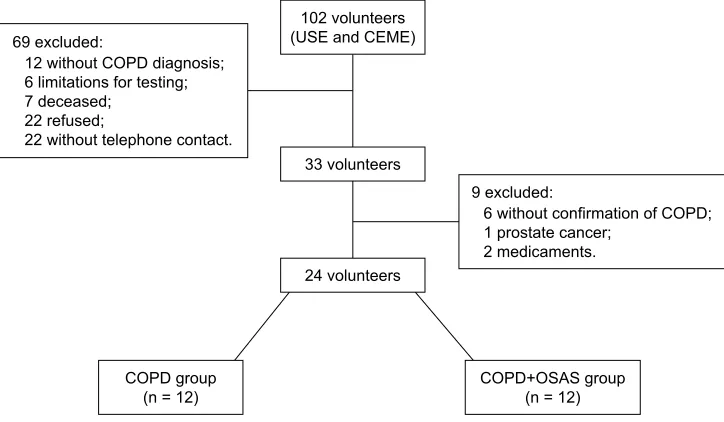
of São Carlos (UFSCar) and from the Municipal Health Specialty Center of São Carlos (CEME) were screened. The groups were screened as described in Figure 1.
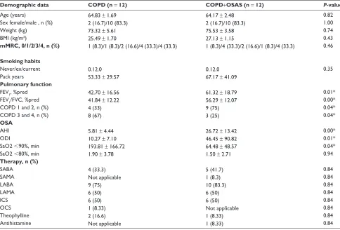
The baseline characteristics of the volunteers are shown in Table 1. The data contained in Table 1 show that the lung function of the COPD group was worse when compared to
the COPD+OSAS group, that is, the degree of respiratory
commitment was higher for the COPD group.
Although the COPD+OSAS group presented higher
levels of AHI and ODI, as expected due to the presence of OSAS, interestingly, the time spent with saturation below
90% (SAT , 90%) and below 80% (SAT , 80%) was much
longer in the COPD group, confirming the great impact of the disease on nocturnal hypoxemia in these patients.
hrV indices at rest
The HRV indices during APM and RSA-m are represented
in Tables 2 and 3, respectively. The COPD+OSAS group
presented higher absolute and normalized LF values when
compared to the COPD group (Table 2, P , 0.05). On the
other hand, the COPD+OSAS group had lower
normal-ized HF values when compared to the COPD group; taken together, data suggest greater sympathetic modulation in the
COPD+OSAS group.
Table 3 shows that both groups had higher SDNN and TINN values during RSA-m when compared to the sitting position, suggesting a greater global autonomic modulation for both groups during the maneuver (P , 0.05). How-ever, there was no interaction between APM values for the two groups.
6MWT variables
The 6MWT parameters are shown in Table 4. Table 4 shows
that the COPD+OSAS group had higher SBP than the COPD
group (P , 0.05). In addition, symptoms of dyspnea and
lower limbs fatigue were worse in the COPD group than in
the COPD+OSAS group (P , 0.05), even though we did not
observe a worse performance in this group. Although there
were no differences in SpO2 between groups during 6MWT
(P = 0.21), the COPD group presented an average value below 90%, which indicates that desaturation was clinically more pronounced in this group.
Strong correlations were observed between the HRV indices during 6MWT and nocturnal desaturation in these patients. We observed that the longer the time spent with saturation below 90%, the greater the parasympathetic modu-lation during walking, represented by the RMSSD and SD1 indices (R = 0.80, P , 0.05).
Discussion
To our knowledge, this is the first cross-sectional study com-paring autonomic responses to APM, RSA-m, and walking, as well as submaximal exercise performance between
subjects with COPD+OSAS versus subjects with COPD.
The main findings of this study showed that individuals in
the COPD+OSAS group presented less severe lung disease
and shorter nocturnal hypoxemia time when compared to the COPD group.
In addition, the autonomic modulation during APM was impaired in both groups; however, the severity of COPD
H[FOXGHG
ZLWKRXWFRQILUPDWLRQRI&23' SURVWDWHFDQFHU
PHGLFDPHQWV H[FOXGHG
ZLWKRXW&23'GLDJQRVLV OLPLWDWLRQVIRUWHVWLQJ GHFHDVHG
UHIXVHG
ZLWKRXWWHOHSKRQHFRQWDFW
YROXQWHHUV 86(DQG&(0(
YROXQWHHUV
YROXQWHHUV
&23'JURXS
Q &23'26$6JURXSQ
Figure 1 Volunteer flowchart.
Abbreviations: CeMe, Municipal health specialty Center of são Carlos; COPD, chronic obstructive pulmonary disease; Osas, obstructive sleep apnea syndrome; Use, Pulmonary ambulatory of the school health Unit.
International Journal of Chronic Obstructive Pulmonary Disease downloaded from https://www.dovepress.com/ by 118.70.13.36 on 22-Aug-2020
Dovepress COPD+Osas: autonomic and functional impact
Table 1 Volunteers’ baseline characteristics
Demographic data COPD (n = 12) COPD+OSAS (n = 12) P-value
age (years)
sex female/male , n (%) Weight (kg)
BMI (kg/m2)
mMRC, 0/1/2/3/4, n (%)
Smoking habits never/ex/current Pack years
Pulmonary function FeV1, %pred
FeV1/FVC, %pred COPD 1 and 2, n (%) COPD 3 and 4, n (%) OSA
ahI ODI
saO2 ,90%, min saO2 ,80%, min Therapy, n (%) saBa saMa laBa laMa ICs OCs Theophylline antihistamine
64.83 ± 1.69 2 (16.7)/10 (83.3) 73.32 ± 5.61 25.49 ± 1.70
1 (8.3)/1 (8.3)/2 (16.6)/4 (33.3)/4 (33.3)
0.12.0 53.33 ± 29.57
42.70 ± 16.56 41.84 ± 12.22 4 (33) 8 (67)
5.81 ± 4.44 10.27 ± 7.10 193.81 ± 166.72 1.90 ± 3.78
4 (33.3) not applicable 9 (75) 6 (50) 6 (50) 1 (8.33) 2 (16.6) not applicable
64.17 ± 2.48 2 (16.7)/10 (83.3) 75.53 ± 3.58 27.13 ± 1.15
1 (8.3)/4 (33.3)/2 (16.6)/1 (8.3)/4 (33.3)
0.12.0 67.17 ± 41.09
61.32 ± 18.79 56.29 ± 12.07 9 (75) 3 (25)
26.72 ± 13.42 46.45 ± 90.82 64.48 ± 48.57 1.50 ± 2.71
5 (41.7) 1 (8.3) 10 (83.3) 6 (50) 6 (50) not applicable 1 (8.33) 1 (8.33) 0.82 1.00 0.74 0.43 0.46 0.35 0.01* 0.00* 0.04* 0.04* 0.00* 0.01* 0.04* 0.94 0.84 0.84 0.84 0.84 0.84 0.84 0.84 0.84
Notes: *Significant difference between COPD group and the COPD+Osas group. The variables are presented as mean ± sD.
Abbreviations: COPD, chronic obstructive pulmonary disease; OSAS, obstructive sleep apnea syndrome; BMI, body mass index; mMRC, modified Medical Research Council dyspnea scale; FeV1, forced expiratory volume in 1 second; pred, predicted; FVC, forced vital capacity; ahI, apnea-hypopnea index; ODI, oxygen desaturation index; saO2 ,90%, saturation time ,90%; saO2 ,80%, saturation time ,80%; saBa, short-acting agonist; saMa, short-acting muscarinic agonist; laBa, long-acting beta2-agonist; laMa, long-acting muscarinic beta2-agonist; ICs, inhaled corticosteroids; OCs, oral corticosteroids.
Table 2 hrV indices before and after active postural change from supine position to orthostatic position
COPD (n = 12) COPD+OSAS (n = 12) Group Position Interaction Supine Orthostasis Supine Orthostasis
Time – – – –
irr sDnn rMssD pnn50 TInn
794 ± 103 13.9 ± 5.3 14.3 ± 7.2 1.7 ± 2.8 67.0 ± 22.2
719 ± 114* 11.7 ± 6.6 11.5 ± 6.5 0.6 ± 1.1 55.4 ± 30.4
818 ± 123 16.1 ± 5.8 15.8 ± 7.9 1.8 ± 3.7 77.9 ± 28.3
764 ± 97* 15.2 ± 7.3 13.7 ± 6.9 0.5 ± 0.9 77.0 ± 34.6
0.27 0.12 0.42 0.98 0.06 0.04 0.38 0.21 0.11 0.46 0.74 0.73 0.78 0.85 0.52
Frequency – – – –
lF_abs hF_abs lF_nu hF_nu lF/hF
79.8 ± 84.8 67.5 ± 47.8 50.1 ± 19.9 49.7 ± 19.8 1.3 ± 1.0
69.2 ± 66.6 60.9 ± 101.2 58.3 ± 19.5 41.2 ± 19.4 3.0 ± 4.7
156.7 ± 107.7+ 106.7 ± 115.1 62.9 ± 14.8+ 36.7 ± 14.8 2.1 ± 1.2
161.3 ± 144.2+ 70.6 ± 61.3 68.6 ± 14.7+ 31.0 ± 14.6 4.6 ± 7.4
,0.01 0.39 0.02 0.02 0.37 0.92 0.33 0.17 0.16 0.11 0.80 0.55 0.80 0.78 0.75
Non-linear – – – –
sD1 sD2
10.4 ± 5.1
16.3 ± 6.6 8.1 ± 4.614.3 ± 8.4 11.2 ± 5.619.7 ± 6.7 9.7 ± 4.918.9 ± 9.5
0.43 0.08 0.21 0.53 0.78 0.78
Notes: *Significant difference between supine position and orthostatic position. +Significant difference between the COPD group and the COPD+Osas group. The variables are presented as mean ± sD.
Abbreviations: hrV, heart rate variability; COPD, chronic obstructive pulmonary disease; Osas, obstructive sleep apnea syndrome; irr, interval between consecutive heart beats; sDnn, standard deviation of all r-r intervals; rMssD, square root of the mean squared differences between adjacent r-r intervals; pnn50, percentage of adjacent r-r intervals with duration difference greater than 50 ms; TInn, triangular interpolation of the irr interval histogram; lF_abs, absolute power in the low frequency band; hF_abs, absolute power in the high frequency band; lF_nu, normalized power in the low frequency band; hF_nu, normalized power in the high frequency band; lF/hF, low frequency to high frequency ratio; sD1, standard deviation of instantaneous beat-to-beat variability; sD2, long-term standard deviation of the continuous r-r interval.
International Journal of Chronic Obstructive Pulmonary Disease downloaded from https://www.dovepress.com/ by 118.70.13.36 on 22-Aug-2020
Dovepress
Zangrando et al
appears to lead to a greater parasympathetic modulation for the COPD group and the association of OSAS to a greater
sympathetic activity for the COPD+OSAS group. During
6MWT, the groups presented similar performance impair-ments despite the greater severity of the COPD group; the time spent with saturation below 90% was associated with marked parasympathetic modulation during sub-maximal exercise.
effects of autonomic tests in subjects
with COPD and in the coexistence of
COPD
+
Osas
In our study, the COPD group presented higher parasym-pathetic modulation during APM when compared to the
COPD+OSAS group. In contrast to findings from this
study, some authors have reported reduced sympathetic and parasympathetic activity for individuals with COPD.12,13
By contrast, Volterrani et al14 reported an increase in
para-sympathetic tone for this population, which is in accordance with the results obtained in this study, where higher normal-ized HF values were found during supine rest and after pos-tural change to the orthostatic position. Stein et al15 related
the changes in the parasympathetic modulation of individuals with COPD to the severity of the disease, which also seems to happen in the present study, where autonomic alteration is present in the group with more severe pulmonary disease.
The COPD+OSAS subjects presented higher sympathetic
modulation during APM when compared to the COPD group.16 Taranto-Montemurro et al5 observed increased
sympathetic modulation in subjects with heart failure + OSAS during sleep. Our results are relevant, as we do not know any other study that has evaluated the coexistence of
COPD+OSAS during postural change. Although marked
sympathetic modulation was observed in both supine and orthostatic positions for the mixed group, none of the groups presented satisfactory postural adjustments (position effect: P . 0.05), demonstrating that postural adjustment is impaired in the presence of OSAS, even with COPD severity being
lower for the COPD+OSAS group. Previous findings have
shown that early neuropathy is common in both COPD and OSA subjects and is closely related to the disease severity.17
Previous findings showed that hypoxemia contributes to autonomic impairment in COPD patients.18 In our study,
Table 3 hrV indices in sitting position and during rsa-m
Time COPD (n = 12) COPD+OSAS (n = 12) Group Maneuver Interaction
Sitting RSA-m Sitting RSA-m
irr sDnn rMssD pnn50 TInn
773 ± 113 14.4 ± 6.1 13.2 ± 5.1 0.4 ± 0.9 70 ± 29.1
757 ± 89 26 ± 16.2* 19.9 ± 19.5 4.1 ± 11.9 123.7 ± 83.0*
815 ± 116 17.3 ± 7.1 17.1 ± 8.1 1.9 ± 3.4 87.5 ± 35.3
802 ± 98 34.8 ± 20.3* 22.7 ± 13.6 4.6 ± 8.5 160 ± 87.2*
0.15 0.15 0.37 0.64 0.15 0.62 0.00 0.10 0.15 0.00 0.95 0.46 0.87 0.81 0.61
Notes: *Significant difference between sitting position and RSA-m. The variables are presented as mean ± sD.
Abbreviations: hrV, heart rate variability; rsa-m, respiratory sinus arrhythmia maneuver; COPD, chronic obstructive pulmonary disease; Osas, obstructive sleep apnea syndrome; irr, interval between consecutive heart beats; sDnn, standard deviation of all r-r intervals; rMssD, square root of the mean squared differences between adjacent r-r intervals; pnn50, percentage of adjacent r-r intervals with duration difference greater than 50 ms; TInn, triangular interpolation of the irr interval histogram.
Table 4 6MWT parameters
COPD (n = 12)
COPD+OSAS (n = 12)
P-value Rest sBP (mmhg) hr (bpm) spO2 BOrg D BOrg P Peaks sBP (mmhg) hr (bpm) spO2 BOrg D BOrg P Covered distance CD predicted (m) CD reached (m) % predicted CDr COPD 1 and 2/ CDr COPD+Osas 3 and 4
CDr COPD 3 and 4/ CDr COPD+Osas 1 and 2
118 ± 10 82 ± 11 93 ± 3 0 ± 1 0 ± 1
147 ± 15 104 ± 17 87 ± 9 3 ± 2 4 ± 3
557.17 ± 38.48 362.12 ± 123.14 64.58 ± 20.03 320 ± 137
394 ± 114
129 ± 9 79 ± 13 95 ± 3 0 ± 0 0 ± 1
159 ± 25 106 ± 13 91 ± 4 2 ± 1 1 ± 1
571.58 ± 41.42 419.04 ± 77.66 73.45 ± 13.54 448 ± 33.05
409 ± 87
0.01* 0.56 0.13 0.10 0.75 0.17 0.72 0.21 0.04* 0.01* 0.38 0.18 0.21 0.18 0.76
Notes: *Significant difference between groups (unpaired t-test). The variables are presented as mean ± sD.
Abbreviations: 6MWT, 6-minute walk test; COPD, chronic obstructive pulmonary disease; Osas, obstructive sleep apnea syndrome; sBP, systolic blood pressure; hr, heart rate; spO2, peripheral oxygen saturation; BOrg D, dyspnea Borg scale; BOrg P, leg discomfort Borg scale; CD, covered distance; CDr, covered distance reach.
International Journal of Chronic Obstructive Pulmonary Disease downloaded from https://www.dovepress.com/ by 118.70.13.36 on 22-Aug-2020
Dovepress COPD+Osas: autonomic and functional impact
the COPD group presented longer desaturation time during sleep and this observation may help explain impaired postural adjustment in this group.
effects of COPD and coexistence of
COPD
+
Osas during rsa-m and 6MWT
In the present study, only RSA-m and 6MWT produced autonomic responses in the subgroups, but without significant differences between the groups. We observed that in both groups RSA-m increased total HRV, as demonstrated by
SDNN and TINN (P , 0.05). Previous findings from our
group showed greater responses to the RSA-m in subjects with COPD and that altered responses were associated
with greater COPD severity and poor diffusion capacity.19
Similarly, in subjects with OSAS, one study showed that the component of respiratory sinus arrhythmia that is mediated by pulmonary vagal feedback remained intact, indicating that subjects presented a response to RSA-m.20 Our findings
showed that both the COPD group and the COPD+OSAS
group presented an increase in total HRV; however, the indices representative of parasympathetic modulation did not change with the maneuver, suggesting possible mismatches of the coexistence of the diseases and the severity of COPD in front of maneuver.
When comparing the two groups, performance during 6MWT was similarly impaired, despite the greater COPD severity of the COPD group; this leads us to suggest that both the coexistence of OSAS and COPD and severe COPD can cause functional losses. In this context, it is known that the severity of COPD, as well as the severity of OSAS, is closely associated with impairments in functional capacity.21
In the present study, only SDNN and SD2 (both representa-tive of total HRV) were sensirepresenta-tive to HRV reduction during exercise. As for the other indexes, we did not observe any differences during walking. Exercise was shown to increase sympathetic modulation and reduce parasympathetic
modu-lation and global HRV.22 In subjects with COPD, physical
training may improve exercise capacity, increase indepen-dence for activities of daily living, and improve cardiac
autonomic control.23 Results showed that in subjects with
OSAS, sympathetic hyperactivity contributes to impairment
in autonomic response during exercise.24 However, we did
not find any studies investigating the coexistence of COPD and OSAS. We believe, therefore, that both parasympathetic hyperactivity (present in COPD) and sympathetic hyperac-tivity (present in OSAS) contribute to impaired autonomic adjustment during exercise. These aspects are physiologically
grounded because the integrity of the parasympathetic modu-lation is fundamental for fast HR adjustments (which occur in the first 30 seconds of exercise, as well as during the first minute of recovery) to allow for greater pulmonary blood flow at this early stage of exercise (cardiodynamic phase). At the same time, the increase of sympathetic modulation during exercise continuation is necessary to maintain cardiac work efficiency and meet exercise demands.
Therefore, the lack of responses during the autonomic tests applied in the present study may indicate great func-tional impairment. Both severity and coexistence of the diseases have a negative impact on cardiac autonomic control. These results are important, because they can lead to the development of important non-pharmacological reha-bilitation strategies in these subjects with the objective to recover the integrity of the autonomic control or to minimize the negative impact of the diseases.25–27
Another interesting result of the present study was the strong correlation found between the time that subjects spent with saturation lower than 90% and the HRV indexes
during the 6MWT (RMSSD and SD1, R = 0.80, P , 0.05),
both representative of parasympathetic modulation. Our data reinforce that higher nocturnal hypoxemia is closely related to greater parasympathetic modulation during walking in both groups. During exercise, normal subjects are expected to pres-ent decreased parasympathetic modulation. It is known that cardiac sympathetic hyperactivity is related to airway vagal hyperactivity in subjects with COPD.14 Therefore, we believe
that the marked desaturation that occurred in COPD subjects in our study was related to greater parasympathetic modula-tion during exercise. These findings suggest that the use of nocturnal oxygen therapy may be appropriate to help minimize
cardiac autonomic maladjustments in these patients.28–30
Study limitations
The present study has some limitations. In relation to the subjects involved in the study, it would be important to have groups with a greater number of individuals, which was not possible due to our rigid exclusion criteria and the difficulty in finding subjects with the coexistence of the diseases. There-fore, future clinical trials with a larger number of individuals are needed to confirm the findings of the present study.
Conclusion
In conclusion, the results of this study indicate that subjects with coexistence of COPD and OSAS have marked sympathetic hyperactivity. In addition, the results suggest that regardless
International Journal of Chronic Obstructive Pulmonary Disease downloaded from https://www.dovepress.com/ by 118.70.13.36 on 22-Aug-2020
Dovepress
Zangrando et al
of the degree of lung involvement caused by COPD, the pres-ence of coexistpres-ence leads to a greater functional impairment than in the presence of pathologies alone. Therefore, it is
suggested that COPD+OSA leads to greater cardiovascular,
autonomic, and functional deficiencies.
Acknowledgment
Katiany Thays Lopes Zangrando was supported by a research grant from Capes (Coordenação de Aperfeiçoa-mento de Pessoal de Nível Superior, Brasília, Brazil) and a research grant from CNPq (Conselho Nacional de Desenvolvimento Científico e Tecnológico – Brasil) (Process Number: 163789/2015-0), and a research grant from FAPESP (Fundação de Amparo a Pesquisa do Estado de São Paulo, São Paulo, Brazil) (Process Number: 2015/26501-1). Luiz Carlos Soares de Carvalho Jr has a CNPq fellowship.
Disclosure
The authors report no conflicts of interest in this work.
References
1. Andreas S, Anker SD, Scanlon PD, Somers VK. Neurohumoral activa-tion as a link to systemic manifestaactiva-tions of chronic lung disease. Chest. 2005;128(5):3618–3624.
2. Park JG, Ramar K, Olson EJ. Updates on definition, consequences, and management of obstructive sleep apnea. Mayo Clin Proc. 2011;86(6): 549–554; quiz 554–555.
3. McNicholas WT, Bonsigore MR; Management Committee of EU COST ACTION B26. Sleep apnoea as an independent risk factor for cardiovascular disease: current evidence, basic mechanisms and research priorities. Eur Respir J. 2007;29(1):156–178.
4. Sociedade Brasileira de Pneumologia e Tisiologia. II Consenso Brasil-eiro sobre Doença Pulmonar Obstrutiva Crônica – DPOC [II Brazilian consensus on chronic obstructive pulmonary disease – COPD]. J Bras
Pneumol. 2004;30(5):1–52. Portuguese [with English abstract].
5. Taranto-Montemurro L, Messineo L, Perger E, et al. Cardiac sympa-thetic hyperactivity in patients with chronic obstructive pulmonary disease and obstructive sleep apnea. COPD. 2016;13(6):706–711. 6. Ioachimescu OC, Teodorescu M. Integrating the overlap of
obstruc-tive lung disease and obstrucobstruc-tive sleep apnoea: OLDOSA syndrome.
Respirology. 2013;18(3):421–431.
7. Global Initiative for Chronic Obstructive Lung Disease. GOLD 2017;
Global Strategy for Diagnosis, Management, and Prevention of COPD.
Available from: htttp://goldcopd.org/gold-2017-global-strategy-diagno-sis-management-prevention-copd/. Accessed April 11, 2018. 8. Pereira CA. Testes de função pulmonar [Pulmonary function tests]. Proj
Diretrizes Assoc Médica Bras e Cons Fed Med. 2001:1–12. Portuguese
[with English abstract].
9. Erman MK, Stewart D, Einhorn D, Gordon N, Casal E. Validation of the ApneaLink for the screening of sleep apnea: a novel and simple single-channel recording device. J Clin Sleep Med. 2007;3(4):387–392. 10. Crapo RO, Enright PL, Zeballos RJ; (Writing Committee Members)
ATS Committee on Proficiency Standards for Clinical Pulmonary Function Laboratories. ATS statement: guidelines for the six-minute walk test. Am J Respir Crit Care Med. 2002;166(1):111–117. 11. Vanderlei LCM, Pastre CM, Hoshi RA, Carvalho TD, Godoy MF.
Noções básicas de variabilidade da frequência cardíaca e sua aplicabili-dade clínica. [Basics of heart rate variability and its clinical applicability]
Rev Bras Cir Cardiovasc. 2009;24(2):205–217. Portuguese.
12. Bédard ME, Marquis K, Poirier P, Provencher S. Reduced heart rate vari-ability in patients with chronic obstructive pulmonary disease indepen-dent of anticholinergic or β-agonist medications. COPD. 2010;7(6): 391–397.
13. Pantoni CB, Reis MS, Martins LE, Catai AM, Costa D, Borghi-Silva A. Estudo da modulação autonômica da freqüência cardíaca em repouso de pacientes idosos com doença pulmonar obstrutiva crônica [Study of the autonomic modulation of resting heart rate in elderly patients with chronic obstructive pulmonary disease]. Rev Bras Fisioter. 2007;11(1):35–41. Portuguese.
14. Volterrani M, Scalvini S, Mazzuero G, et al. Decreased heart rate vari-ability in patients with chronic obstructive pulmonary disease. Chest. 1994;106(5):1432–1437.
15. Stein PK, Nelson P, Rottman JN, et al. Heart rate variability reflects severity of COPD in PiZ alpha1-antitrypsin deficiency. Chest. 1998; 113(2):327–333.
16. Chhabra SK, De S. Cardiovascular autonomic neuropathy in chronic obstructive pulmonary disease. Respir Med. 2005;99(1):126–133. 17. Resta O, Rana L, Procacci V, Guido P, Picca V, Scarpelli F. Autonomic
dysfunction in normotensive awake subjects with obstructive sleep apnoea syndrome. Monaldi Arch Chest Dis. 1998;53(1):23–29. 18. Stewart AG, Waterhouse JC, Howard P. Cardiovascular autonomic
nerve function in patients with hypoxaemic chronic obstructive pul-monary disease. Eur Respir J. 1991;4(10):1207–1214.
19. Mazzuco A, Medeiros WM, Sperling MP, et al. Relationship between linear and nonlinear dynamics of heart rate and impairment of lung function in COPD patients. Int J Chron Obstruct Pulmon Dis. 2015;10: 1651–1661.
20. Jo JA, Blasi A, Valladares E, Juarez R, Baydur A, Khoo MC. Deter-minants of heart rate variability in obstructive sleep apnea syndrome during wakefulness and sleep. Am J Physiol Heart Circ Physiol. 2005; 288(3):H1103–H1112.
21. Cholidou KG, Manali ED, Kapsimalis F, et al. Heart rate recovery post 6-minute walking test in obstructive sleep apnea: cycle ergometry versus 6-minute walking test in OSA patients. Clin Res Cardiol. 2014; 103(10):805–815.
22. Roque AL, Valenti VE, Massetti T, et al. Chronic obstructive pulmonary disease and heart rate variability: a literature update. Int Arch Med. 2014;7:43.
23. Borghi-Silva A, Mendes RG, Trimer R, et al. Potential effect of 6 versus 12-weeks of physical training on cardiac autonomic function and exercise capacity in chronic obstructive pulmonary disease. Eur J Phys
Rehabil Med. 2015;51(2):211–221.
24. Cepeda FX, Toschi-Dias E, Maki-Nunes C, et al. Obstructive sleep apnea impairs postexercise sympathovagal balance in patients with metabolic syndrome. Sleep. 2015;38(7):1059–1066.
25. Camillo CA, Laburu Vde M, Gonçalves NS, et al. Improvement of heart rate variability after exercise training and its predictors in COPD.
Respir Med. 2011;105(7):1054–1062.
26. Borghi-Silva A, Reis MS, Mendes RG, et al. Noninvasive ventilation acutely modifies heart rate variability in chronic obstructive pulmonary disease patients. Respir Med. 2008;102(8):1117–1123.
27. Sin DD, Wong E, Mayers I, et al. Effects of nocturnal noninvasive mechanical ventilation on heart rate variability of patients with advanced COPD. Chest. 2007;131(1):156–163.
28. Gong X, Huang L, Liu X, et al. Correlation analysis between polysom-nography diagnostic indices and heart rate variability parameters among patients with obstructive sleep apnea hypopnea syndrome. PLoS One. 2016;11(6):e0156628.
29. Aeschbacher S, Bossard M, Schoen T, et al. Heart rate variability and sleep-related breathing disorders in the general population. Am J
Cardiol. 2016;118(6):912–917.
30. Lewis MJ, Annandale J, Lewis KE. Influence of long-term oxygen therapy on heart rate and QT time-series in hypoxic patients with chronic obstructive pulmonary disease. Clin Physiol Funct Imaging. 2009; 29(6):431–439.
International Journal of Chronic Obstructive Pulmonary Disease downloaded from https://www.dovepress.com/ by 118.70.13.36 on 22-Aug-2020
International Journal of COPD
Publish your work in this journal
Submit your manuscript here: http://www.dovepress.com/international-journal-of-chronic-obstructive-pulmonary-disease-journal The International Journal of COPD is an international, peer-reviewed
journal of therapeutics and pharmacology focusing on concise rapid reporting of clinical studies and reviews in COPD. Special focus is given to the pathophysiological processes underlying the disease, intervention programs, patient focused education, and self management protocols.
This journal is indexed on PubMed Central, MedLine and CAS. The manuscript management system is completely online and includes a very quick and fair peer-review system, which is all easy to use. Visit http://www.dovepress.com/testimonials.php to read real quotes from published authors.
Dovepress
Dove
press
COPD+Osas: autonomic and functional impact
International Journal of Chronic Obstructive Pulmonary Disease downloaded from https://www.dovepress.com/ by 118.70.13.36 on 22-Aug-2020