BETA-AMYLOID / PLASM A LIPOPROTEIN
INTERACTIONS: IM PLICATIONS FOR VASCULAR
DAMAGE.
by
LEE STANYER
A THESIS PRESENTED FOR THE DEGREE OF
DOCTOR OF PHILOSOPHY
of the
UNIVERSITY OF LONDON
2002
DEPARTMENT OF MEDICINE
CENTRE FOR DIABETES, ENDOCRINOLOGY AND METABOLISM
All rights reserved
INFORMATION TO ALL USERS
The quality of this reproduction is dependent upon the quality of the copy submitted.
In the unlikely event that the author did not send a complete manuscript and there are missing pages, these will be noted. Also, if material had to be removed,
a note will indicate the deletion.
uest.
ProQuest 10014366
Published by ProQuest LLC(2016). Copyright of the Dissertation is held by the Author.
All rights reserved.
This work is protected against unauthorized copying under Title 17, United States Code. Microform Edition © ProQuest LLC.
ProQuest LLC
789 East Eisenhower Parkway P.O. Box 1346
Abstract
Abstract.
Cardiovascular (CV) risk factors may be implicated in the pathogenesis of
Alzheimer’s disease (AD). The aim of this project was to examine potential
pathological mechanisms whereby a common CV risk factor, hypercholesterolaemia,
may modulate the onset and development of AD,
A pathological hallmark of AD is the deposition of the cytotoxic peptide, beta
amyloid (AP), in the walls of cerebral blood vessels. Platelets release AP upon
activation and therefore subjects with conditions associated with platelet hyperactivity
may be at increased risk of developing vascular amyloid deposits,
Hypercholesterolaemia is associated with increased platelet activity, and affected
individuals were investigated to establish if abnormal Aj3 status was evident. Platelet
Ap release under resting and stimulated conditions was increased in
hypercholesterolaemics in comparison with normocholesterolaemic individuals and
correlated positively with plasma total cholesterol and low-density hpoprotein (LDL)
cholesterol These data indicate that abnormal platelet Ap release occurs in
hypercholesterolaemia.
Amyloid deposition in cerebral vessels is accompanied by smooth muscle cell
degeneration, endothelial dysfunction and narrowing of the vessel lumen. Although
soluble AP may produce pathological changes, it is generally accepted that AP
toxicity relates to its aggregation state. Factors that promote AP aggregation are
influence of plasma lipoproteins upon Aj3 fibrillogenesis was examined. In view of
evidence that oxidative stress plays a role in the pathogenesis of both cardiovascular
disease and AD, the influence of lipoprotein oxidation upon A|3 fibrillogenesis was
also investigated. The chemical interactions between native and oxidised plasma
lipoproteins and AP were studied extensively and therapeutically relevant strategies
which may prevent lipoprotein mediated fibrillogenesis, including antioxidant and
beta-sheet breaker peptide treatments, examined.
Evidence for functional and pathophysiological roles for AP, including actions
upon platelet and endothelial function as well as the modulation of vessel tone has
been reported. In the present study, to establish if the biological effects of Ap are
potentiated by native and oxidised plasma lipoproteins, the actions of soluble, fibrillar
and lipoprotein-treated AP preparations upon platelet function (aggregation and
serotonin release), human aortic endothelial cell viability and rat aorta vasoactivity
were studied. The results of these studies were compared with respect to the
Table o f Contents
Table of Contents.
Abstract... 2
Table of Contents... 4
Acknowledgements... 14
Abbreviations... 15
List of Figures... 19
List of Tables... 22
Chapter 1 General Introduction... 24
1.1. Alzheimer’s disease (AD)... 25
1.1.1. Neuropathology of AD... 25
1.1.2. Neurofibrillary tangles... 27
1.1.3. Senile plaques... 28
1.1.4. Cerebral blood vessel deposits... 30
1.2. P-amyloid precursor protein (P-APP)... 31
1.2.1. P-APP isoforms... 33
1.2.2. P-APP function... 34
1.2.3. Proteolytic processing of p-APP... 37
1.2.4. a-secretase... 39
1.2.5. P-secretase... 40
J.3. P-amyJoid (A3)... ... ... ... 44
1.3.1. Cellular origins of A3... 45
1.3.2. Platelets and A3 release... 47
1.3.3. General structural characteristics of A 3... 48
1.3.4. Polymerisation of monomeric A3 to amyloid fibrils ... 52
1.3.5. Factors influencing A3 fibrillogenesis... 56
1.4. Apolipoprotein E (apo E)... 58
1.4.1. Apo E polymorphisms... 59
1.4.2. Apo E and AD... ... ... ... 60
1.4.3. Apo E and A3... 61
1.5. A3 function and cytotoxicity... 63
1.5.1. Vascular A3 function and cytotoxicity... 6 6 1.5.2. Receptor mediated mechanisms of A3 toxicity... 69
1.6. Cardiovascular disease and AD... 71
1.6.1. Hypercholesterolaemia and AD... 73
1.6.2. Cholesterol - influence on 3-APP processing and A3 cytotoxicity.. 74
1.6.3. Hypercholesterolaemia, oxidative stress and AD... 76
Table o f Contents
C h ap ter2 Materials and Methods... ... .. 80
2.1. Platelet Ap release... 81
2.1.1. Subjects... 81
2.1.2. Blood collection and platelet-rich plasma (PRP) preparation 81 2.1.3. Incubation of PRP samples... 81
2.1.4. Extraction and determination of Aj3 immunoreactivity in platelet releasates... 82
2.1.5. Plasma lipids... 82
2.2. A3 fibrillogenesis... 84
2.2.1. Synthetic peptides... 84
2.2.2. Lipoprotein preparation... 84
2.2.2a. Isolation of lipoproteins... 84
2.2.2b. Oxidation of lipoproteins... 86
2.2.3. Characterisation of lipoproteins... 87
2.2.3a. Protein measurement... 87
2.2.3b. Total cholesterol and triglyceride measurements... 87
2.2.3c. Free cholesterol measurement... 88
2.2.3d. Phospholipid measurement... 88
2.2.3 e. Assay for thiobarbituric-acid reactive substances... 89
2.2.3f. Conjugated diene determination... 89
2.2.3g. Agarose-gel electrophoresis... 90
2 2 .5, Congo red spectrophotometry... 93
2.3. Platelet aggregatory responses to Ap peptides... 93
2.3.1. Isolation and preparation of gel-filtered human platelets... 93
2.3.2. Preparation of Ap peptides... 95
2.3.3. Preparation of fibrinogen solution, ...,,,, 96
2.3.4. Platelet incubation and aggregation... 96
2.3.5. Determination of supernatant serotonin (5-hydroxytryptamine, 5-HT) concentration... 98
2.4. Endothelial cell culture... 98
2.4.1. Experimental treatment... 100
2.4.2. Cell viability assays... 101
2.4.2a. Trypan blue exclusion... 101
2,4,2b, 3-(4,5-dimethylthiazole-2yl)-2,5-diphenyl tétrazolium bromide (MTT) reduction assay... 102
2.4.2c. Lactate dehydrogenase release... 103
2.4.3. Proteasome activity assay... 103
2.5. Rat aorta vasoactivity... 104
2.5.1. Tissue preparation... 104
Table o f Contents
2,6. Statistical Analysis... 106
Chapter 3 A comparison of platelet Ap secretion in hypercholesterolaemic and normal individuals... 108
3.1. Introduction... 109
3.2. Aim of the Study... 109
3.3. Study Design... 110
3.4. Results... I l l 3.4J. Platelet Ap release, ... ... I l l 3.4.2. Plasma lipids... 113
3.5. Discussion... 116
Chapter 4 A study of the effects of native and oxidised plasma lipoproteins upon APi-4o fibrillogenesis in vitro... 119
4.1. Introduction... 120
4.2. Aims of the Study... 121
4.4. Results... 123
4.4. Ap 1 . 4 0 polymerisation... 123
4.4.1. Fluorescence of Th-T in the presence of amyloid fibrils... 123
4.4.2. Influence of purified apo E isoforms upon AP%^ fibrillogenesis.. 123
4.4.3. Co-incubation of native / oxidised plasma lipoproteins and Api. 4 0... 127
4.4,3a. Very-low-density lipoprotein (VLDL)... 127
4.4.3b. Low-density lipoprotein (LDL)... 131
4.4.3c. High-density lipoprotein (HDL)... 134
4.4.4. Characterisation of lipoproteins... 136
4.4.5. Investigation of the interaction between oxidised lipoproteins and Api. 4 0... 140
4.4.5a. Free radicals... 140
4.4.5b. Lipid peroxidation derived aldehydes (LPDA’s)... 143
4.4.5b. Sodium borohydride reduction of aldehydes... 143
4.4.5b. Lipoprotein oxidation in the presence of aminoguanidine... 147
4.4.5b 4-Hydroxynonenal-induced modification of hpoproteins... 151
4.4.5c. Protein-protein interactions... 156
4.4.6. Inhibtion of A g i^ polymerisation by g-sheet breaker peptides.... 164
4.5. Discussion... 172
4.5.1. Plasma lipoprotein-AJ3 interactions... 172
Table o f Contents
4.5.3. Lipoprotein oxidation and AJ3 fibrillogenesis... 178
4.5.4. Inhibition of lipoprotein-induced fibrillogenesis by iA(35... 181
Chapter 5 A study of platelet aggregatory responses to A P i^ peptides... 183
5.1. Introduction... 184
5.2. Aim of the Study... 186
5.3. Study Design... 186
5.4. Results... 188
5.4.1. ADP-induced platelet aggregation and 5-HT release... 188
5.4.2. The effect of native and oxidised plasma lipoproteins upon ADP-induced platelet aggregation and 5-HT release... 18.9 5.4.3. The effects of soluble and fibrillar APi. 4 0 upon ADP-induced platelet aggregation and 5-HTrelease... 197
5.4.4. The effects of plasma lipoprotein / APi- 4 0 co-incubations upon ADP-induced platelet aggregation and 5-HT release... 200
5.5. Discussion... 203
5.5.2. Platelet aggregatory responses to
lipoprotein / A p i^ co-incubations... 207
Chapter 6 Human endothelial cell responses to soluble and fibrillar A p... 212
6.1. Introduction... 213
6.2. Aim of the Study... 215
6.3. Study Design... 215
6.4. Results... 217
6.4.1. Effects of Ap %_4o preparations upon endothelial cell viability 217 6.4.1a. Trypan blue exclusion and LDH release... 217
6.4.1b. MTT reduction... 218
6.4.2. Proteasome activity assay... 222
6.5. Discussion... 227
6.5.1. Api_4o-induced cellular toxicity... 227
6.5.2. APi-4 0-induced inhibition of proteasome activity... 231
Table o f Contents
7.1. Introduction... 238
7.2. Aims of the Study... 239
7.3. Study Design... 239
7.4. Results... 240
7.4J , NoradrenaJine induced vasoconstriction... 240
7.4.2. Vasoconstrictive responses to noradrenaline in Api- 4 0 treated vessels... 240
7.5. Discussion... 244
Chapter 8 General discussion and concluding remarks... 248
8.1. Platelet Ap secretion in hypercholesterolaemia... 250
8.2. Effects of plasma lipoproteins on A p i^ fibrillogenesis... 251
8.3. Lipoprotein-AP interactions; biological effects... 252
8.3a. Platelet aggregatory responses to AP... ... 252
8.3b. Endothelial cell responses to AP... 253
8.4. Summary... 255
Publications pertaining to the thesis... 259
Appendix : Tables and Figures (not included in text)... 261
A cknowledgements
Acknowledgements.
I would like to thank Dr. Christopher Smith and Professor John Betteridge for
their continued advice, encouragement and enthusiastic supervision during the course
of this research, and for their valuable remarks when deciding research direction. I
would also like to thank Dr. Christopher Smith for his guidance and constructive
comments on the preparation of this thesis and for his technical assistance with
several experimental systems used whilst carrying out this research.
I am incredibly grateful to Dr. Michael Cooper for his helpfid advice and
useful instruction, particularly in relation to lipoprotein preparation / characterisation
and platelet aggregation. I would also like to thank Dr. Michael Cooper for the
countless discussions relating to this work.
I am gratefiil to Miss Jean De Luca for her help in typing the manuscripts of
publications pertaining to this thesis, and for her advice when formatting this
document.
I would like to thank Parke Davis / Pfizer for providing the financial support
that has made this research possible.
Finally, I would like to thank Miss Dawn Preston for her support, devotion
Abbreviations.
5-HT 5-hydroxytryptamme, serotonin
ACh Acetylcholine
AD Alzheimer’s disease
ADAM A disintegrin and metalloproteinase
ADP Adenosine diphosphate
AG Aminoguanidine
AGE Advanced glycation end-products
AMC 7-amino-4-methyl-coumarin
ANOVA Analysis of Variance
Apo B Apolipoprotein B
Apo E Apolipoprotein E
ATP Adenosine triphosphate
AP Beta-amyloid
BACE Beta-site APP cleaving enzyme
BBB Blood brain barrier
CAA Cerebral amyloid angiopathy
CD Circular dichroism
CHD Coronary heart disease
CNS Central nervous system
CSF Cerebrospinal fluid
Abbreviations
DMSO Dimethyl sulphoxide
ECso Effective concentration (interpolated from concentration-response
curve) producing 50% response
EDTA Ethyl diamine tetra acetic acid
EM Electron microscopy
FTIR Fourier transform infra-red spectroscopy
GTP Guanosine triphosphate
HAEC Human aortic endothelial cells
HDL High-density lipoprotein
Hm G-CoA P-hydroxy-p-methylglutaryl-CoA
HNE 4-hydroxy-2-/ran’>s-nonenal
HODE 9-hydroxyoctadecadienoic acid
HPLC High-performance liquid chromatography
HSPG Heparin sulphate proteoglycan
iAP5 5 amino acid peptide inhibitor of beta-amyloid polymerisation
H)L Intermediate-density lipoprotein
Hj-1 Interleukin-1
IL-6 Interleukin-6
KPI Kunitz protease inhibitor
LDH Lactate dehydrogenase
LDL Low-density lipoprotein
LPDA Lipid peroxide derived aldehydes
M TT 3-(4,5-din]et_hyJt]LiazoJe-2yJ)-2,5-dipherjyJ tetrazoliurn bromide
NA Noradrenaline
NFT Neurofibrillary tangle
NMR Nuclear magnetic resonance spectroscopy
NO Nitric oxide
PBS Phosphate buffered saline
PPP Platelet-poor plasma
PRP Platelet-rich plasma
QLS Quasi elastic light scattering
RAGE Receptor for advanced glycation end-products
RNA Ribose nucleic acid
SAP Serum amyloid P
SD Standard deviation
SDS Sodium dodecyl sulphate
SEM Standard error of means
SOD Superoxide dismutase
SR Scavenger receptor
TACE Tumour necrosis factor-alpha converting enzyme
TEARS Thiobarbituric acid reactive substances
TFE Trifluoroethanol
TGF-a Transforming growth factor-alpha
ThT Thioflavin T
Abbreviations
VLDL Very low-density lipoprotein
VSMC Vascular smooth muscle cells
azM Alpha-2 macroglobulin
List of Figures.
Figure Page
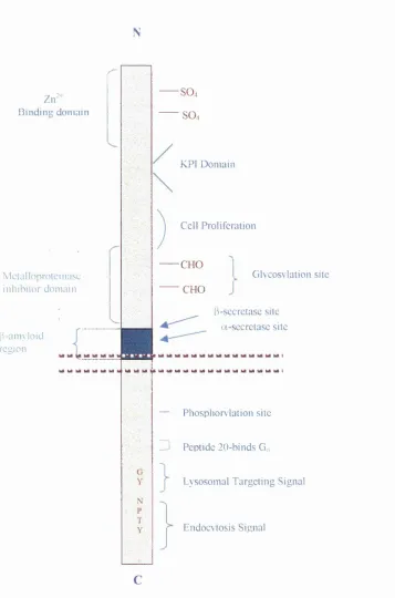
1.1. General structure of PAPP-770... 36
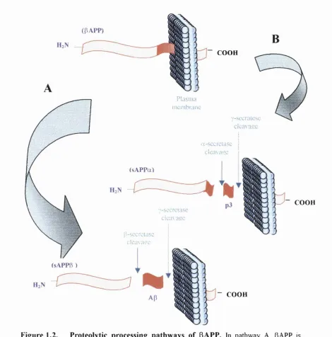
1.2. Proteolytic processing pathways of PAPP... 38
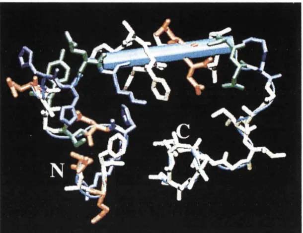
1.3. Structure of the A(3i_4o peptide in water-SDS micelle medium
obtained by NMR... 49
1.4. Prediction of the secondary structure of the A(3 peptide... 50
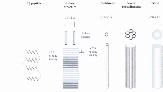
1.5. Schematic representation of Ap polymerisation... 55
3.1 Collagen stimulated platelet release of Ap in normal
and hypercholesterolaemic subjects... 112
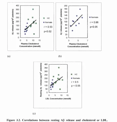
3.2. Correlations between resting Ap release and cholesterol or LDL... 114
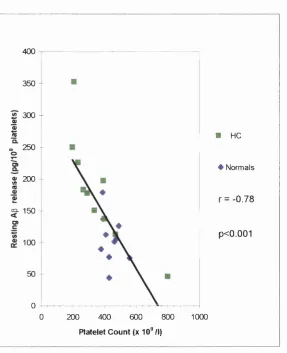
3.3. Correlation between resting platelet AP release and platelet count 115
4.1. Thioflavin-T assay of APmo fibril formation... 124
4.2. Congo Red assay of ApMo amyloid fibril formation... 124
4.3. Influence of apoE isoforms upon A P i^ fibril formation... 126
4.4. Promotion of APi. 4 0 fibrillogenesis by native and oxidised
plasma hpoproteins... 129
4.5. Dose dependent promotion of APi_4o fibrillogenesis by native
and oxidised plasma lipoproteins... 133
4.6. Agarose gel electrophoretic mobility of native and oxidatively
modified VLDL, LDL and HDL... 137
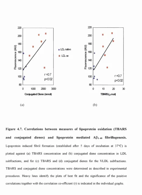
4.7. Correlations between measures of lipoprotein oxidation
List o f Figures
Ap 1^ 0 fibrillogenesis... 138
4.8. Efifect of the presence of antioxidants upon lipoprotein
induced Ap MO fibrillogenesis... 142
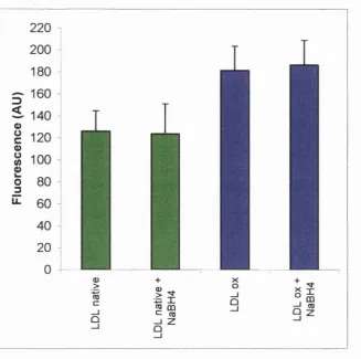
4.9. Effect of aldehyde reduction on lipoprotein mediated
Ap MO fibrillogenesis... 146
4.10. The effect of aminoguanidine treatment upon native and
oxidised LDL induced ApMo polymerisation... 148
4.11. The effect of aminoguanidine on the agarose gel electrophoretic
mobility of native / oxidised VLDL and LDL... 150
4.12. Effect of HNE modification upon the agarose gel electrophoretic
mobility of plasma lipoproteins... 152
4.13. Effect of HNE modification of plasma lipoproteins on
ApMo fibrillogenesis... 155
4.14. Effect of lipoprotein protein removal upon lipoprotein-mediated
Ap MO fibril formation... 162
4.15. Inhibition of ApMO fibrillogenesis by iAp5 in vitro... 166
4.16. Inhibition of VLDL-induced ApMO fibrillogenesis by iAp5 in vitro 167
4.17. Inhibition of LDL induced A P i^ fibrillogenesis by iAp5 in vitro 169
4.18. Effect of iAp5 addition on HDL induced Ap ifibrillogenesis... 171
5.1. Platelet aggregation induced by ADP in the absence or presence
of (a) native and oxidised VLDL, (b) native and oxidised LDL,
(c) native and oxidised HDL... 191
of (a) native and oxidised VLDL, (b) native and oxidised LDL,
(c) native and oxidised HDL... 193
5.3. Platelet responses to ADP stimulation (1 jimol.l'^) in the
presence or absence of soluble or fibrillar A p i^ ... 199
5.4. Platelet responses to ADP stimulation in the presence
of native and oxidised LDL/Ap 1 - 4 0 coincubates... 202
6.1. ApMo induced endothelial toxicity as assessed using the MTT assay 219
6.2. Chymotrypsin-like activity of the 20S proteasome detected
in HAEC lysates... 223
6.3. Effect of APmo treatment on 20S proteasome chymotrypsin-like
activity in HAECs... 225
7.1. Noradrenaline-induced constriction of rat aortic rings
in the presence or absence of soluble APi_4o... 241
7.2. Noradrenaline-induced constriction of rat aortic rings in the
presence or absence of fibrillar Api_4o preparations ... 242
7.3. Vasoactive actions of fibrillar APi- 4 0 preparations on aortic
responses to Inmol.l'^ NA... 243
App 1. Determination of optimal excitation and emission
wavelengths for the ThT assay... 262
App 2. Determination of optimal buffer pH for the ThT assay... 263
List o f Tables
List of Tables.
Table Page
3.1. Plasma lipid profiles in normal subjects and
hypercholesterolaemics... 113
4.1. Characterisation of lipoprotein classes... 136
4.2.
Characterisation of lipoproteins treated or untreated\vithNaBH4... 145
4.3. Characterisation of lipoproteins oxidised in the presence
or absence of AG... 149
4.4. Characterisation of HNE modified lipoproteins... 154
4.5. Characterisation of native/oxidised lipoproteins and
lipid-extracted samples... 158
4.6. Measures of oxidation for complete lipoproteins and
lipid-extracted samples... 159
5.1. ADP stimulated platelet aggregation responses
and 5-HT release... 189
5.2. Platelet responses to ADP stimulation in the presence or
absence of native and oxidised lipoproteins... 196
5.3. Platelet responses to ADP stimulation (Ipmol.f^) in the
presence or absence of soluble or fibrillar APmo... 198
6.1. Extents of survival of endothelial cells treated with soluble or
fibrillar APi. 4 0 or combinations of native or oxidised lipoproteins
App 4. Extents of survival of endothelial cells treated with soluble / fibrillar
Api- 4 0 or combinations of native / oxidised lipoproteins and APi^o,
As determined using the trypan blue excusion assay... 267
App 5. Supernatant LDH activity from endothelial cell cultures treated with
soluble / fibrillar ApMo or combinations of native / oxidised
lipoproteins and APmo... 269
App 6 . Extents of survival of endothelial cells treated with soluble / fibrillar
Api- 4 0 or combinations of native / oxidised lipoproteins and Api-4 0,
Chapter 1: General Introduction
Chapter 1.
1.1. Alzheimer’s disease.
Alzheimer’s disease (AD) is the fourth major cause of death in the developed
world after heart disease, cancer and stroke. It is also the most common form of adult
dementia (Bums et al, 1995). In countries such as Great Britain and the United
States, AD affects approximately 1-6% of the population over the age of 65 and as
many as 20% of individuals over the age of 80. The dramatic rise in life expectancy
during the last century, primarily through improvements in health care and nutrition,
combined with demographic changes, has resulted in a disproportionately large
number of elderly people in the population. Furthermore, in many industrialised
countries, individuals of 85 years of age and older constitute the fastest growing
segment of the population over the age of 65 (Hardy and Allsop, 1991). As a
consequence the incidence of AD is rapidly increasing and it is estimated that by the
year 2025 there will be approximately 23 million AD sufferers globally. The financial
cost to society for diagnosing and managing AD, primarily for custodial care, is
already staggeringly high, and therefore with the ageing of society the economic
burden will continue to increase unless preventative strategies can be formulated.
1.1.1. Neuropathology of AD.
Typically AD is characterised by a loss of memory, perception, judgement and
emotional stability which progressively worsens, usually leading to death in a severely
debilitated, immobile state between four and twelve years after onset. The gradual and
inexorable decline in cognitive function is a result of several major neuropathological
Chapter 1: General Introduction
brain lesions first described by Alois Alzheimer in 1907, neurofibrillary tangles and
senile plaques (Alzheimer, 1907). Since Alzheimer's original description, a number of
other neuropathological features have been recognised in AD such as granulovascular
degeneration, Hirano and Lewy bodies and neuropil threads (Bums et al, 1995).
These degenerative changes, as well as the plaques and tangles, do not occur
uniformly throughout the brain, but are found predominantly in the hippocampus and
amygdala and also in the temporal and parietal cortex (Braak and Braak, 1991). The
neuropathological changes are accompanied by neurochemical imbalances, with
deficits having been reported in cholinergic, noradrenergic, serotonergic and
dopaminergic systems, as well as with respect to amino acid and peptide
neurotransmitters (Bowen et al, 1979; Palmer et al, 1987a; Palmer et al, 1987b;
Procter et al, 1988; Walker et al, 1988; Lowe et al, 1988; Fisher et al, 1992). There
is considerable evidence to suggest, however, that most of these pathological changes
are secondary to the proteinaceous lesions originally described by Alzheimer.
Abnormal fibrous proteins are found in the brains of AD patients not only in
the neurofibrillary tangles and senile plaques but also in the walls of cerebral blood
vessels. All of these lesions are also found, to a lesser degree, in the ‘normal’ elderly.
Most of us who live into our late seventies will develop at least a few senile plaques
and neurofibrillary tangles, particularly in the hippocampus and other brain regions
important for memory. For the most part, the distinction between normal ageing and
AB-'is. quantitative rather than- qualitative. Usually- patients with Alzheimer’s type
dementia have moderately or markedly n>ore mature neuritic plaques, neurofibrillary
molecular biological techniques and other technologies during the 1990’s have aided
our understanding of the processes that distinguish the pathological steps that result in
AD from those present in the ‘normal’ ageing process. Knowledge of the specific
proteins comprising these lesions has led to substantial progress in deciphering the
mechanisms that underlie the development of the AD phenotype.
1.1.2. Neurofibrillary Tangles.
Neurofibrillary tangles (NFTs) are dense bundles of abnormal fibrillar
aggregates that accumulate in the cytoplasm of dying neurons. The aggregates are
formed as a response to the accumulation of an excessive number of phosphate
moieties on the microtubular associated protein, tau (Goedert et al, 1988; Harrington
et al, 1991). In their normal functional state, tau proteins facilitate the formation and
stabilisation of microtubules, structures that in neurons, are primarily involved in
neurotransport. In the phosphorylated state, however, tau is no longer able to
promote the assembly and stability of microtubules (Clark et al, 1998). The
hyperphosphorylated tau dissociates fi'om the microtubules, assumes a paired helical
filament configuration and forms insoluble aggregates, eventually forming NFTs.
Cytoplasmic organelles, as well as RNA, become entrapped in the NFT, possibly
contributing to the dysfunctional state of the neuron and to its eventual death.
NFTs are not unique to AD. Intraneuronal NFTs and tangle like inclusions in
glial cells are also found in a number of seemingly unrelated neurodegenerative
disorders and in ‘normal’ ageing. In many respects, the abnormal tau in these
Chapter 1: General Introduction
The formation of NFTs is quite clearly a characteristic feature of AD, possibly
resulting in neuronal cell death and a consequential decline in cognitive function.
However, the fact that neurofibrillary tangles arise in a variety of etiologically
unrelated diseases seems to suggest that these lesions most probably represent a
response of neurons to a range of insults, and not those specific to AD. It is widely
thought that NFT formation is a relatively late event in the pathogenesis of AD and is
a consequence of the pathological processes that result in senile plaque formation and
cerebrovascular amyloidosis.
1.1.3. Senile Plaques.
Senile plaques are the major neuropathological lesions present in AD. They
are extracellular proteinaceous structures, 50-200pm in diameter, which consist
predominantly of a core of amyloid fibrils (Wisniewski and Terry, 1973). The term
amyloid refers to a group of chemically heterogenous proteins found in a number of
tissues and disease states. A common feature of all types of amyloidosis is that
normally innocuous, soluble proteins polymerise to form insoluble fibrils. Such fibrils
eventually form amyloid deposits that invade the extracellular spaces of organs
destroying normal tissue architecture and function. The amyloid protein specific to
AD is referred to as beta-amyloid (Ap), a 39-42 amino acid peptide derived from a
larger beta amyloid precursor protein (pAPP).
The typical or classical senile plaque is composed of a central, dense core of
radiating Ap fibrils surrounded by a rim of dystrophic neurites together with two
plaques one usually observes microglial cells; scavenger cells capable of responding to
inflammation or the destruction of neuronal tissue. Around the outside of the plaque
are so called reactive astrocytes, glial cells often found in injured brain areas. Deposits
of Ap first appear in the brain parenchyma as loose accumulations, which are referred
to as diffuse plaques (Yamaguchi et al, 1988). These deposits contain very few
dystrophic neurites, microglial cells or astrocytes and the amyloid contained within
these lesions is of a non-fibrillar nature (Yamaguchi et al, 1990a). As the plaques
mature, the amyloid deposits become more dense and filamentous and become closely
associated with a number of other proteins including heparan sulphate proteoglycans
(HSPGs), alphai-antichymotrypsin, various complement components and
apolipoprotein E. All of these molecules have also been shown to influence the
aggregation state of the peptide (Eikelenboom and Stam, 1982; Snow et al, 1988;
Abraham et al, 1988, Namba et al, 1991; Rogers et al, 1992a & 1992b). The
progressive accumulation of fibrillar Ap also coincides with an increasing presence of
dystrophic neurites around the periphery of the amyloid mass and it is the ability of
filamentous Ap to induce neurotoxicity which has implicated the peptide as the key
aetiological factor in the pathogenesis of AD. The majority of amyloid deposits
however, are of the diffuse variety and their presence in normal non-demented
individuals may represent the lesions in a pre-clinical, non-cytotoxic state (Yamaguchi
Chapter 1: General Introduction
1.1.4. Cerebral blood vessel deposits.
Cerebrovascular amyloid deposition is highly prevalent in AD and has been
reported to be present in 62-95% of AD subjects (Vinters, 1987; Premkumar ei al,
1996). This lesion is characterised pathologically by the deposition of Ap in the media
of cortical and meningeal arteries and arterioles, as well as in parenchymal arterioles
and capillaries (Vinters, 1987). Unlike difiuse plaques which are largely composed of
non-fibrillar 42 residue AP peptide, vascular Ap deposits are predominantly of a 40
residue AP species and are assembled into fibrils (Murphy et al, 1994; Gravina et al,
1995). Amyloid deposition in the walls of these blood vessels is accompanied by
several morphological and biochemical alterations. The first recognisable stage in
vascular amyloidosis is the presence of small foci of amyloid at the junction of the
media and adventitia. These early deposits are formed by the attachment of clusters of
fibrillary material to the basement membrane at the abluminal pole of smooth muscle
cells (Wisniewski et al, 1992; Coria and Rubio, 1996). As the amount of amyloid
material increases, it encircles and separates individual smooth muscle cells causing
cellular degeneration (Wisniewski and Wegeil, 1994). Collapse and degeneration of
the endothelium (Kalaria and Hedera, 1995a) as well as thinning of the basement
membrane and narrowing of the vessel lumen are also prominent features (Kalaria and
Hedera, 1995b). Advanced stages of amyloid deposition are accompanied by the
replacement of the media by fibrillary material, with the complete disappearance of
smooth muscle cells (Yamaguchi et al, 1992). Vessels that are particularly heavily
laden with amyloid may be accompanied by the spread of amyloid fibrils into the
membrane in a process referred to as dysphoric angiopathy (Wisniewski et al, 1992).
Indeed, cerebrovascular deposits of AP often coexist with senile plaques in aged
brains and examination of serial sections of AD brains has shown that the core of
senile plaques may be continuous with the microvascular amyloid deposits (Miyakawa
et al, 1982). Furthermore, immunolocalisation of highly specific basement membrane
and endothelial cell components, such as collagen IV and the perlecans in many
extravascular Ap deposits, have suggested a link between vascular amyloid
components and the formation of cortical amyloid deposits (Kalaria, 1992; Kalaria et
al, 1996b). In addition, the presence of vascular amyloid significantly correlates with
senile plaque density in the temporal lobe of demented subjects, although it does not
correlate well with the severity of dementia (Mountjoy et al, 1982). Taken together
these observations suggest that the two types of lesion may, in certain circumstances,
be linked pathologically or could even contribute to each other’s development.
Alternatively, the detrimental actions of Ap upon the vasculature, which essentially
result in vessels that are structurally brittle and are unable to sustain trauma or blood
pressure changes, could have implications for cerebral perfusion and permeability with
consequences for localised neuronal populations.
1.2.
p-amyloid precursor protein.
It is apparent that there is considerable neuropathological evidence to indicate
that deposition of Ap is a central event in the spectrum of brain changes associated
Chapter 1: General Introduction
includes genetic linkage studies which have shown that a subset of familial AD cases
are linked to several different mutations in the P-APP gene (Selkoe, 1994a and
1994b) and in the genes that encode presenilins (Levy-Lahad et al, 1995a;
Sherrington et al, 1995). The P-APP gene consists of 19 exons, is located on
chromosome 21 and encodes for a type I integral membrane protein that is
proteolytically cleaved to different fragments, including the Ap peptides. Most p-APP
gene mutations cause an increase in the proteolytic processing of p-APP, increasing
production of the Ap peptide (Citron et al, 1992; Suzuki et al, 1994; Haass et al,
1994). Furthermore, individuals with Down’s syndrome, who possess an additional p-
APP gene, develop insoluble Ap deposits early in life followed in later years by
neurofibrillary pathology. Significantly, the AD causing mis-sense mutations in the
genes on chromosome 14 and 1, which encode presenilin 1 and presenilin 2
respectively, also affect p-APP processing (Scheuner et al, 1996; Citron et al, 1997).
These gene mutations increase the production of a 42 residue form of the Ap peptide
that is particularly prone to precipitation and aggregation (Lansbury, 1997b).
Abnormal processing of P-APP and increases in the production of AP therefore
appear to be central events in the pathogenesis of the hereditary forms of AD. Since
the pathological lesions in sporadic forms of AD are similar to those in the familial
forms, it is possible that abnormal or alternative processing of p-APP also plays an
important role in these more frequent sporadic forms of the disease. A complete
understanding of the structure, function, cellular expression and proteolytic
processing of P-APP is therefore essential when considering possible pathological
1.2.1. P-APP Isoforms.
The p-amyloid precursor protein has a number of different isoforms that are
generated by the alternative splicing of p-APP mRNA, principally from exons 7, 8 and
15 of the P-APP gene (Kang et al, 1987; Kitaguchi et al, 1988; Ponte et al, 1988;
Golde et al, 1990; Sandbrink et al, 1994a). The most abundant transcripts are p-APP-
695, P-APP-714, p-APP-751 and P-APP-770. All of these spliced forms of mRNA
encode for multidomain proteins with a single membrane-spanning region, a small
cytosolic carboxy-terminal tail and a large extracellular N-terminal domain. The Ap
peptide domain has its carboxy terminus about halfway through the membrane bilayer
and extends into the extracellular or intraluminal domain (Beyreuther and Masters,
1991). The difference between these isoforms occurs in the N-terminal domain and
results from alternative splicing of exons that encode a Kunitz-type protease inhibitor
(KPI) domain and a surface glycoprotein found on neurons, platelets and endothelial
cells called MRC OX-2. While P-APP-695 contains neither of these sequences, p-
APP-714 possesses the MRC OX-2 domain, p-APP-751 contains the KPI domain and
P-APP-770 contains both. These differences are likely to reflect the function and
cellular expression of the various isoforms. For example p-APP-695 predominates in
brain tissue, while the other forms are most abundant in peripheral tissues. Two
secretory variants of P-APP that do not contain the AP peptide sequence have also
been described, namely P-APP-365 and P-APP-563, and these proteins are referred to
as amyloid precursor like proteins (/\pLP) in order to distinguish them from the
Chapter 1: General Introduction
1.2.2. p-APP Function.
The functions of the p-APP isoforms remain poorly understood despite
intensive research efforts. The overall structure of the protein however, suggests that
p-APP functions as a receptor or growth factor (Rossjohn et al, 1999). Indeed the C-
terminal domain of p-APP has been reported to interact with a number of key proteins
that are linked to molecular signalling pathways, including the GTP binding protein Go
(Nishimoto et al, 1993), the adaptor protein Fe65 (Russo et al, 1998) and the
microtubule-associated protein PATl (Zheng et al, 1998). p-APP has also been
implicated in pro- and anti-apoptotic actions (Yamatsuji et al, 1996; Wolozin et al,
1996; Xu et al, 1999) which, it must be noted, does not contradict the growth
factor/receptor hypothesis, since such molecules are known to be intimately involved
in apoptosis. However, APP knockout mice display only subtle neurological deficits
and so a role for P-APP in apoptosis seems unlikely. Studies using p-APP knockout
mice have revealed deficiencies to be displayed in neurite outgrowth and survival
(Perez et al, 1997) suggesting an involvement of p-APP in sustaining neuronal
viability, in axonogenesis and in dendritic arborisation.
A number of functions have also been ascribed to the large extracellular N-
terminal domain, primarily due to the identification of several functional domains on
P-APP. A sequence of 5 amino acids in the N-terminal domain (RERMS sequence)
for example, has been shown to have certain growth promoting properties, stimulating
proliferation in non-neuron,^ ce% %nd neurite outgrowth in neuronal cells (Ninomiya
et al, 1993; Ninomiya et al, Jii) f t a/, 1994). The N-terminus also contains two
proteoglycans (Williamson et al, 1996) (Figure 1.1). It has been proposed that this
region of p-APP may be involved in cell-cell and cell-substratum adhesion (Breen et
al, 1991; Breen, 1992). P-APP is also known to be involved in other binding
interactions including Cu (II) and Zn(II) binding activities. The Zn(II) binding is
assumed to play mainly a structural role (Bush et al, 1993b), whereas the ability of P-
APP to catalyse the reduction of Cu(II) to Cu(I) (Multhaup et al, 1996) is evidence,
perhaps, of antioxidant properties (Goodman and Mattson, 1994).
P-APP isoforms containing the KPI insert in the N-terminal domain (p-APP-
751, p-APP-770) have been reported to affect blood coagulation involving platelets.
This action is believed to be directly mediated by the KPI domain, which acts to
inhibit factor XIa of the coagulation cascade (Van Nostrand et al, 1995; Mahdi et al,
1995). The p-APP-751 form was shown to be identical to protease nexin II, a platelet
a-granule constituent, providing further evidence for an involvement of P-APP in
blood coagulation and platelet aggregation activities (Oltersdorf et al, 1989; Van
N
Chapter I: General Introduction
Z ir" B inding dom ain
M ctalloprolcinasc inhibitor dom ain
b -a n n lo id region
SO4 SO4
KPI D om ain
C ell Proliferation
■CHO
CHO
G lv co s\la tio n site
fCsecretasc site
a-sccrctasc site
— Phosphory lation site
Z) Peptide 20-binds Go
L ysosom al Targeting Signal
E ndocvtosis Signal
Figure 1.1. General structure of PAPP-770. A number o f the proposed functional
1.2.3. Proteolytic processing of p-APP.
Newly synthesized P-APP is subject to post-translational modifications in the
endoplasmic reticulum and the Golgi apparatus, where it acquires N- and O- linked
carbohydrates, tyrosine sulphates (Weidemann et al, 1989) and phosphates (Walter et
al, 1997). p-APP is then taken from the trans-Go\g\ network, inserted into surface-
destined vesicles and transported to the cell surface. Following maturation, the
various forms of p-APP can be subjected to further intracellular processing by two
independent proteolytic pathways. Proteolysis by either of these pathways generates a
number of secreted and membrane bound P-APP derivatives. The principal proteolytic
cleavage of p-APP is performed by a protease designated a-secretase. This enzyme
cleaves p-APP within the Ap peptide sequence, between residues 612 and 613,
releasing a large, soluble N-terminal domain (designated APPsa) and leaving a lOkD
membrane bound carboxyl-terminal fragment termed C83. Alternatively, to initiate the
generation of the amyloidogenic Ap peptide fragment, P-secretase cleaves p-APP at
the NHz-terminus of Ap to release a distinct lOOkD soluble NH2-terminal domain
(APPsP) and leaving a 12kD membrane-bound carboxyl-terminal fragment, C99. Both
C99 and C83 can be further cleaved by one or more y-secretases leading to the release
Chapter I: General Introduction
(PAPP)
COOH
Plasma membrane
■/-sccralesc
dca^asc
A-sccrctasc
dcn\ame
(sAPPa)
7-sccrctasc
cicm a u c
p-sccrctasc
cicaxacc
(sAPPB )
COOH
m m
COOH
Figure 1.2. Proteolytic processing pathways of (3APP. In pathway A, pAPP is
cleaved at the amino terminus o f the Ap sequence by P-secretase, releasing a large N-terminal
derivative termed sAPPp. Subsequent cleavage o f the C-terminal fragment by y-secretase
generates Ap. Alternatively, in pathway B, pAPP is cleaved within the Ap sequence by a
-secretase to generate sAPPa and subsequent cleavage by y--secretase generates a truncated Ap
Initially it was believed that the reaction catalysed by the a-secretase cleavage
constituted a physiological mechanism, since p-APP cleavage within the AP peptide
sequence precludes the formation of the amyloidogenic fragment (Esch et al, 1990;
Sisodia et al, 1990). This proposed mechanism may, however, be an
oversimplification of what occurs physiologically, since in most cell types, very little
P-APP is cleaved by a-secretase, with most of the P-APP protein remaining intact.
Also, a number of studies have shown that both P- and y-secretase processing of P-
APP occurs under physiological conditions, indicating that all the fragments of p-
APP, including the Ap peptide, are normal products (Haass et al, 1992b; Seubert et
al, 1992).
1.2.4. a-secretase.
The a-secretase mediated processing o f P-APP is similar to the processing of
a number of other integral membrane proteins, such as TNF-a, TGF-a and L-selectin
(Werb and Yan, 1998). With all of these proteins, processing consists of a constitutive
component, which allows continuous proteolysis, and a regulatory element that can be
activated via protein kinase C (Nitsch et al, 1992; Buxbaum et al, 1993; LeBlanc et
al, 1998) and other second messenger cascades (Mills and Reiner, 1999). A number
of enzymes have been proposed as candidates for the constitutive and regulatory
components of a-secretase. These proposed enzymes belong to the ADAM (a
disintegrin and metalloproteinase) family of proteases. The first of these enzymes to
be identified was a protease known to be responsible for TNF-a release called tumor
Chapter 1: General Introduction
showed a number of defects in the processing of integral membrane proteins (Peschon
et al, 1998), including defects in fibroblast APP secretion. These defects were only
evident however, under PKC stimulated conditions (Buxbaum et al, 1998). TACE
therefore plays a role only in the regulatory component of the a-secretase processing
of P-APP in fibroblasts.
Recently, two other members of the ADAM family have also been implicated
in a-secretase processing of p-APP, ADAM 10 (Lammich et al, 1999) and MDC9
(Koike et al, 1999). Co-transfection of ADAMIC or MDC9 with p-APP results in the
increased regulatory and constitutive secretion of APPsa. Furthermore, in vitro
studies have shown that ADAMIC can specifically cleave synthetic substrates that
contain the a-secretase cleavage site. A dominant negative form of ADAMIC was
also shown to interfere with endogenous a-secretase processing of P-APP (Lammich
et al, 1999). The presence of ADAMIC at the cell surface but also in the Golgi
apparatus and in surface-destined vesicles agrees with previous reports providing
evidence for a-secretase processing of p-APP in these subcellular compartments
(Sisodia et al, 1992; Kuentzel et al, 1993; De Strooper et al, 1993; Haass et al,
1995a). Conclusive identification of candidate a-secretase enzymes, such as
ADAMIC, will not be possible until the in vivo biological effects of these proteases
have been thoroughly examined.
1.2.5. P-secretase.
The identification of candidate enzymes for the P-secretase cleavage has
the amyloidogenic Ap peptide fragment. Recently a number of investigators have
identified a promising candidate for P-secretase, referred to as BACE-1 (beta-site
APP-deaving enzyme-1) (Hussain et al, 1999; Sinha et al, 1999; Vassar et al, 1999;
Yan et al, 1999). BACE-1 is a type I integral membrane protein with an extracellular
domain that contains an aspartyl proteinase catalytic site. Crucially, this protease has
been shown to exhibit the properties expected of P-secretase, cleaving synthetic
peptides that contain the p-secretase cleavage site. Furthermore, transfection of
BACE-1 into cells increases P-secretase processing of P-APP, as recognised by the
generation of APPsp and C99 fragments, whereas specific inhibition of BACE-1 by
antisense oligonucleotides has been shown to reduce P-secretase activity.
Interestingly, forms of p-APP that contain a missense mutation linked to a familial
form of AD (Swedish double mutation), and which are known to cause enhanced p-
secretase processing of p-APP (Citron et al, 1992), are preferentially cleaved by
BACE-1 over wild type APP forms. This provides further evidence to support the
notion that BACE-1 is a true p-secretase candidate.
In addition to matching the biochemical properties of P-secretase, BACE-1
also shows the expected cellular expression and subcellular localisation patterns.
Most, if not all cells express P-APP and generate Ap, and p-secretase is therefore
expected to have a widespread tissue distribution. In agreement with this view,
BACE-1 mRNA has been reported to be present in most peripheral tissues, with a
moderately higher expression in the brain, particularly in neuronal cells (Vassar et al,
Chapter 1: General Introduction
subcellular compartments of the secretory pathway including the Golgi apparatus,
trans-Go\%i network, secretory vesicles and endosomes (Haass et al, 1995a; Stephens
and Austen, 1996; Skovronsky et al, 2000). BACE-1 functions optimally at low pH
and localises within intracellular compartments of the secretory pathway, particularly
the Golgi apparatus and endosomes. Furthermore BACE-1 overexpression induces P-
secretase cleavage in these compartments. These characteristics of BACE-1 are
consistent with previous findings that P-secretase activity is found in such locations
only. The identification of candidates for both a - and p-secretase enzymes in the same
subcellular compartment, i.e. in the trans-Go\g\ network, may therefore suggest that
these two proteases compete for the same substrate in this location. Further evidence
to support BACE-1 as a candidate for p-secretase comes from studies which have
shown that the predicted membrane topology of BACE-1 places the active site in
correct topological orientation relative to the p-secretase cleavage site in p-APP
(Vassar et al, 1999). This evidence avoids any need to invoke unusual mechanisms to
explain how the enzyme gains access to its substrate.
Genome searches have indicated that at least one other BACE homologue
exists, and indeed recent studies have identified BACE-2, a p-secretase believed to be
important in a familial form of AD known as the Flemish mutation (Farzan et al,
2000). This may suggest that multiple forms of BACE exist, possibly representing a
1.2.6. y-secretase.
The y-secretase activity is the final cleavage event that releases the Ap peptide
from the p-secretase cleaved carboxyl terminal fragment of P-APP. One of the more
unusual aspects of y-secretase cleavage is that its cleavage site lies within the
hydrophobic environment of the cell membrane, a region not normally associated with
the hydrolysis of peptide bonds. Proteolysis of integral membrane proteins in or close
to the transmembrane domain however, does appear to be important in several other
signalling pathways. The best studied examples of such intramembranous cleavage
mechanisms include the processing of the sterol receptor element binding protein
(SREBP), Notch and several mitochondrial inner membrane proteins (Sakai et al,
1996; Brown and Goldstein, 1996; Leonhard et al, 1996; Schroeter et al, 1998).
Various mechanisms have been proposed for the transmembrane domain cleavage of
these proteins, each including the presence of co-factors that allow y-secretase to gain
access to its substrate. Such theories rely on the presence of co-factors that create
membrane microdomains which allow entry of water for hydrolysis reactions,
cofactors that aid transport of the substrate to intracellular compartments in which y-
secretase resides, or factors that may be involved in the transport or maturation of y-
secretase itself. Candidates for the y-secretase enzyme have yet to be identified,
although several have been proposed. These include the presenilin proteins, although
their role as co-factors is more likely, prolyl endopeptidase, calpain and the
proteasome (Mundy, 1994; Shinoda et al, 1997; Yamazaki et al, 1997). Further
Chapter 1: General Introduction
the identity of y-secretase, although the idea of a multifactorial y-secretase is certainly
feasible.
1.3.
P-amyloid.
Following processing by p- and y-secretase, the N-terminal derivatives of P-
APP, including Ap, are secreted into an aqueous, extracellular environment in a
soluble monomeric form. Numerous AP species exist in biological fluids and in the
tissues associated with their deposition, these peptides having extensive amino- and
carboxyl-terminal heterogeneity (Golde and Younkin, 1996). The major species
secreted by cells and which can be detected in human plasma and CSF is APi^o (>60-
70%), although some Api^i (-15%) is also present along with minor amounts of
other peptide species, i.e. Api-2 8, Api-3 3, Api.3 4, AP3-3 4, Api.3 7, Api- 3 8 and APi. 3 9
(Seubert et al, 1992; Suzuki et al, 1994; Ida et al, 1996; Wang et al, 1996). Although
ApMo is the major species produced, the principal species deposited in the
parenchyma of the AD brain is APi- 4 2 (Iwatsubo et al, 1994; Gravina et al, 1995).
Thus, species ending at AP42, which constitute a relatively minor component of the
Ap that is normally secreted by cells (with the exception perhaps of neuronal cells) in
many cases make up the majority of the AP that is deposited in the AD brain.
However, significant amounts of APwo are deposited in the typical late onset AD
brain, and in most studies, ApMo appears to be the predominant form associated with
1996). Both of these A(3 species therefore seem to be intimately involved in the
pathogenesis of AD. The main difference between these two forms of the Ap peptide
however, is their propensity to polymerise into fibrils, an event that possibly leads to
amyloid deposition and ultimately AD. The mechanisms that are involved in the
polymerisation of Ap peptides has been a focus for many research groups during the
past decade, and this has lead to a greater understanding of the factors that are
involved in the development of the AD phenotype.
1.3.1. Cellular origins of Ap.
The cellular origins of the AP that is deposited in cerebral blood vessels and
parenchymal tissues remains a contentious issue despite numerous studies. As outlined
above, pAPP, the source of the amyloidogenic Ap fragment, is widely expressed in a
number of cell types throughout the body. Most, if not all, of these cells are capable of
producing the amyloidogenic Ap fragment through proteolytic cleavage of the
precursor, particularly cells of a neuronal and vascular origin (smooth muscle cells
and endothelial cells). These cells have been reported to show unusually high
expression levels of PAPP and to produce significant quantities of Ap (Ciallella et al,
1994; Davis-Salinas et al, 1995; Wisniewski et al, 1995; Vassar et al, 1999). This has
led many authors to advocate either a neuronal or circulatory source as that
responsible for deposition of Ap within the AD brain. Evidence seems to suggest that
neuronal and circulatory sources may both make a significant contribution to the
deposition of Ap, although the peptide from these two sources may be deposited in a
Chapter 1: General Introduction
with blood vessel amyloid (Glenner and Wong, 1984) and is the predominant form
detectable in plasma, whereas the longer Ap 1 . 4 2 species, which is less soluble and more
cytotoxic, is the dominant form found in plaques (Masters et al, 1985) and is thought
to originate from neuronal cells. Differences in Ap peptide length between vessels and
plaques may be due to different processing of the precursor protein in vascular and
neural cells and/or post-depositional processing due to local tissue factors. This would
seem to indicate that Ap of a circulatory origin contributes to cerebral amyloid
deposits and ultimately CAA, whereas neuronal Ap contributes to parenchymal
deposits and senile plaque formation. Since cerebral blood vessel deposits of Ap tend
to coexist with senile plaques, however, it is possible that amyloid from a circulatory
source could influence or contribute to deposition within the parenchyma, and vice
versa. A recent study in which male rats were intravenously infused with soluble APi_
4 0 demonstrated that soluble circulating Ap can cause disturbances in the blood brain
barrier (BBB) with resultant pathological events becoming evident in the CNS and
periphery (Su et al, 1999). Furthermore, this study reported that several parenchymal
brain regions important for cognition also showed pathological deterioration. All of
these brain regions were associated with extensive glial cell activation, evidence
consistent with previous observations that AP activates cultured astrocytes (Pike et
al, 1994) and which suggests a reactionary response to damaged cerebral vessels,
perhaps as an attempt to maintain BBB integrity. Considerable evidence has implied
that perturbation of the BBB is involved in the pathogenesis of AD, mainly through
studies that have detected serum proteins in the parenchyma of AD brain tissue
support the hypothesis that one pathogenic mechanism for A(3 deposition in AD may
be via disruption and deterioration of the cerebrovasculature. A recent study
supporting this view reported that the distribution of A(3 in white matter correlated
with the distribution of blood vessels, suggesting that its origin may be vascular rather
than neuronal (Iwamoto et al, 1997). Additional support for this theory comes from a
report that plasma Ap levels are elevated in familial AD as well as in 13% of sporadic
AD individuals (Scheuner et al, 1996), indicating that circulatory Ap may influence
the development of AD in certain individuals.
1.3.2. Platelets and Ap release.
Apart from smooth muscle cells and the endothelium, a significant amount of
Ap has been detected in human platelets. These cells have been shown to account for
approximately 90% of the total anti- Ap immunoreactivity that is detectable in whole
blood (Chen et al, 1995) and are thus the primaiy source of Ap in the human
circulation. Furthermore, Ap has been suggested to reside within the secretory
granule components of platelets and has been shown to be released upon activation by
agonists that result in degranulation (Smith C.C.T, 1997; Li et al, 1998). Exactly how
pAPP is processed within platelets to yield amyloidogenic fragments that are
subsequently released upon activation is unknown. It has however been suggested
that Ap may be released from secretory granules into the cytosol upon proteolysis of
pAPP, possibly as a consequence of platelet activation (Smith, C.C.T., 1997). It is
Chapter 1: General Introduction
concentrations of Ap, which ultimately lead to enhanced amyloid deposition within
the cerebral vasculature. Several studies have shown that platelet hyperactivity is
associated with AD and normal ageing (Gleerup and Winther, 1988; Sevush et al,
1998) and that elevated concentrations of ApMo are present in the plasma of these
individuals (Shinkai et al, 1995). Conditions that are otherwise associated with
platelet hyperactivity, such as hypertension, hyperlipidaemia and arterial injury (Smith,
C.C.T. et al, 1989; Smith, C.C.T. et al, 1992; McAuliffe et al, 1993), may therefore
result in increased plasma concentrations of AP which may present these individuals
with an increased risk of developing AD.
1.3.3. General structural characteristics of Ap.
The first partial amino acid sequence of Ap was reported by Glenner and
Wong (1984) with the complete sequence later being described by Kang et al (1987).
The Ap peptide contains two hydrophobic domains comprising residues 17-21 and
residues 29-40/42, the latter sequence representing the C-terminus of the peptide that
is reputedly embedded in the plasma membrane. As one might expect, this sequence is
rich in the hydrophobic amino acids valine and isoleucine, residues often found in
proteins with p-sheet structure (Chou and Fasman, 1978). Structural prediction
studies confirm this assumption showing a high probability for P-sheet structure in the
C-terminus from residue 28 (Soto et al, 1994) whereas the probability is lower
between residues 9 and 21. This latter sequence may show either an a-helical or P-
sheet structure. Two P-turns are also predicted between residues 6 and 8, and
Structure that consists o f tw o (3-strands separated by an a -h elical (3-tum (Figures 1.3
and 1.4).
Figure 1.3. Structure of the Api-40 peptide in water-SDS micelle medium
obtained by NMR. N and C indicate the N and C termini, respectively and a short a
-helical segment is indicated by the blue rod. Acidic and basic amino acid residues are shown
m blue and orange, respectively, whereas green and white denote the positions o f polar and
non-polar residues (taken from Roher et al, 2000, with permission).
The structural determination o f soluble Ap in aqueous buffers under physiological
conditions has been fraught with problems due to the propensity o f the AP peptides to
aggregate. Substantial structural information has been obtained, how ever, by using
Chapter I: General Introduction
and detergents such as S D S to maintain the peptide in its soluble formation. U sing
nuclear m agnetic resonance spectroscopy (N M R ), circular dichroism (C D ) and
Fourier transform infra-red sp ectroscopy (FTIR), it has been reported that Ap
generally adopts an a -h elica l conform ation in organic solvents, w hereas in aqueous
buffers it is predominantly in the p-sheet conform ation (Hilbich et al, 1991a; Fraser et
al, 1992b, Zagorski et al, 1992, Shen et al, 1994; S oto et al, 1995; Lansbury et al,
1995; El A g n a f et al, 1998). The presence o f helices, loops and kinks in various
regions o f the AP peptide have also been described, how ever these structures seem to
be largely dependent upon external environmental influences, such as pFl and the
solvent em ployed (Sticht et al, 1995, C oles et al, 1998; Shao et al, 1999). It is likely
that Ap exists in a variety o f different conform ations that are dependent upon
environmental conditions, interactions with other m olecules and metal binding ability
(S o to et al, 1994, Bush et al, 1994) The variability in secondary structures reported
may therefore represent a transition o f A p from a -h elix to random coil to p-sheet in
the process o f fibrillogenesis.
High probabilit>
^ or h for (1-sheet
conformation
1 2 3 4 5 6 7 8 9 10 11 12 13 14 15 16 17 18 19 20 21 22 23 24 25 26 27 28 29 30 31 32 33 34 35 36 37 38 39 40 41 42
J J
: 1■-...
P-tum Central P-tum Highly hydrophobic
hydrophobic region
cluster
Ap p-sheet content is tightly linked to insolubility, fibril formation and
cytotoxicity (Halverson et al, 1990; Lansbury, 1999). It is thought that the Ap
peptide undergoes a conformational change during fibrillogenesis from a-helical to P-
sheet structure. Studies using synthetic fragments of the Ap sequence have been
fruitful in determining the regions and peptide sequences of the Ap molecule
responsible for such conformational changes. These studies have shown that the
markers of P-pleated sheet conformation are present within a stretch of 15 amino
acids, between residues 14 and 28 (Gorevic et al, 1987). A recent study supporting
this prediction has reported that the smallest region of the N-terminus of AP capable
of forming amyloid fibrils stretches over residues 14-23 (Tjemberg et al, 1999).
Deletions or substitutions in this region result in the loss of fibril forming ability,
suggesting that this sequence forms the core of amyloid fibrils (Hilbich et al, 1991a;
Wood et al, 1995). This becomes particularly apparent when making substitutions
within the hydrophobic domain of residues 17-21 (Hilbich et al, 1992). It has been
suggested that this hydrophobic sequence may be important in the process of
dimérisation, an event that forms the basic building block of the amyloid fibril (Chaney
et al, 1998; Roher et al, 2000). In sharp contrast, the C-terminal hydrophobic domain,
residues 29-40/42, has been shown not to be necessary for fibril formation, but may
be important in aiding insolubility (Gorevic et al, 1987). This region has been reported
to determine the rate of AP fibril formation rather than the stability and structural
properties of the fibrils (Jarrett et al, 1993a, 1993b and 1993c). This may explain the
differences in solubility and rates of fibril formation observed between A P i^ and APi.