Review
1
Marine
carbohydrate-based
compounds
with
2
medicinal properties
3
Ariana Vasconcelos1,2, and Vitor Pomin1,2,3,*
4
1 Program of Glycobiology, Institute of Medical Biochemistry Leopoldo de Meis, Federal University of Rio de
5
Janeiro, Rio de Janeiro 21941-590, Brazil;
6
2 University Hospital Clementino Fraga Filho, Federal University of Rio de Janeiro, Rio de Janeiro 21941-913,
7
Brazil;
8
3 Department of BioMolecular Sciences, and Research Institute of Pharmaceutical Sciences, School of
9
Pharmacy, University of Mississippi, MS 38677-1848, USA.
10
* Correspondence: [email protected], [email protected] or [email protected]; Tel.: +1 (662)
11
915-3114.
12
13
Abstract: The oceans harbor a great diversity of organisms, and have been recognized as an
14
important source of new compounds with nutritional and therapeutic potential. Among these
15
compounds, carbohydrate-based compounds are of particular interest because they exhibit
16
numerous biological functions associated with their chemical diversity. This gives rise to new
17
substances for the development of bioactive products. Many are the known applications of
18
substances with glycosidic domains obtained from marine species. This review covers the
19
structural properties and the current findings on the antioxidant, anti-inflammatory, anticoagulant,
20
antitumor and antimicrobial activities of medium and high molecular-weight carbohydrates or
21
glycosylated compounds extracted from various marine organisms.
22
Keywords: marine organisms; carbohydrate; glycoside; antioxidant; anticoagulant;
23
anti-inflammatory; antitumor; antimicrobial.
24
25
1. Introduction
26
The oceans cover about 70% of the earth's surface, and harbor a great diversity of living beings,
27
ranging from unicellular bacteria to large multicellular mammals [1]. The large biodiversity of the
28
marine environment is also accompanied with great chemical variety, which makes this habitat a
29
promising source of new biomedically active molecules [2, 3]. Currently some products obtained
30
from marine sources are in the clinical trials phase for possible use as analgesics [4], anti-cancer
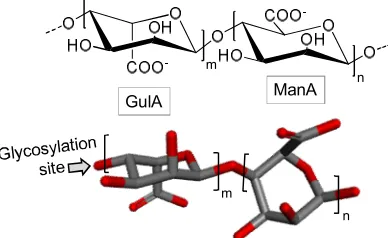
31
drugs [5], and for treatments against viruses [6-8]. Despite these studies, marine potential remains
32
largely unknown.
33
Among the promising but poorly explored marine molecules are carbohydrates, which stand
34
out for their varied structural and chemical characteristics. Besides participating in energy storage
35
and as structural component (especially in exoskeletons of invertebrates), carbohydrates play also
36
many other key biological roles such as fertilization signaling [9-11], pathogen recognition [12],
37
cellular interactions [13], tumor metastasis [14], in addition to important pharmacological activities
38
such as antitumor [15, 16], antiviral [17, 18], anticoagulants [19], antioxidants [20] and
39
anti-inflammatory [2, 21, 22].
40
In this review we will discuss about the structural and biological aspects of the various
41
carbohydrate-based compounds of marine origin endowed with potential biomedical and
42
biotechnological applications. The main goal of this report is to reinforce to the scientific community
43
the great value of marine-derived carbohydrates and glycosylated compounds of medium- and high
44
molecular-weight (MW) to drug discovery and development. Although these molecules can present
45
actions on multiple systems, attention is made more on their antioxidant, anti-inflammatory,
46
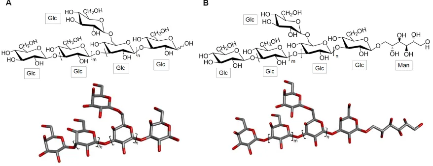
anticoagulant, antitumor and antimicrobial properties.
47
2. Diversity of carbohydrates from marine sources
48
Carbohydrates are the most abundant biomolecules on Earth considering cellulose and chitin as
49
the main representatives. These organic compounds act not only as the main energy source (as seen
50
in starch and glycogen) but also as biologically functional structural players in events of cellular
51
recognition, especially when present at the cell surface [23]. Carbohydrates are also the most
52
complex biomolecules in terms of structure. The enhanced dynamic behavior, large conformational
53
fluctuations, diversity of monomeric units accompanied by various enantiomers, multiple types of
54
glycosidic bonds, and extensive post-polymerization modifications are factors that contribute to
55
increase the structural complexity of these molecules. The carbohydrate classes are also vast. It can
56
include both neutral and negatively charged saccharides with variable lengths. Famous examples
57
are the N-linked or O-linked oligosaccharides in glycoproteins, glycosaminoglycan (GAG) in
58
proteoglycans, glycolipids, sulfated fucans, sulfated galactans, among many other highly
59
glycosylated products [24].
60
3. Structure and function
61
3.1. Alginic acid
62
Alginic acid is a polysaccharide obtained from brown algae. It has linear structure and consists
63
of β-D-manuronic acid (ManA) and α-L-guluronic acid (GulA) in repeating building blocks. These
64
building blocks may be composed of consecutive GulA residues [GulA-GulA-GulA-GulA]n,
65
consecutive ManA residues [ManA-ManA-ManA-ManA]n, or alternating ManA and GulA residues
66
[GulA-ManA-GulA-ManA]n (Figure 1) [25]. This polysaccharide has a wide spectrum of application
67
in medicine, in food industry, in biotechnology and in other industrial sectors [26].
68
69
GulA ManA
n m
n m
70
Figure 1: Chemical structure of alginate. It is composed of building blocks of α-L-guluronate
71
(GulA) and β-D-mannuronate (ManA) units.
72
In the study of So and colleagues, alginic acid has been presented as a promising antioxidant
73
Sarithakumari and coworkers investigated the antioxidant and the anti-inflammatory potential of
75
alginic acid isolated from the brown algal species Sargassum wightii by in vivo assays using rats with
76
induced arthritis [28]. Histopathological analysis of the animal paw tissue showed that treatment
77
with alginic acid has the capacity to decrease the paw edema as well as the inflammatory infiltrates
78
in the studied animal models. The polysaccharide was also able to reduce the activity of enzymes
79
such as cyclooxygenase, lipoxygenase and myeloperoxidase, besides reducing the levels of
80
C-reactive protein, ceruloplasmin and rheumatoid factor. Reduction of lipid peroxidation and
81
increased antioxidant enzyme activity was also reported [28].
82
Supportively Endo and associates showed in a separate work two years later that alginic acid is
83
able to eliminate free radicals and reduce the ferrous ion in stored pork [29]. The antioxidant activity
84
of alginic acid was attributed to its capacity to chelate metal, to scavenge free radicals and to reduce
85
ferric ions in the tissue. This last ability is quite useful in light of the elevated levels of Fe ions in pork
86
meat. The literature also reports the antimicrobial activity of this polysaccharide [30]. For instance, in
87
the work of Neettoo and collaborators, an alginate-based coating was tested in order to increase the
88
microbiological safety in digestions of cold-smoked salmon. This study demonstrated the efficacy of
89
alginate to control the growth of Listeria monocytogenes, a bacterium responsible for serious
90
infections, mainly those caused by salmon uptake [30].
91
3.2. Sulfated polysaccarides
92
3.2.1. Fucoidan
93
Of complex structure fucoidans are obtained from brown algae. They generally consist of a
94
backbone mostly 3-linked α-L-fucopyranose (Fuc) (Figure 2A) or alternating α-L-Fuc residues with 3-
95
and 4-glycosidic linkages (Figure 2B). Either case can be replaced with sulfate or acetyl groups,
96
and/or side branches containing Fuc or other glycosyl units [31]. In addition to Fuc residues, they
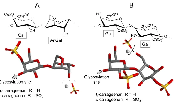
97
may contain small amounts of several other monosaccharide types, such as glucose (Glc), galactose
98
(Gal), xylose and/or mannose [32].
99
100
Glycosylation site
Fuc
B
Glycosylation site Fuc
A
101
Figure 2. Fucoidans are polymers mostly composed of α-L-fucopyranose (Fuc) residues either
102
(A) mostly 3-linked or (B) 3- and 4-linked.
103
One of the first attempts to propose the fucoidan structures was made in 1950 by Percival and
104
understand some of the fucoidan’s biological action, Patankar and coworkers have revised the
106
fucoidan structure four decades later and described it as a polysaccharide consisted mainly of
107
3-linked α-L-Fuc units (Figure 2A) [34]. More recent papers stated that Fuc units in fucoidan
108
backbone can occur in the α-1,2 linkage type besides the α-1,3 and/or α-1,4 bonds [35]. It was also
109
stated that sulfation can occur at positions 2, 3 and/or 4 as well [35]. Despite the many published
110
works regarding fucoidan, the relationship between structure and biological activities is not clearly
111
and easily established because of the obstacles in full structure determination [36, 37]. However, the
112
scientific interest on fucoidans is so appealing because of the large spectrum of application - much
113
research has annually been carried out in terms of both structure and biomedical function [38].
114
The highly cited review of Fitton covered potential applications of fucoidans in several types of
115
therapies in which it was observed that the anti-inflammatory potential of fucoidan lies on its
116
pleiotropic effects. These include selectin inhibition, complement inhibition and enzyme inhibitory
117
activity [39]. In a comparative study of the anticoagulant property of fucoidans extracted from
118
various species of algae, Laminaria saccharina was the one presenting the fucoidan with the highest
119
level of activity [40, 41]. In vitro and in vivo assays are capable to evaluate the safety and clinical
120
effects of fucoidan ingestion on hemostasis. Very strong in vitro anticoagulant activity has been
121
presented as opposed to a modest effect on the in vivo assay [42]. Investigations on the antioxidant
122
activity of fucoidans conclude that oral administration of fucoidan may lower serum parameters
123
such as triacylglycerides, total cholesterol, low-density lipoprotein cholesterol and plasma Glc
124
levels, and improve the anti-oxidation and innate immunity of catfish Pelteobagrus fulvidraco [43].
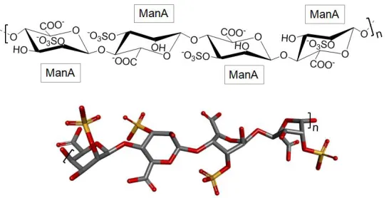
125
Other works have reported the anti-cancer activity of fucoidans extracts. An example is the
126
well-cited paper from Cumashi and collaborators in which nine different fucoidans have been
127
screened in terms of their multiple biomedical properties [40]. It has been shown that fucoidans from
128
L. saccharina, Laminaria digitata, Fucus serratus, Fucus distichus and F. vesiculosus have the capacity to
129
block adhesion of MDA-MB-231 breast carcinoma cells, resulting in potential beneficial therapeutics
130
against tumor metastasis [40]. Following the same rational, other works have demonstrated that
131
fucoidans of other seaweed species such as Ecklonia cava, Sargassum hornery and Costaria costata can
132
present positive effects on human melanoma and colon cancer [44]. Fucoidans from other brown
133
seaweeds like Saccharina japonicus and Undaria pinnatifida possess high antitumor activity and can
134
inhibit proliferation and colony formation of breast cancer and melanoma cancer cell lines [45].
135
Fractions of native fucoidan and its derivatives have shown activity against the formation of colonies
136
of two colorectal carcinoma cells, DLD-1 and HCT-116 [46].
137
The literature also mentions the antimicrobial properties of fucoidans [8, 47]. An example is the
138
publication of Thuy and associates, where the anti-human immunodeficiency virus (HIV) activity of
139
fucoidans extracted from three brown algae Sargassum mcclurei, Sargassum polycystum and Turbinara
140
ornata is reported [47]. All these fucoidan types tested in this work exhibited anti-HIV effects. The
141
mechanism of action has been attributed to the capacity of the fucoidan in blocking the first steps of
142
HIV entry into the target cells [8, 47]. A very recent study illustrates the synthesis of silver
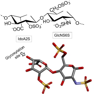
143
nanoparticles (AgNPs) using fucoidan extracted from the alga Padina tetrastromatica as part of the
144
coating material [48]. The focus of this work was on the increased antibacterial activity of antibiotics
145
coated with AgNPs and fucoidan against antibiotic resistant bacteria. The synergistic effect of the
146
combined antibiotics and fucoidan in nanoparticles resulted in a two-fold increase of the
147
anti-bacterial activity as compared to these molecules in separate treatments.
148
3.2.2. Laminaran
151
The main chain of laminarans, also found in brown alga, is mostly consisted of 3-linked β-D- Glc
152
residues (Figure 3) with a small proportion (usually less than 10%) of branches of single β-D-Glc
153
residues attached to the C6 position of the Glc residues of the backbone [49]. According to the
154
reducing terminal ends, laminarans may be of two types: one type with chains which are terminated
155
by D-Glc residues (type G) (Figure 3A) and the second type with chains ending with D-mannitol
156
(Man) residues (type M) (Figure 3B) [50]. The proportions of the two types of laminaran, and their
157
consequent structures, vary according to the seaweed species. Environmental factors such as
158
seasonal periods, salt concentration and frond age are additional influencing factors on chemical
159
structures of laminaran [51]. Other environmental factors, including water temperature, salinity,
160
waves, sea currents and depth of immersion (maybe pressure) have been also reported to influence
161
on laminaran chemical composition [52]. Laminarans exist in either highly or poorly soluble forms.
162
The first form is characterized by complete solubility in cold water, while the other is only soluble in
163
hot water. The different solubility levels are influenced by the presence and number of branching
164
residues. The higher the branching content, the greater the solubility in cold water [50].
165
166
167
Figure 3. Representative chemical structure of laminaran which is composed of a backbone of
168
3-linked β-D-glucose (Glc) units with possible 6-linked branches of Glc residues and with reducing
169
terminal ends with (A) Glc units (laminaran type G) or (B) D-mannitol (Man) residues (laminaran
170
type M).
171
Laminaran exerts many bioactivities such as anti-cancer, anti-inflammatory, anticoagulant and
172
antioxidant effects [52]. A recent review was published discussing the anticancer effects of two
173
brown algal polysaccharides - emphasis was given on laminaran [49]. In this review it was stated
174
that laminaran can enhance the therapeutic effects of commercial anticancer drugs [49]. For instance,
175
laminaran can exhibit in vitro inhibiting effect on the formation of colonies of colon cancer cells
176
DLD-1. This polysaccharide also showed a synergistic effect with X-ray irradiation against this same
177
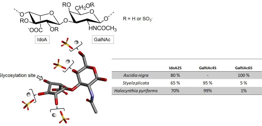
cancer cell line by decreasing the amounts and size of the colonies [53]. In the study of Malyarenko et
178
al., lamellar sulfates of Fucus evanescens showed the capacity to decrease the migration ability of
179
cancer cells in vitro by inhibiting the activity of certain metalloproteinases such as MMP-2 and
180
MMP-9 [54].
181
According to the publication of Lee and coworkers, laminaran shows also the capacity to
182
enhance the release of some inflammatory mediators [55]. This makes laminaran a potential
183
antimicrobial activity, this marine glycan shows also the inhibitory capacity on both gram-positive
185
and gram-negative bacteria such as Salmonella typhimurium, Listeria monocytogenes and Vibrio
186
parahaemolyticus to adhere on HT-29-Luc cells of human enterocytes, besides inhibiting the invasion
187
of S. typhimurium in this cell line [56]. The literature survey has shown that laminaran is able to
188
prevent HIV activity by decreasing (a) the adsorption of the HIV particle in human lymphocytes and
189
(b) the efficiency of the HIV reverse transcriptase, which plays an important role in the proliferation
190
of the virus during the infection cycle [57]. This study suggests that laminaran acts as an efficient
191
inhibitor of HIV replication and proliferation [57].
192
3.2.3. Carrageenans
193
Carrageenans are sulfated galactans found in red seaweeds and composed of linear chains of
194
alternating 3-linked D-Gal (conventionally ascribed as A units) and 4-linked α-D-Gal or
195
D-3,6-anhydrogalactose (anGal) (B units), forming thus disaccharide repeating building blocks [58].
196
Carrageenans are classified according to the presence of the 3,6-anhydrous bridge at the
197
4-linked AnGal residues and the positions and numbers of sulfate groups (Figure 4). Carrageenans
198
are traditionally identified by a Greek prefix accordingly to their structures. Structures vary in terms
199
of sulfation pattens and the presence of AnGal units. The International Union of Pure and Applied
200
Chemistry (IUPAC) establishes a nomenclature based on a code for the carrageenans: G = 3-linked
201
β-D-Gal; D = 4-linked α-D-Gal; DA = 4-linked α-D-3,6-AnGal and S = sulfate ester (SO3-) [59, 60].
202
203
Glycosylation site
ҡ-carrageenan: R = H ɩ-carrageenan: R = SO3
-Gal
AnGal
ξ-carrageenan: R = H
λ-carrageenan: R = SO3
-Glycosylation site
Gal
Gal
HO
O
O
OSO3
-O CH2OH
O
HO
OSO3
-O CH2OR
A B
204
Figure 4. Representative chemical structures of carrageenans. These polymers are made up of
205
alternating 3-linked galactose (Gal) units and (A) 4-linked anydrogalactose (AnGal) as seen in kappa
206
(κ) and iota (ɩ) carrageenans or (B) Gal units as seen in zeta (ξ) and lambda (λ) carrageenans. These
207
polymers also contain sulfation as their major substituent.
208
In Figure 4, four illustrative structures are shown: (A) one composed of AnGal units, and (B)
209
other composed of Gal units, both either in their sulfated or non-sulfated forms of occurrence. The
210
three most commercially exploited carrageenans are kappa (κ), iota (ɩ) and lambda (λ). Their
211
corresponding names, based on the IUPAC nomenclature and on the letter codes are carrageenan
212
2,4-disulfate (DA2S-G4S), carrageenan 4-sulfate (DA-G4S), and carrageenan 2,6,2-trisulfate
213
(D2S,6S-G2S), respectively[59, 60]. In addition to these three major types of carrageenan, two other
214
types, called carrageenan ѵ and μ are frequently found in carrageenan commercial samples. They are
215
In the food industry, carrageenans are widely explored because of their physicochemical
217
properties like emulsifying, thickening, gelling and stabilizing effects. These additives give textural
218
properties and protective effects to a wide range of food products [61]. Carrageenans are also widely
219
used in the pharmaceutical and cosmetic industries [62].
220
Carrageenan-derived oligosaccharides produced by γ-irradiation exhibited antioxidant
221
properties seen through multiple assays such as the hydroxyl radical scavenging, the power
222
reduction and the 1,1-diphenyl-2-picrylhydrazyl (DPPH) radical scavenging ability [63]. The effect
223
observed was dose-dependent and the carrageenan types were also observed to differently impact
224
on the antioxidant activity, following the order of λ <ɩ <κ[63].
225
Talarico et al. analyzed the action of λ and ɩ carrageenans against dengue virus serotypes. In this
226
study both carrageenans were shown to be potent inhibitors of the multiplication of dengue virus
227
type 2 (DENV-2) and 3 (DENV-3) in Vero and HepG2 cells, with effective concentration values of
228
50% (EC50) of 0.14 to 4.1 μg [18].
229
Still with respect to antiviral activity, Diogo and colleagues evaluated the action of
230
λ-carrageenan against two viral pathogens of veterinary interest, bovine herpesvirus type 1
231
(BoHV-1) and herpesvirus suid type 1 (SuHV-1) viruses [64]. λ-carrageenan was able to reduce the
232
infectivity of both types of virus. The concentration required to inactivate 50% of virus, virucidal
233
concentration (VC50) was 0.96 ± 0.08 μg/mL for BoHV-1 and 31.10 ± 2.28 μg/ml for SuHV-1. The
234
antiviral activity of λ-carrageenan for BoHV-1 expressed in inhibitory concentration (IC50) was 0.52 ±
235
0.01 μg/ml, whereas for SuHV-1 was 10.42 ± 0.88 μg/mL.
236
In vitro tests have shown that ɩ-carrageenan is a potent inhibitor of influenza A (H1N1) virus
237
infection [65]. From this information Leibbrandt and coworkers decided to test a commercially
238
available nasal spray containing ɩ-carrageenan in a model of influenza A infection in mice.
239
Treatment of mice infected with a lethal dose of influenza A PR8/34 H1N1 virus and administered
240
with ɩ-carrageenan at concentration of 60 μg/ml repeated twice daily starting within 48 hours
241
post-infection resulted in strong protection of the mice, in a similar fashion as those treated with
242
oseltamivir [65].
243
Studies on the cytotoxic effects of κ and λ-carrageenans on human cervical carcinoma cells
244
(HeLa) and human umbilical vein endothelial cells (HUVEC) have shown that both carrageenans
245
have no significant effect on HUVEC (normal cells). However, both carrageenans were cytotoxic to
246
HeLa, although λ-carrageenan has stronger cytotoxicity properties compared to κ-carrageena. In
247
addition, λ-carrageenan has been shown to have a stronger effect on suppression of tumor cell
248
proliferation and cell division compared to κ-carrageenan [66].
249
3.2.4. Sulfated polymannuronate
250
Sulfated polymannuronate (SPM), also referred as polymannuroguluronate, is a sulfated
251
polysaccharide extracted from brown algae, rich in 4-linked β-D-ManA with a mean MW of 10,000
252
254
Figure 5. Structural representation of the brown algal sulfated polymannuronate (SPM). It is
255
composed of 4-linked β-D-mannuronate (ManA) units in polymers with mean MW of 10 kDa.
256
Sulfation can occur at either C2 or C3 positions.
257
SPM has presented anti-HIV properties [68]. SPM entered into the Phase II clinical trial in
258
China, becoming the first marine sulfated polysaccharide with the potential to become a real
259
anti-AIDS drug [69]. Several authors have focused on the elucidation of the molecular mechanism
260
involved in the anti-HIV activity of SPM and their beneficial effects on the human cells of the
261
immune system [69]. The particular study of Miao and coworkers has reported that CD4 is one of the
262
possible targets for the specific binding of SPM on lymphocytes [69]. SPM-derived oligosaccharides
263
have been shown the capacity to interact, in multiple ways, with gp120 and present therefore an
264
anti-HIV outcome [68]. SPM can also inhibit the adhesion of the HIV Trans-activator of transcription
265
(Tat) on SLK cells by direct binding to the KKR site (high-affinity heparin binding region) of Tat [67].
266
This structural information facilitates the elucidation of the structure-activity relationship of sulfated
267
polysaccharides in the fight against HIV-1 infection.
268
3.2.5. GAGs
269
GAGs are linear and heterogeneous sulfated glycans. Although structurally complex, the
270
skeletons of these polysaccharides are simply constituted by repeated building blocks of
271
disaccharides composed of alternating uronic acid (UroA) or Gal and hexosamine. The hexosamine
272
may be glucosamine (GlcN) or N-acetylgalactosamine (GalNAc) and its differently substituted
273
(mostly sulfated) derivatives. UroA can be either glucuronic acid (GlcA) or iduronic acid (IdoA) [70].
274
Heparin, heparan sulfate (HS), chondroitin sulfate (CS), dermatan sulfate (DS), keratan sulfate
275
(KS) and hyaluronan (HA) are the major classes of GAGs found in animals. Although GAGs are all
276
composed of repeating disaccharide units, the patterns of sulfation and the alternating
277
monosaccharides that make up these units within the polymers vary significantly. The GAG
278
classification is conventionally set on these structural variations. Interestingly, GAGs of marine
279
organisms can present distinct structures of those from terrestrial animals, even considering the
280
same class of GAGs [71]. Structural variations and heterogeneities of GAG chains (from either
281
marine or terrestrial sources) especially in terms of sequence domains and the common occurrence
282
in the extracellular matrix or on the surface of cells are all relevant contributing factors to the
283
diversity of their biological and medical functions.
284
3.2.5.1. Heparin
286
Heparin is mostly composed of alternating N-sulfated 6-sulfated α-D-GlcN (GlcNS6S) and
287
2-sulfated α-L-IdoA units (IodA2S), both 4-linked (Figure 6). Among its occurrence in marine
288
invertebrates, heparin is found in several phyla such as mollusks, crustaceans, annelids,
289
echinoderms, tunicates and other urochordates [72]. In some of these invertebrates the heparin-like
290
structures have presented structural peculiarities which are unique and not commonly found in the
291
commonest and well-known mammalian-derived heparins. These unique properties may comprise
292
low-levels of N- and 6-sulfation content and high-levels of N-acetylation on the GlcN units together
293
with consistent amounts of GlcA units [73]. Naturally occurring low MW heparins are also found in
294
marine invertebrates [74]. Works have also suggested that marine heparin structures are related to
295
the species of occurrence and the chemical differences lie mostly on the relative abundance of the
296
various composing disaccharide units or different chains [75]. In addition to these structural
297
variations, the marine invertebrate heparin-like compounds show also variable biological functions
298
[72].
299
IdoA2S
GlcNS6S
300
Figure 6. The heparin structure is mostly composed of alternating N-sulfated 6-sulfated
301
α-D-glucosamine (GlcNS6S) and 2-sulfated α-L-IdoA (IdoA2S) units, both 4-linked.
302
Dietrich and coworkers reported the presence of a heparin in the crustacean Penaeus brasiliensis
303
[74]. Of particular importance were the findings that this low MW heparin (LMWH) is enriched with
304
non-sulfated UroA residues and exhibits potent antithrombotic activity. In vitro anticoagulant
305
activities have shown that its effect is exerted on the inhibition of factor Xa and inhibition of
306
thrombin (IIa) mediated mainly by cofactor heparin II (HCII) as opposed to mammalian heparins
307
which exert their anticoagulant activity mainly through the inhibition of IIa and factor Xa mediated
308
by antithrombin (AT). This shrimp-derived heparin has also presented potent in vivo antithrombotic
309
activity as compared to the mammalian LMWH. Oppositely to the shrimp heparin, another heparin
310
isolated from the crab Goniopsis cruentata has showed insignificant in vitro anticoagulant activity and
311
low bleeding potency [72].
312
The heparin-like compound extracted from the shrimp Litopenaeus vannamei has shown capacity
313
to reduce the influx of inflammatory cells in the lesion sites of a model of acute inflammation
314
because this marine GAG is able to reduce the activity of the MMP in the peritoneal lavage of
315
inflamed animals [73]. This molecule has been reported to reduce almost 90% the activity of MMP-9
316
study has shown that this shrimp “heparinoid” is capable of suppressing neovascularization process
318
[76].
319
An analogous of heparin isolated from the ascidian Styela plicata was investigated in a model of
320
colitis in rats [77]. The result observed was a decrease in the production of TNF-α, TGF-β and VEGF,
321
as well as reduced activation of NF-β and MAPK kinase. At the cellular level, this tunicate heparin
322
analogue can attenuate the recruitment of lymphocytes and macrophages and reduce apoptosis
323
levels on epithelial cells. A drastic reduction in collagen-mediated fibrosis has also been observed
324
[77].
325
3. 2.5.2. HS
326
Heparin and HS are structurally related GAGs since both are composed of GlcN units in their
327
backbones, although with different occurring concentrations [78]. HS is typically considered a
328
less-modified heparin version. Among the sulfated GAGs, HS is the one with the greatest structural
329
variability. Depending on the tissue and species of origin, such polysaccharide may be composed of
330
several distinct disaccharide units, containing either GlcA or IdoA and GlcN with different extents
331
of N- and/or 6-O-sulfation besides N-acetylation and 3-O-sulfation [78].
332
For example, the HS isolated from shrimp Artemia franciscana includes a high degree of
333
N-sulfation and a relatively low degree of 6-O-sulfation of the GlcN residues. This compound
334
exhibited high anticoagulant activity mediated by HCII [79]. In a study by Gomes and associates, a
335
novel HS structure with unique characteristics was isolated from the bivalve mollusk Nodipecten
336
nodosus (Figure 7). This HS was reported to be formed by GlcA and GlcN units and rare types of
337
sulfation which can occur on carbons 2 or 3 of the GlcA units [80]. This mollusk HS can inhibit
338
thrombus growth without inducing the provoking hemorrhage. The same group reported later the
339
action of this HS in inhibiting P-selectin mediated events such as metastasis and recruitment of
340
inflammatory cells [81].
341
342
R = H or SO3 -Rn= SO3-or Acetyl Glycosylation
site GlcA
GlcN
343
Figure 7. The heparan sulfate structure from the bivalve mollusk Nodipecten nodosum composed
344
of alternating glucuronic acid (GlcA) and glucosamine (GlcN), both 4-linked. This molecule has a
345
rare sulfation pattern on carbons 2 or 3 of the GlcA units. The C6 position of GlcN can also be
346
sulfated. The substituents of Rn can be either acetyl or sulfate.
347
3.2.5.3. DS
349
DS is a linear variable-length polysaccharide composed of alternating disaccharide building
350
blocks of 4-linked α-L-IdoA and 3-linked β-D-GalNAc units. These alternating disaccharide units
351
can be variably sulfated at position C2 position of IdoA (IdoA2S) and/or C4 (GalNAc4S) and/or C6
352
(GalNAc6S) or both carbons (GalNAc4S6S) in the GalNAc unit, giving rise to different sulfated
353
disaccharides [70].
354
In addition of being present in mammalian tissues, DS with high sulfation content was also
355
found in different species of clam and tunicates [82-84]. The work of Pavão et al. [84] raises an
356
interesting discussion about the structure-function relationship of DS extracted from different
357
species of ascidians (Figure 8). For example, DS isolated from Ascidia nigra is fully sulfated at the C6
358
position of the GalNAc unit (100%) and at the C2 position of IdoA (80%) (Figure 8). The DS from S.
359
plicata however is less sulfated at the C2 position (65%) and widely sulfated at the C4 position of
360
GalNAc (Figure 8). The DS isolated from Halocynthia pyriforms is similar to that one seen in S. plicata
361
(Figure 8).
362
363
364
Figure 8. Representative chemical structure of dermatan sulfate (DS) which is composed of a
365
backbone of 4-linked α-L-Idorunate (IdoA) and 3-linked β-D-N-acetylgalactosamine (GalNAc) units.
366
The different radicals represent different patterns of sulfate substitutions. Ascidian DS are highly
367
sulfated at the 2-position of IdoA, but differ in the sulfation pattern at GalNAc. The insert table
368
displays the sulfation rates of the ascidian species Ascidia nigra, Styela plicata and Halocynthia
369
pyriforms.
370
In terms of biological activities, the different sulfation patterns in ascidian DS seem to
371
collaborate differently to the outcome. In terms of anticoagulation, the DS molecules from S. plicata,
372
H. pyriforms which bear more GalNAc4S units have presented significant HCII-mediated IIa
373
inhibition as opposed to A. nigra DS which did not present considerable anticoagulant activity [84,
374
85]. With the exception of A. nigra, the two ascidian DSs presented 10- and 6-fold more activity for
375
HCII-related inhibition than the mammalian-derived native and oversulfated DS, respectively [84].
376
DS from S. plicata was also investigated regarding its anti-inflammatory activity in a model of
377
colitis in rats [77]. This GAG exhibited a superior anti-inflammatory effect to that of mammalian
378
epithelial cells. It is important to note that no hemorrhagic propensity has been pointed out after
380
treatment with the ascidian glycan [77].
381
Kozlowski and colleagues investigated the effect of two DS isolated from S. plicata and Phallusia
382
nigra in events of thrombosis, inflammation and metastasis [86]. The study showed that both GAGs
383
can reduce thrombus size in a model of arterial thrombosis induced by FeCl3. In addition, they can
384
also attenuate metastasis of MC-38 colon carcinoma, B16-BL6 melanoma cells and the infiltration of
385
inflammatory cells in a mouse model of thioglycollate-induced peritonitis. The authors suggested
386
that the observed effects are related to the inhibition of P-selectin [86].
387
3.2.5.4. Fucosylated chondroitin sulfate
388
Fucosylated chondroitin sulfate (fCS) is a distinct marine GAG found exclusively in sea
389
cucumber (Echinodermata, Holothuroidea). This GAG is composed of the regular CS backbone with
390
branches of α-L-Fuc units attached to the 3-position of the GlcA residues (Figure 9). The lateral units
391
of Fuc can show different patterns of sulfation according to the holoturian species [87, 71].
392
393
394
Figure 9. Structural representation of the holoturian fucosylated chondroitin sulfate (fCS). The
395
structure is composed of α-L-fucose (Fuc), β-D-glucuronic acid (GlcA) and N-acetyl
396
β-D-galactosamine (GalNAc).
397
398
With regard to the fCS’ therapeutic properties, this glycan exhibits a wide range of applications:
399
anticoagulant [88], anti-metastasis [89] anti-inflammatory [90] and antiviral activities [91]. For this
400
reason several papers have focused on the study of fCS. One of the works investigated samples of
401
fCS extracted from three species of sea cucumbers: Apostichopus japonicus, Stichopus chloronotus and
402
Acaudina molpadioidea in order to carry out a structural comparison between the three molecules and
403
their antioxidant and anti-inflammatory properties. Analysis of 1H and 13C nuclear magnetic
404
resonance (NMR) of the polysaccharide identified three patterns of sulfation of the fucose branches:
405
4-O-, 2,4-di-O and 3,4-di-O-sulfation. In addition, their activities were affected by the sulfation
406
patterns of the Fuc branches, revealing that sulfation in O4 is particularly important [92].
407
In the work of Ustyzhanina and colleagues, the fCS isolated from sea cucumber Cucumaria
408
japonica inhibited platelet aggregation in vitro, and demonstrated significant anticoagulant activity.
409
AT as well to influence von Willebrand factor activity. This latter property significantly
411
distinguishes fCS from the LMWH [93]. fCS isolated from sea cucumber Holothuria Mexicana
412
exhibited high affinity with fibroblast growth factors 1 and 2. These factors are important in the
413
neovascularization event. In addition, it presented intrinsic anticoagulant activity and inhibited the
414
activation of IIa and factor Xa by AT [94]. Still regarding anticoagulant properties, a new fCS isolated
415
from the sea cucumber Holothuria scabra, was tested in comparison to heparin, and was shown to
416
prolong activated partial thromboplastin time [95].
417
As mentioned above, the fucosylated sulfated polysaccharide also presents some antiviral
418
properties, including against HIV. The anti-HIV action of holothurians fCS has generated a patent
419
filed in the European patent bank [96]. Recent studies have reported the anti-HIV activity of the fCS
420
obtained from the sea cucumber Thelenota ananas, which inhibited several strains of HIV-1
421
replication with different potencies. This study also reported that T. ananas fCS can bind potently to
422
the recombinant HIV-1 gp120 protein, but did not inhibit recombinant HIV-1 reverse transcriptase
423
[91].
424
3.3. N-acetylated sugars
425
3.3.1. Chitin and chitosan
426
Chitin is an important constituent of the exoskeleton of many organisms such as crustaceans
427
and insects. In the marine environment chitin is certainly the most abundant biopolymer, being
428
structurally composed GlcNAc and GlcN bound by β1,4 glycosidic bonds (Figure 10). In chitin, the
429
GlcNAc content is higher than 70% of the total monosaccharide, making this polysaccharide highly
430
N-acetylated. This, in turn, significantly decreases its water solubility property [97, 98].
431
Chitosan is a cationic polysaccharide composed of the same units and the glycosidic linkage of
432
chitin (Figure 10). However, low amounts of GlcNAc are found in chitosan, usually less than 50%.
433
Physicochemical characteristics such as hydrophobicity and inter-chain interactions depend on the
434
amount and distribution of the acetylated groups [97, 98].
435
436
437
Figure 10. The chemical structures of chitin and chitosan. Chitin is consisting mainly of
438
2-acetamido-2-deoxy-D-β-glucopyranose (GlcNAc) units and partially of
439
(DA) is less than 50% (GlcNAc content), the polymer is then named chitosan, otherwise, it is named
441
chitin. DA is defined as the average number of N-acetylation per 100 monomers expressed as a
442
percentage.
443
The chitosan molecule is non-toxic and has many biomedical applications, including bone
444
tissue regeneration [99] and effects against a wide variety of pathogenic microorganisms [100-102].
445
Its proper use depends on many physicochemical factors and these factors can be managed
446
accordingly to the levels of activity aimed for the chitosan. Examples of these factors are MW, degree
447
of deacetylation, degree of substitution, length and position of a substituent in the GlcN units and
448
pH [98].
449
The antineoplastic activity of chitin/chitosan and low MW chitin was evaluated using a human
450
monocyte leukemia cell line, THP-1. Chitin and chitosan suppressed 100% growth of THP-1 tumor
451
cells at concentrations equal to or greater than 1.5 mg/mL. The low MW chitin exhibited the same
452
EC50 of 250 μg/mL [103].
453
Antioxidant properties of chitosan were also investigated. Trung and Bao studied the molecule
454
extracted from L. vannamei [104]. Their study suggested that the antioxidant effect observed was
455
based on the free radical scavenging activity and the reduction of potency. Another study related to
456
the antioxidant effect of this marine glycan was carried out by Sarbon et al. [105]. In their work the
457
chitosan was extracted from the ladle shells species Scylla olivacea. The chitosan of S. olivacea
458
exhibited a dose-dependent effect, where at the concentration of 10 mg/mL, the natural chitosan
459
showed a greater reduction effect than the commercial chitosan.
460
Given the versatile applicability of this acetylated glycan, Divya and associates tested the
461
antifungal and antioxidant activity of chitosan nanoparticles (ChNP) [106]. ChNP was tested in
462
comparison to Amphotericin B, and showed good antifungal activity against all selected pathogens.
463
The ChNP also exhibited significant antioxidant activity [106]. Previous work by the same group
464
showed that chitosan nanoparticles inhibited the growth of clinically important microorganisms
465
such as Staphylococcus aureus, Pseudomona aeruginosa, Escherichia coli and Klebsiella pneumoniae besides
466
exhibiting antibiofilm activity with an inhibition rate of up to 98% [102].
467
A recent study was conducted with chitin/chitosan obtained from the shrimp shell Penaeus
468
monodon [107]. These polysaccharides showed inhibitory effects on the proliferation of the human
469
ovarian cancer cell line, PA-1. Chitin and chitosan can suppress 100% growth of PA-1 tumor cells at
470
the respective concentrations of 50 μg/mL and 10 μg/mL [107].
471
3.4. Triterpene glycosides
472
The glycosides consist of amphiphilic compounds which contain a sugar bound to another
473
functional group through a glycosidic bond (Figure 11). While the sugar can be a simple unit
474
(monosaccharide) or various units (oligosaccharide) and the aglycone (functional group) may be a
475
O
O O
O HO
O
R2
OH R1
Monosaccharide R1 R2
D-xylose H H
D-quinovose H CH3
D-glucose H CH2OH
3-O-methyl-D-xylose Me CH2OH
6-O-acetyl-D-glucose H CH2OAc
3-O-methyl-D-quinovose Me CH3
3-O-methyl-D-xylose Me H
3-O-methyl-D-glucuronic acid Me CO2H
477
Figure 11.Chemical structures of glycoside with triterpenic backbone of holostane type bound to a
478
sugar unit (glucose, Glc, in the case) and the possible substituents.
479
Glycosides of marine organisms can be isolated from sea cucumbers [109], starfish [110], sponge
480
[111], algae [112] and corals [113]. Due to the great diversity of this class of molecule, many studies
481
have focused on the investigation of their therapeutic properties. For example, glycosides isolated
482
from the edible red seaweed Laurencia undulata, called Floridoside or D-isofloridoside, have their
483
antioxidant properties investigated by Li and colleagues. The two compounds showed significant
484
antioxidant activity and are presented as potential inhibitors of MMP-2 and MMP-9 [114].
485
Aurantoside K (a tetramic acid glycoside isolated from a sponge belonging to the genus
486
Melophlus) exhibited a broad spectrum of antifungal activity against strains of Candida albicans, with
487
the minimum inhibitory concentration (MIC) of 31.25 μg/mL for a strain resistant to amphotericin,
488
and 1.95 μg/mL for a wild-type strain. It also showed a zone of inhibition of 14 mm of diameter in
489
the concentration of 100 μg/disc for yeast Cryptococcus neoformans, 28 mm for Aspergillus niger, 31 mm
490
Penicillium sp., 21 mm Rhizopus sporangia and 29 mm Sordaria sp. at the same concentration of 100
491
μg/disc [115]. Another study carried out with a class of triterpene glycosides, called variegatusides,
492
isolated from the sea cucumber Stichopus variegates Semper (Holothuriidae), showed that these
493
compounds have potent antifungal activities in biotestes in vitro [116].
494
Wang and collegues verified the cytotoxic effects of thirteen purified triterpenic glycosides of
495
Holothuria scabra Jaeger and Cucumaria frondosa Gunnerus (Holothuroidea) against four human cell
496
lines in order to advance the structure-activity relationship of these molecules [117]. The results
497
showed that the number of glycosyl residues in the sugar chains and the aglycone side chain may
498
affect their cytotoxicity to tumor cells and selective cytotoxicity in neoplastic versus normal cells.
499
Works like this arouse interests in the use of these glycosides for the development of new antitumor
500
drugs [105].
501
Given the vast number of actions that these compounds can present, it is worth also to
502
understand the underlying mechanisms by which these molecules function. A good option to
503
uncover their molecular mechanisms of action of marine glycosides is by identifying the
504
relationships between their structures and activities. In a review of Park and co-authors the
505
For example, Stichoposide C and Stichoposide D, both isolated from the holothurian Stichopus
507
chloronotus, exert anticancer activity [118]. However, the activity of the compounds occurs by
508
distinguished mechanisms due to differences in the sugar content. Stichoposide C has quinovose,
509
and induces apoptosis through the generation of ceramide by the activation of acidic
510
sphingomyelinase (SMase) and neutral SMase. While Stichoposide D possesses Glc as the second
511
monosaccharide unit induces apoptosis by the activation of ceramide 6 synthase leading to the
512
increase of cellular levels of ceramide.
513
Following the same thought, a recent study compared the effects of three frondosides (A, B and
514
C) extracted from Cucumaria frondosa and its aglycone against pancreatic cancer cells. What can be
515
observed was that frondoside A potentially inhibited the growth of pancreatic cancer cells with an
516
EC 50 of ~ 1 μM. Frondoside B was less potent with an EC 50 of ~ 2.5 μM. Frondoside C and aglycone
517
had no effect [119]. Frondoside A has potent antiproliferative, anti-invasive and antiangiogenic
518
effects on a variety of cancers [120-122].
519
Cyclic steroid glycosides isolated from the starfish Echinaster luzonicus, identified as
520
luzonicoside A (LuzA) and luzonicoside D (LuzD), were tested for their potential inhibitory capacity
521
against RPMI-7951 and SK-Mel melanoma cell lines -28. LuzA inhibited proliferation, colony
522
formation and migration of SK-Mel-28 cells more significantly than LuzD. The authors suggested
523
that molecular mechanism of action is related to the regulation of the activity of cleaved caspase-3
524
and poly (ADP-ribose) polymerase (PARP), together with the levels of Survivin, Bcl-2, p21 and
525
cyclin D1 [110].
526
3.5. Glycoproteins
527
Glycoproteins are glycoconjugates in which various sugar monosaccharides are covalently
528
attached to the protein backbone. Two major types of sugar chains (N and O-linked) are found in
529
glycoproteins. N-linked sugar chains contain a GlcNAc residue at its reducing end which is, in turn,
530
attached to the amide group of an asparagine (Asn) residue of the polypeptide backbone. The
531
O-linked acid chains contain a residue of GalNAc at its reducing terminus which is, in turn, attached
532
to a serine (Ser) or threonine (Thr) residue of a polypeptide backbone (Figure 12) [123].
533
534
535
Figure 12. Chemical representatives of glycoproteins. (A) O-linked glycoprotein, which binds to
536
the peptide by the amide group of a serine residue (Ser) or threonine (Thr). (B) N-linked
537
Glycoproteins represent a large class of biomolecules. Many of the proteins that are components
539
of cell membranes are glycosylated (giving rise to glycoproteins). Glycoproteins may have essential
540
functions as receptors that capture various ligands into the cell such as transport proteins that are
541
involved in the ingestion of various compounds, or as structures that mediate molecular recognition,
542
signaling and interactions between cells [124].
543
Among the glycoproteins of marine systems, it is important to consider the role of lectins.
544
Lectins are recognition proteins of non-immune origin that bind to carbohydrates. They play many
545
varied biological functions including regulation of cell adhesion, recognition of molecules in cell-cell
546
and cell-molecule interactions, and are also known to have vital immune functions [125]. Lectins are
547
isolated from a variety of marine organisms including algae [126, 127], sponges [128], mollusks [129]
548
and echinoderms [130].
549
Many studies have reported the therapeutic effects of glycoproteins, especially lectins. For
550
example, a study by Silva and collaborators has aimed on the potential anti-inflammatory action of
551
the lectin extracted from the red alga Pterocladiella capillacea [131]. The authors have observed a
552
reasonable anti-inflammatory effect through both the paw edema model and the neutrophil
553
migration model, based on the injection of carrageenan as inflammation stimulus [131]. In a different
554
work, the antinociceptive and anti-inflammatory effects of the lectin extracted from the red alga
555
Solieria filiformis were evaluated [132]. In this work, the animals were pretreated with lectin by 30
556
min before receiving the nociceptive or inflammatory stimuli. The S. filiformis lectin significantly
557
reduced the number of abdominal writhes and reduced the paw licking time in the formalin test.
558
The lectin of S. filiformis also reduced neutrophil migration in a peritonitis model, in addition to
559
reducing paw edema induced by carrageenan, dextran and serotonin [132]. In a recent work,
560
Fontenelle and colleagues investigated the lectin extracted from the red seaweed Bryothamnion
561
triquetrum, and reported its anti-inflammatory effect in mice [133].
562
Reports of anticancer activity of lectins have also been found in the literature. In one of the
563
collected works, besides investigating the biological activity the authors also dealt with structural
564
aspects of a lectin of the sea mollusk Crenomytilus grayanus. Cell viability assays have shown that C.
565
grayanus lectin recognizes Gb3 globotriose on the surface of breast cancer cells, leading to cell death
566
[134]. Also regarding anticancer activity, Liu et al. investigated the in vivo antitumor activity of
567
hemocyanin (multifunctional glycoprotein) of the shrimp L. vannamei in Sarcoma-180 (S180) model
568
of tumor-bearing mice [135]. After 8 days of treatment, the dose of 4mg/kg significantly inhibited the
569
growth of S180 to 49% compared to untreated animals [135].
570
In terms of antimicrobial activity a new lectin was isolated from the green algae Halimeda
571
renschii. The mannose-specificity lectin showed potent activity against influenza virus in NCI-H292
572
cells at half maximal effective dose (ED50) of 2.45 nM. Antiviral action occurred through high affinity
573
binding to hemagglutinin from the viral envelope [127].
574
3.6. Glycolipids
576
Glycolipids comprise a large and diverse group of lipids that serve numerous cellular functions
577
[136]. They are amphipathic lipids, containing a hydrophilic portion, which is composed of units of
578
carbohydrates, from which gives its name (the prefix "glyco"). Its lipid moiety is referred to as the
579
hydrophobic tail, generally constituted of aliphatic fatty acid chains [137].
580
Among the classes of glycolipids are glycosphingolipids that are constituents of cell membranes
581
in a wide variety of organisms (fungi, plants, animals and marine organisms) [138]. These
582
compounds have biotechnological potential and play important physiological role due to variations
583
in their sugar chains.They are classified into cerebrosides, ceramide oligohexosides, globosides and
584
gangliosides based on the constituent sugars (Table 1). In recent years, some glycosphingolipids
585
have been isolated from marine invertebrates such as echinoderms, porifera and mollusks [139].
586
587
Table 1. Classification of sphingolipids according to their sugar content.
588
Sugar moiety Glycosphingolipid
monosaccharide cerebrosídeo
disaccharide ceramide dihexoside
oligosaccharide ceramide oligohexoside
oligosaccharide + amino sugar globoside
oligosaccharide + sulfate sulfatide
oligosaccharide + sialic acid ganglioside
589
Marine algae synthesize three major types of glycolipids, they are monogalactosylcyglycerides
590
(MGDGs), digalactosyldecylglycerides (DGDGs) and sulfoquinovosylcyglycerides (SQDGs) (Figure
591
13). These glycoglycerolipids are present in the chloroplasts of any eukaryotic algae. MGDGs and
592
DGDGs are the most abundant lipids of the thylakoid membrane and appear to play a crucial role in
593
photosynthesis [140].
594
595
596
Figure 13. Representation of the general structure of the three main types of seaweed
597
glycolipids. (A) Monogalactosylcyclic glycerides (MGDGs), (B) digalactosyldecylglycerides
598
Many papers have sought biologically active glycolipids from marine organisms to elucidate
600
the structure-function relationships of glycolipids and to develop new medicinal resources. A good
601
example of this was the study where eight new cerebrosides named Renierosides were isolated from
602
an extract of the marine sponge Haliclona (Reniera) sp. The isolated compounds exhibited cytotoxicity
603
of five human tumor cell lines, including human lung cancer (A549), human ovarian cancer
604
(SK-OV-3), human skin cancer (SK-MEL-2), cancer cell line of the human central nervous system
605
(XF498) and human colon cancer (HCT15) [141].
606
Plouguerné et al. identified SGDGs in fractions obtained after the purification of the organic
607
extract of the Sargassum vulgare brown alga [142]. These metabolites exhibited antiviral activity
608
against the herpes simplex virus type 1 (HSV1) and 2 (HSV2) viruses. The main SQDG responsible
609
for anti-HSV1 and anti-HSV2 activities was characterized as 1,2-di-O-palmitoyl-3-O-
610
(6-sulfo-α-D-quinovopyranosyl) glycerol [142]. Two SQDGs isolated from the red alga Palmaria
611
palmata showed potent anti-inflammatory activity. Bioactive compounds were identified as (2S)
612
-1-O-eicosapentaenoyl-2-O-myristoyl-3-O-(6-sulfo-α-D-quinazopyranosyl)-glycerol and (2S)
613
-1-O-eicosapentaenoyl- 2-O-palmitoyl-3-O-(6-sulfo-α-D-quinovopyranosyl-glycerol and
614
demonstrated nitric oxide inhibitory activity with IC50 values of 36.5 and 11.0 μM, respectively [143].
615
In the paper by Reyes and coworkers the first characterization of the MGDGs, DGDGs and
616
glycosylceramides of Isochrysis galbana (Haptophyte) was described together with a study of their
617
anti-inflammatory properties as inhibitors of tumor necrosis factor α (TNF-α), a protein of cell
618
signaling involved in the inflammatory response of the acute systemic phase [144]. In a recent paper,
619
Che and colleagues have described that sea cucumber cerebrosides have improved learning and
620
memory deficits, protecting against oxidative stress in vivo, and increasing the survival rate of PC12
621
cells, a rat pheochromocytoma cell line [138].
622
Overall, the bioactivities of the glycoglycerides are directly related to the sugar moiety. The
623
position of the glycerol binding to the sugar, the length and the location of the acyl chain and the
624
anomeric sugar configuration are all key structural contributors [145].
625
3.7. Iminosugar
626
Naturally occurring iminos or azasugars are monosaccharides with nitrogen-substituted
627
heterocyclic oxygen. In 1960 the first member of this class of compounds was isolated and
628
characterized, a 5-amino-5-deoxyglucose antibiotic called Nojirimycin. Subsequently, more than 25
629
additional Nojirimycin analogs were described from plant and microbial sources [146, 147].
630
Iminosugar is commonly obtained from terrestrial sources or through chemical synthesis [146].
631
However, the work of Segraves and Crews described at the first time iminosugars from the marine
632
environment [147]. In this work, three compounds were extracted from the sea sponge Batzella sp.
633
and presented as iminosugar nucleus with a long chain of alkyl linked, indicating to be alkylated
634
imino sugars. They were identified as Batzellasides A, B and C (Figure 14). The identification of
635
these compounds occurred by comparison with the known natural and synthetic iminosugars
636
638
Batzellaside A n = 8 Batzellaside B n = 7 Batzellaside C n = 9 5-amino-5-deoxy-Glc
639
Figure 14. Representation of a nucleus of alkylated imino sugar. Batzellasides vary according to
640
the size of the alkyl chain: Batzellasides A (8 carbon chain), Batzellasides B (7 carbon chain),
641
Batzellasides C (9 carbon chain).
642
The iminosugars have shown important therapeutic importance such as antiviral [148],
643
insecticides [149], and nematicidal activities [150]. These potentials are associated with the ability of
644
these molecules to selectively inhibit enzymes that degrade carbohydrates (glycosidases). An
645
example of the antiviral activity of iminosugars is seen from its capacity to interfere with the
646
glycoprotein processing [151].
647
Regarding the investigation of these activities, the study by Segraves and Crews, evaluated the
648
antimicrobial action of the three iminosugars studied therein (Batzellasides A, B and C) against the
649
bacteria Staphylococcus epidermidis [147]. The three structures were able to inhibit the growth of the
650
microorganism with lower MICs to 6.3 μg / mL [147].
651
The work of Sayce et al. has shown that the 1-deoxynojirimycin iminosugars bear Glc and
652
inhibit the production of infectious virus in vitro including dengue virus (DENV), hepatitis B virus,
653
virus hepatitis C virus, HIV and influenza A virus. Inhibition of endoplasmic reticulum
654
α-glycosidases prevents virus release and is the main antiviral mechanism of action of iminosugars
655
against DENV [151].
656
4. Concluding Remarks
657
It is not today that the marine environment arouses the interest of researchers in exploring their
658
riches. The great diversity of this environment has led to the level of important source of obtaining
659
novel molecules, especially with respect to molecules with biomedical potential such as those with
660
glycosidic nature.
661
Throughout this work we have seen a miscellany of investigations into the effects of marine
662
glycans or glycoconjugate on human health. However, it is still necessary to understand the
663
molecular mechanisms behind these activities in order to understand the structural aspects of these
664
molecules and what this diversity of structures may represent in the final effect.
665
Despite advances in study techniques that have allowed a reasonable understanding of the
666
structure-activity relationship and some underlying mechanisms of action of these compounds,