ABSTRACT
PERKINS, PATRICK JOSEPH. Investigation of Novel Methods and Tools for Quality Control and Analysis of Ribo-seq Data. (Under the direction of Steffen Heber).
Translation is the process by which cells synthesize proteins from mRNA, and is a vital part of gene expression. During translation, ribosomes move along strands of mRNA, processing codons and transferring amino acids from tRNA to growing polypeptide chains. Ribosome profiling, or Ribo-seq, has emerged as a powerful method for quantifying translation by taking a global snapshot of the position of actively translation ribosomes. In this dissertation I present four novel methods and tools for quality control, visualization, and analysis of Ribo-seq data.
In chapter 1, I provide a review of the background and supporting information for each of the subsequent chapters, and highlight seminal studies within each of the respective fields. I then discuss the motivation, objectives, and value of my research in respect to the current landscape of Ribo-seq data analysis.
In the second chapter, we present riboStreamR, an R shiny web application for Ribo-seq and RNA-seq quality control. The app is constructed from using various tools in the Bioconductor suite for next generation sequencing data analysis. The platform accepts Bam files as input, from which the aligned reads are annotated with a set of descriptive features. These features are used to dynamically filter and subset reads, facilitating customized visualizations. The various tools in riboStreamR allow for the inspection of read length distributions, feature mapping distributions, trinucleotide periodicities, meta-gene read distributions, differential translation efficiency, and more. Users can also produce R markdown reports containing their graphics.
abnormally high numbers of ribosomes. Parameter optimization for the three scans is performed by selecting for parameter sets which identify peaks with higher conservation within biological replicates. This peak conservation metric was also used to compare the performance of our algorithm to competing methods. Additionally, we analyze codon and amino acid biases, and perform a motif analysis at peak sites.
Chapter 4 describes riboSimR, a tool for simulation of Ribo-seq count data and exploration of experimental design parameters. Ribo-seq and RNA-seq count tables are simulated using a negative binomial model, with mean count and dispersion parameters being drawn from real datasets. Users may choose to simulate count tables based on their own datasets, or from two separate datasets provided within the tool. The platform allows users to explore the effect of experimental design decisions such as level of sample replication, sequencing depth, and choice of differential analysis tool. Additionally, we explore the effect of imbalances between replication and sequencing depth levels between experiment types.
Investigation of Novel Methods and Tools for Quality Control and Analysis of Ribo-seq Data.
by
Patrick Joseph Perkins
A dissertation submitted to the Graduate Faculty of North Carolina State University
in partial fulfillment of the requirements for the degree of
Doctor of Philosophy
Bioinformatics
Raleigh, North Carolina 2019
APPROVED BY:
_______________________________ _______________________________ Steffen Heber Jose Alonso
Committee Chair
_______________________________ _______________________________ Dahlia Nielsen David Reif
BIOGRAPHY
ACKNOWLEDGMENTS
I would primarily like to sincerely thank my graduate advisor Dr. Steffen Heber for his guidance and expertise during my graduate career at NC State and in preparation of the work described in this dissertation. Steffen provided me a wealth of opportunities to succeed as a graduate student, and always set a great example as an enthusiastic and dedicated research scientist.
I would also like to thank Jose Alonso, Anna Stepanova, and the members of the Alonso/Stepanova PGRP group that helped guide my research and provided me with amazing feedback.
Additionally, I would like to thank the other members of my graduate committee, Jung-Ying Tzeng, Dahlia Nielsen, and David Reif, for their valuable time and support, as well as for their scheduling flexibility.
I would also like to acknowledge the extremely helpful staff and faculty in the Bioinformatics Research Center and Computer Science Department.
TABLE OF CONTENTS
LIST OF TABLES ... vii
LIST OF FIGURES ... viii
Chapter 1: Introduction ... 1
1.1. Background ... 2
1.1.1. Translation ... 2
1.1.2. Ribo-seq ... 4
1.1.3. Processing and Quality Control of Data ... 5
1.1.4. Experimental Design, Simulation, and Power Analysis ... 7
1.1.5. Ribosome Pausing ... 9
1.1.6. Identification of Peaks in Ribosome Density ...11
1.1.7. Anomaly Detection ...12
1.1.8. Negative Selection Algorithm ...14
1.2. Research Objectives ...16
1.2.1. Motivation ...16
1.2.2. Objectives and Value ...18
Chapter 2: RiboStreamR: A web application for quality control, analysis, and visualization of Ribo-seq data ...21
2.1. Background ...22
2.2. Implementation ...25
2.2.1. Environment ...25
2.2.2. Data Processing and Platform Design ...26
2.3.1. Functionality ...27
2.3.2. Graphic Customization ...30
2.3.3. Reference Datasets and Anomaly Detection ...31
2.3.4. Report Generation ...33
2.3.5. Performance ...34
2.4. Discussion ...36
2.4.1. Need for a web application based Ribo-seq analysis platform ...36
2.4.2. Future Work ...36
2.5. Conclusion ...37
Chapter 3: Identification of Ribosome Pause Sites Using a Z-Score Based Peak Detection Algorithm ...38
3.1. Introduction ...38
3.2. Data Processing ...40
3.3. Smoothed z-score Algorithm ...41
3.3.1. Algorithm Basics...41
3.3.2. riboSmoothR ...42
3.3.3. Peak Conservation ...43
3.4. Algorithm Performance ...45
3.4.1. Parameter Optimization ...45
3.4.2. Method Comparison ...46
3.5. Analysis of Peak Regions ...47
3.5.1. Codon and Amino Acid Usage ...48
3.5.3. GO Enrichment and Motif Analysis ...50
3.6. Conclusion ...52
Chapter 4: RiboSimR: a tool for simulation and power analysis of Ribo-seq experiments ...54
4.1. Introduction ...54
4.2. Methods...57
4.2.1. Simulation Strategy ...58
4.2.2. Differential Analysis ...60
4.2.3. Power Assessment ...61
4.2.4. Implementation ...62
4.3. Results ...63
4.3.1. Data Preparation ...63
4.3.2. Simulation Results ...64
4.4. Conclusion ...70
Chapter 5: Using a Novel Negative Selection Inspired Anomaly Detection Algorithm to Identify Corrupted Ribo-seq and RNA-seq Samples...72
5.1. Introduction ...73
5.2. Properties of BDUNN...76
5.2.1. Detector Generation ...76
5.2.2. Boundary Set Determination ...77
5.2.3. Boundary Set Reduction ...79
5.2.4. Testing ...80
5.3.1. Synthetic Datasets ...81
5.3.2. Outlier Detection Datasets ...84
5.3.3. Analysis of Ribo-seq and RNA-seq quality ...87
5.4. Implementation ...90
5.5. Conclusion ...91
Chapter 6: Conclusions, Future Work, and Tool Availability ...93
6.1. Conclusions and Future Work ...93
LIST OF TABLES
Table 2.1. Description of alignment attributes ...29
Table 2.2. Description of tools within riboStreamR ...32
Table 3.1. Cohen’s kappa values between sample pairs ...46
Table 3.2. GO term and motif analysis ...52
Table 4.1. Differential scenarios, assuming effect size ...62
Table 4.2. Differential scenarios, assuming D ...63
Table 5.1. Properties of the datasets used for performance evaluation ...85
Table 5.2. Results for Iris, Glass, and Haberman’s survival datasets ...86
Table 5.3. Results for Skin Pigmentation dataset ...87
Table 5.4. Results for artificial RNA-seq dataset ...90
LIST OF FIGURES
Figure 1.1. Structure of mRNA ... 3
Figure 1.2. Overview of Ribo-seq ... 5
Figure 1.3. Overview of ribosomal pausing ...10
Figure 1.4. Description of negative selection algorithm ...15
Figure 2.1. Ribo-seq experimental procedure and data analysis ...24
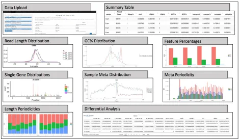
Figure 2.2. RiboStreamR data analysis pipeline...28
Figure 2.3. Examples of riboStreamR tool output ...30
Figure 2.4. Examples of riboStreamR’s graphical output customization options ... 35
Figure 3.1. Visualization of determining P-site offsets for Ribo-seq reads ...42
Figure 3.2. Graphical depiction of the smoothed z-score scans used by riboSmoothR ...44
Figure 3.3. Method comparison using agreement between samples, calculated using ICC ...47
Figure 3.4. Amino acid representation at peak sites identified by riboStreamR ... 48
Figure 3.5. Codon representation at peak sites ...49
Figure 3.6. Strings of amino acids at peak sites, length 2 ...51
Figure 3.7. Strings of amino acids at peak sites, length 3 ...51
Figure 4.1. Effect of replicate number and sequencing depth on statistical power ...65
Figure 4.2. Effect of altering depth and replication at different levels between experiment types ...67
Figure 4.3. Power stratification for DTE by average count value ...68
Figure 4.4. Tool comparison results ...69
Figure 4.5. FDR threshold comparison results ...69
Figure 5.1. Visual comparison of different negative selection inspired algorithms ...77
CHAPTER 1: INTRODUCTION
The breakthrough discoveries in molecular biology and genetics in the latter half of the 20th
century, along with the subsequent development of advanced techniques for DNA sequencing, have led to a drastic rise of experimental techniques capable of producing high resolution whole-genome data (Muir et al. 2016). Among these techniques are RNA-seq, which measures the number of mRNA transcripts within a cell, and Ribosome Profiling, which produces a snap-shot of the positions of all the actively translating ribosomes in a cell at a nucleotide-specific resolution (Mortazavi et al. 2008, Ingolia et al. 2009). Together, these technologies can be leveraged to quantify the two primary mechanisms of gene expression: translation and transcription. Ribo-seq offers the ability to quantify protein synthesis throughout a cell, as well as for investigating the dynamics and activity of ribosomes during translation (Ingolia et al. 2009).
bioinformaticians with a platform for end-to-end processing of data. The rest of this manuscript is organized as follows: in the background section, I provide supporting information and literature review of the fundamental principles behind Ribo-seq, the analysis of Ribo-seq data, experimental design of Ribo-seq studies, ribosome pausing, and anomaly detection; in the research objectives section, I will go more into the motivation for my research and the value that it provides to the scientific community; the subsequent four chapters consist of research papers that describe our novel methods and tools for Ribo-seq analysis, data quality control, data visualization, identification of ribosome pause sites, and identification of low quality samples; and lastly, the conclusion will address the contributions of this work, recommendations for future research, and tool availability.
1.1 Background
1.1.1 Translation
ribosomal subunit from prematurely binding. This complex scans the 5’UTR until it comes upon a methionine start codon, which causes the dissociation of factors from the small subunit.
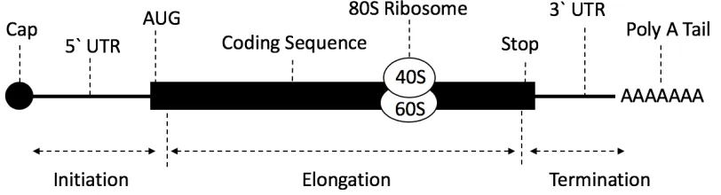
Arrival at the start codon initiates the binding of the large 60S subunit, and the formation of the complete 80S ribosome (Sonenberg and Dever 2003). During elongation, aminoacyl tRNAs, which are tRNA that are chemically bonded to their related amino acid, deliver amino acids to the ribosome for incorporation into the growing peptide chain, in coordination with a set of elongation factors (Dever e al. 2012). Aminoacyl tRNA are bound to their corresponding codon in the A-site of the ribosome, and are then moved to the P-site, where a peptidyl bond is formed between the new amino acid and the growing polypeptide chain. Deacylated tRNA then dissociate from the ribosome at the E-site. Elongation continues until the ribosome reaches a stop codon. During termination, release factors facilitate the cleavage of the peptidyl-tRNA bond at the P-site, releasing the protein (Dever et al. 2012). The ribosome and other translational machinery are released from the mRNA and recycled for successive use. See Figure 1.1 for a diagram of mRNA.
Figure 1.1 Structure of mRNA and process of translation. The mRNA has a 5’ cap made from
methylated guanosine triphosphate. Ribosomes scan through the 5′ untranslated region (UTR) until
they reach the first AUG codon. The coding region contains the sequence of codons that code for amino
acids. In order to actively translate, both 40S and 60S ribosomal subunits are required, which interact
to make the 80S ribosome. The 80S ribosome moves along the mRNA coding region in 5′ to 3′ direction
1.1.2 Ribo-seq
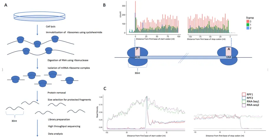
Ribo-seq was first proposed for investigating the dynamics of translating ribosomes by Ingolia et al. in 2009 (Ingolia et a. 2009). In a Ribo-seq experiment, the cells are lysed and the mRNA molecules containing ribosomes are isolated. The mRNA is subsequently digested using ribonucleases. The sections of mRNA which are bound to translating ribosomes are protected from digestion, producing ribosome ‘footprints’ of around 30nt in length. A density gradient or cushion separation technique can be used to isolate the ribosomes and their associated mRNA fragments, and phenol/chloroform purification removes the ribosomal proteins (Ingolia et al. 2012). As the length of the protected regions is generally known, a size selection of the resulting mRNA fragments is performed to select for fragments which likely represent ribosome footprints, instead of contaminants such as ribosomal or transfer RNAs. Adapters are then ligated to the 3’ end of the fragments, and reverse transcriptase is used to reverse transcribe them from RNA to cDNA. See Figure 1.2 for a depiction of the Ribo-seq process.
1.1.3 Processing and Quality Control of Data
In order to perform downstream analysis of Ribo-seq data, the data must first be processed, and a quality analysis must be performed to verify that the data meets certain standards. Fastq files containing raw Ribo-seq reads are processed by removing all low quality reads and reads which are too short, and subsequently trimming read adapters and barcodes. Once raw data has been processed, the reads are mapped to their respective genomes using a mapping tool and a genome annotation file. Popular tools for aligning reads to a reference genome include Bowtie, Tophat, STAR, and Kallisto (Trapnell et al. 2009, Dobin et al. 2013, Bray 2016). These alignment methodologies of these programs differ in multiple aspects, such as how they handle mapping junctions and whether they map to transcriptomes or genomes. A mapping tool should be chosen through an evaluation of how the its methods may potentially impact the goals of a project.
The resulting alignments can be used to evaluate relative sample quality. In order to evaluate the quality of a Ribo-seq sample, we need to define a set of descriptive features which summarize the quality. The following list provides examples of such features that can be represents as single values:
§ Percentage of uniquely mapped reads, using a standardized read mapping procedure. § Summary statistics for read length distribution, including mean, median, and standard
deviation. Could include a measure of what percentage of reads lie outside of a standard ribosome fragment size range, like 25-32nt.
§ Summary statistics for GC percentage distribution, including mean, median, and standard deviation.
§ Percentage of reads mapping to each of the available features in an annotation, such as coding sequence, 5 and 3’UTR, rRNA, tRNA, intergenic, etc…
§ Sample tri-nucleotide periodicity, calculated as percentage of coding sequence reads in the major frame, after performing a p-site adjustment. Could also include periodicity values for each specific read length.
§ Sample complexity, calculated as the number of unique start/end read positions divided by the total number of reads. Gives a generalization of read stacking and spread.
which are available as web based platforms (Michel et al. 2015, Carja et al. 2017). RiboGalaxy hosts and provides a graphical user interface for various standalone tools, while Riboviz enables browser based comparisons of published ribosome profiling datasets. A comprehensive Ribo-seq analysis can typically involve multiple of these software packages, and often requires a considerable amount of expert knowledge, as well as experience in programming or command line usage.
1.1.4 Experimental Design, Power Analysis, and Simulation
affected by this counting error, so our ability to detect differential genes is therefore dependent on gene counts themselves. Determining appropriate levels of sample replication and sequencing depth for Ribo-seq and RNA-seq are critical aspects of experimental design and must be considered prior to conducting any experiment.
Our ability to detect differential genes is also largely dependent on the statistical methods and tools we choose to use for our analysis. As most experiments have limited sample sizes, empirical Bayes methods are often used to estimate the biological variation of genes by shrinking variance estimates towards the average trend across all genes (Anders and Huber, 2010, Robinson et al. 2010). It is essential that researchers understand the importance of choosing a tool for differential analysis, as they often contain different variance estimation procedures and multiple testing correction methods, both of which can have noticeable effects on the results of the tests. Other seemingly simple decisions, such as choosing a threshold for statistical significance to use when determining differential genes, can be of great importance. The level of change that is considered biologically significant can vary based on the purpose of an experiment, as can trends in the relationship between p-values and effect sizes (McCarthy and Smyth, 2009).
observe large differences in the level of sampling depth and replication between transcript and ribosome footprint sequencing. Problems can also be introduced when testing for statistical significance, as evident by the discrepancies amongst methodologies which employ more complex two factor tests to calculate differential translation efficiency (Eastman et al. 2018, Larsson et al. 2011, Olshen et al. 2013, Xiao et al. 2016).
1.1.5 Ribosome Pausing
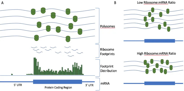
The rate of translation along an mRNA strand can be estimated by assessing the cumulative distribution of ribosomal positions within all coding regions. The density of ribosomes at a position corresponds to the probability of randomly sampling an active ribosome at that position on the mRNA. This information can be used to estimate the rate of ribosomes during translation. Using Ribo-seq data, researchers have verified that the distribution of ribosomes along mRNA transcripts is not uniform due to inconsistencies in the rates of translating ribosomes (Varenne et al. 1984). We can assume that the probability of randomly sampling a ribosome footprint at a position on a fragment is inversely proportional to the speed of the ribosome at that region.
of various RNA binding motifs and the occurrence of ribosome pauses (Zhang et al, 2017). RNA binding proteins can potentially interfere with ribosomal activity, and ribosomes may show affinity for some motifs, causing potentially impactful hybridization events. The presence of mRNA secondary structures, such as stems, loops, and hairpins can also affect the rate of elongating ribosomes (Zhang et al, 2017). See Figure 1.3 for a depiction of ribosome pausing.
As discussed previously, pausing of ribosomes during translation is indicated in multiple biological processes that effect gene expression. In viruses and bacteria, stalling can affect growth rate and initiate abandonment of translation (Burgess-Brown et al. 2008). Premature release of ribosomes from mRNA leads to the production of incomplete polypeptides which are later destroyed, lowering the overall expression of the gene (Fluman et al. 2014). A similar process, called no-go decay, occurs in eukaryotes, in which ribosome pausing triggers endonucleolytic degradation of mRNA (Pelechano et al. 2015).
Pausing can also affect the co-translational folding of nascent peptide chains, as slower translation allows proteins more time to fold (Burgess-Brown et al. 2008). Another possible consequence of pausing is ribosomal frameshifting, in which ribosomes become unsynchronized with the major frame (Fluman et al. 2014). This causes the production of alternative and often dysfunctional proteins. While connections between ribosome pausing and these various phenomena have been verified, the extent of the effect that pausing has on gene expression is not yet fully known.
1.1.6 Identifying Pause Sites
rely on generating a global distribution of pausing scores, which represent the positional ribosome densities normalized using both the mean number of reads within the gene and the transcript density determined by RNA-seq. Once such a score distribution has been generated, a threshold can be used to determine positions of significantly high ribosome density. Other researchers have used position specific metrics to determine pause sites, which account for ribosome density directly upstream and downstream of potential pause sites (Perkins and Heber 2018). Identified pause sites can be evaluated using multiple techniques, such as comparison to known peak sites, by peak site correlation with features such as sequence context and mRNA secondary structure, and by calculation of peak site conservation between biological replicates (Zhang et al, 2017, Perkins and Heber 2018).
1.1.7 Anomaly detection and Artificial Immune Systems
normal and anomalous behavior in a manner which is meaningful within your problem space. The means by which anomalies are introduced into a dataset can vary greatly between disciplines, and therefore it is necessary that the methods for identifying them be context-specific.
Artificial immune systems (AIS) represent a class of biologically inspired algorithms that are based on the characteristics of the immune system (Read et al. 2012, Hofmeyr and Forest 2000). The study of AIS can be subdivided into two separate fields: the act of attempting to solve problems in engineering or data science using algorithms or mathematical methods inspired by immunological mechanisms, or attempting to model and simulate the activity of the immune system using computational techniques (Read et al. 2012). In this dissertation, I will be focusing on the former of these two areas of research. Twentieth century advances in the fields of immunology, microbiology, and molecular biology have revealed the complex biological properties of the immune system; properties which have numerous applications to common problems in engineering. These include the ability of the body to maintain a memory bank of immune responses, to self-organize and accurately regulate responses with no central controller, to recognize a wide range of patterns in the form of receptors, and to classify substances as either a part of the organism’s own ecosystem or a potentially harmful pathogen (Read et al. 2012).
foreign bodies or suppress their effect, and the adaptive immune system, which adapts a pathogen-specific response that can be stored and recalled if the pathogen is encountered later. Adaptive immune response is primarily due to the activity of two classes of white blood cells originating from the bone marrow called B-cells and T-cells. B-cells are responsible for the production of antibodies, which bind to antigens located on the surface of pathogens (Medzhitov and Janeway 1997). The binding of an antibody to an antigen acts as a signal for the destruction of the foreign body. The various types of T-cells in the body aid in the activation and maturation of B-cells, bind to and destroy pathogens and infected host cells, and regulate the activity of other immune cells through suppression. Communication between, and recruitment of, immune cells is mediated by the release of various types of signaling cytokines by both immune cells and infected cells (Kawai and Akira 2006). It is these advanced coordinated mechanisms of the immune system which are of great interest to engineers and computer scientists.
1.1.8 Negative Selection Algorithm
between self and non-self instances, we need to generate detectors that act as T and B-cells. Most algorithms in this field, including that of the seminal paper by Forrest et al., take as input a set of normal self instances, and build a set of detectors that can be used to identify the non-self instances (Forrest et al. 1994). This is essentially a one-class classification problem, with a detector generation (training) stage and a monitoring (testing) stage. In the detector generation stage, detectors are randomly generated and compared to the set of self instances by some matching rule. Detectors that match a self instance are removed, while those that do not match are added to a final detector set. This process is iterated until some pre-established end criteria is met. Once the list of detectors is finalized, new instances are compared to the detectors to test whether they are self or non-self. While this algorithm type was originally designed to work with sets of binary strings, it has more recently been applied to real-value problem spaces (Gonzalez and Dasgupta 2003, Ji and Dasgupta, 2009). See Figure 1.4 for a diagram of the training and testing phases of NSA.
Methods based on the properties of V-detector, which introduced variable detector radii to real-value negative selection algorithms, have proven to be reliable systems for anomaly detection (Ji and Dasgupta, 2009). Application of negative selection algorithms have proven over time to be somewhat limited due to issues in efficiency and accuracy. In some instances, generating a reasonable definition of self points which accurately represents their characteristics can be difficult. Cases with complex or high dimension self definitions can result in expansive problem spaces that require a cumbersome number of detectors (Read et al. 2012). Additionally, random detector generation can lead to large detector sets and an inefficient number of iterations if attempting to ensure high coverage of the problem space.
1.2 Research Objectives 1.2.1 Motivation
Technological advances in the field of next generation sequencing in the last 20 years have led to a significant reduction in the cost of sequencing and a massive surge in throughput possible throughput. In a 2016 study, Muir et al. reported the sequence read archive (SRA) hosted nearly 5 petabases (5 x 10^15 bases) of sequence data, and had doubled in less than two years (Muir et al. 2016). Additionally, the National Human Genome Research Center (genome.gov) estimates that sequencing costs per genome are currently roughly 1/10,000th of what they were only a decade
As such, these techniques have become accessible to researchers around the world. A query of abstracts and paper titles on NCBIs Pubmed (ncbi.nlm.nih.gov/pmc/) yielded 34,380 hits for RNA-seq and 2,093 hits for Ribo-seq, with approximately 88% of these combined results originating from papers published in the last five years.
As the scale and complexity of modern NGS technologies increase, the gap between biologists and their data becomes larger. In the current era of ‘big data’, it is common that biologists and wet-lab researchers have limited interaction with their data between the time their samples are sent to be sequenced, and when bioinformaticians delivers a set of finalized results. This is a worrisome scenario for multiple reasons. Most importantly, their experience and expertise can often be extremely valuable during data processing, QC, and analysis. Additionally, they are likely the party that will be responsible for interpreting their results from a biological perspective, and therefore they need to understand the steps that went into producing a set of results. Lastly, biologists must make important decisions pertaining to the design of future experiments based on their understanding of the data. For these reasons, collaboration and communication between biologists and computational scientists is vital for achieving the goals of a study, as is the availability of tools which are accessible to and interpretable for users with limited knowledge or experience in computer science or statistics.
applications which must be locally downloaded and installed, and 25 are command line tools (Wang et al. 2016). This demonstrates that an overwhelming fraction of bioinformatics tools in the Ribo-seq community are likely daunting for users with limited computer science experience or computational resources.
1.2.2 Objectives and Value
Data quality control is a vital step when working with next generation sequencing data, as any biological assumptions that are drawn from genomic data are only as valuable as our confidence that the data accurately reflects the biological system under investigation. We aim to establish a tool for in-depth quality analysis of RNA and Ribo-seq, which can be used by researchers to validate their experimental procedures and to ensure that their downstream analyses are based off of reliable data. Our goal is to make data quality control both an informative and painless process, and to give users from any field or experience level a means to learn from their sequencing data. We look to accomplish this by providing a user-friendly point-and-click platform and a of a set of standardized methodologies built from reliable tools in the Bioconductor R suite. Another one of the primary objectives of my work is to maximize the opportunity for a user to meaningfully interpret their data. This can be accomplished, for example, by intelligently highlighting aspects of a user’s data which are of some interest, such as abnormalities or specifically interesting comparisons.
In order to leverage our Ribo-seq quality metrics to make actionable decisions, we need a method to evaluate which features are abnormal. Anomaly detection is a field dedicated to identifying observations which vary significantly from what is expected. We can use expert knowledge of Ribo-seq quality to identify samples which we trust as high-quality, and deploy an anomaly detection algorithm, such as a negative selection algorithm, to classify a user’s unseen samples as high or low quality. This method can be used to help researchers identify quality abnormalities in user samples, and, as noted previously, can be leveraged to aid in interpretation of complicated quality metrics.
researcher must make many choices that can have drastic downstream consequences, and there is often a fear of ‘leaving something on the table’. We therefore aim to develop a tool which allows users to test important experimental design decision using simulated RNA-seq and Ribo-seq data. These include decisions such as the amount of sample replication, level of sequencing depth, and choice of differential analysis tool and significance thresholds. This tool has the potential to save researchers valuable time and money throughout the research process, and provides yet another way for users to meaningfully interact with their data.
CHAPTER 2: RiboStreamR: A web application for quality control, analysis, and visualization of Ribo-seq data
Ribo-seq is a popular technique for studying translation and its regulation. A Ribo-seq experiment produces a snap-shot of the location and abundance of actively translating ribosomes within a cell’s transcriptome. In practice, Ribo-seq data analysis can be sensitive to quality issues such as read length variation, low read periodicities, and contaminations with ribosomal and transfer RNA. Various software tools for data preprocessing, quality assessment, analysis, and visualization of Ribo-seq data have been developed. However, many of these tools require considerable practical knowledge of software applications, and often multiple different tools have to be used in combination with each other. We present riboStreamR, a comprehensive Ribo-seq quality control (QC) platform in the form of an R Shiny web application. RiboStreamR provides visualization and analysis tools for various Ribo-seq QC metrics, including read length distribution, read periodicity, and translational efficiency. Our platform is focused on providing a user-friendly experience, and includes various options for graphical customization, report generation, and anomaly detection within Ribo-seq datasets. RiboStreamR takes advantage of the vast resources provided by the R and Bioconductor environments, and utilizes the Shiny R package to ensure a high level of usability. Our goal is to develop a tool which facilitates in-depth quality assessment of Ribo-seq data by providing reference datasets and automatically highlighting quality issues and anomalies within datasets.
2.1 Background
The rapid developments of next-generation sequencing technologies have made it possible to probe gene transcription reliably on a genome-wide level (Wang et al. 2009), However, transcript abundance is often an insufficient proxy for protein abundance (Greenbaum et al. 2003). Ribosome profiling, also known as Ribo-seq, has been developed to close this gap (Ingolia et al. 2009, Guo et al 2010). In a Ribo-seq experiment, the mRNA- ribonucleoprotein complexes formed by translating ribosomes are isolated and subjected to nuclease digestion. The mRNA fragments that are associated with ribosomes are protected from digestion and can be isolated and sequenced. Each of the resulting sequences correspond to the position of an active ribosome on the translated transcript, see Figure 2.1. Typically, a Ribo-seq experiment also includes an accompanying RNA-seq component where the abundance of all transcripts is measured. Having access to both types of data allows users to estimate the number of ribosomes that are associated with an individual transcript. Hence, Ribo-seq can be used to infer high-resolution information about ribosome occupancy, translation initiation, elongation, and termination, as well as translational efficiency and translational regulation. Ribo-seq has become a popular research tool, and the amount of publicly available Ribo-seq data is rapidly increasing. Comprehensive data sets are now available for all major model organisms including yeast, bacteria, human, mouse, worm, fly, zebrafish, and Arabidopsis (Ingolia et al. 2009, Li et al. 2009, Michel et al. 2012, Ingolia et al. 2011, Stadler et al. 2012, Dunn et al. 2013, Bazzini et al. 2012, Merchante et al. 2011).
multiple locations. The remaining reads are then mapped to a genome or transcriptome using mapping tools such as Tophat or STAR (Trapnell et al. 2009, Dobin et al. 2013). After preprocessing and mapping, a typical Ribo-seq analysis begins with multiple quality control steps, including investigating read length distributions (RLDs), assessing trinucleotide footprint periodicity, and computing read counts for various feature types, such as coding sequences, 3’ and 5’ UTRs, and non-coding RNAs. Often, meta-gene plots are computed as well. Meta-gene plots are visualizations of the aggregated read densities over a set of genes, and can aid in investigating ribosome occupancy patterns during ribosome initiation, elongation, and termination.
Once a thorough quality control analysis has been performed, users often annotate translated features (ORFs, translation start and stop sites) and search for differentially translated genes. Such genes show significant changes in ribosome occupancy across different treatments, tissues, genotypes, or time-points relative to their RNA-seq levels. Additionally, Ribo-seq data can be used to investigate various aspects of translational control, such as ribosome pausing, codon usage, alternative splicing and nonsense mediated decay (Li et al. 2012, Juntawong et al. 2014, Smith and Baker 2015).
show a large peak around the start codon, as well as larger percentages of sequencing reads in frame with the start codon.Riboviz is another tool which allows browser based comparative analyses of published ribosome-profiling datasets (Carja et al. 2017). See Wang et al. (2017) for a detailed review of computation resources for ribosome profiling. A comprehensive Ribo-seq analysis can typically involve multiple of these software packages, and often requires a considerable amount of expert knowledge, as well as experience in programming or command line usage. Hence, there is a distinct need for a flexible and user-friendly environment for comprehensive Ribo-seq analyses which is accessible to mainstream biologists who lack training in bioinformatics. The goal of the riboStreamR platform is to provide such an environment. RiboStreamR is a web application written in R that hosts a suite of custom tools for Ribo-seq data analysis. These tools aid in data processing, quality control, and visualization of Ribo-seq and RNA-seq data, and facilitate user customization and reproducibility.
The rest of this paper is organized as follows: the implementation section discusses the design of the riboStreamR environment, including data processing steps and platform design. Next, the results section discusses the basic functionality and performance of riboStreamR, including graphic customization and anomaly detection. In the discussion section, we describe advantages and shortcomings of our web platform, as well as our planned future work. A worked example of a riboStreamR data quality analysis is also provided in Additional file 1.
2.2 Implementation
2.2.1 Environment
programming language for applications in the field of bioinformatics. The Bioconductor project within R provides various previously established functions and packages for next generation sequencing (NGS) data analysis (Huber et al. 2017). In addition, it also offers vast options for customization of graphics and visualizations, and it provides easy access to a wealth of statistical and machine learning tools. The R code for the application is available at https://github.com/pjperki2/riboStreamR.
In order to make riboStreamR an accessible and user-friendly web application, the Shiny package was used. Shiny is an R package which supports the development of interactive web applications by converting R code into CSS and HTML. The web interface of riboStreamR gives the user the ability to interact with their data via a streamlined graphical user interface instead of a command line or programming language. This supports our aim to make data analysis more manageable for researchers that lack experience in programming. Additionally, a server-based platform places the computational load on the host server instead of the user’s computer and removes the burden of the user having to download and install multiple different pieces of software or R packages.
2.2.2 Data processing and platform design
GRangesList object (Lawrence et al. 2013). When initially generated, these matrix-like objects contain information about each individual alignment in the BAM files, including the chromosome, strand, and start/end position. A set of additional descriptive attributes (see Table 2.1) are then computed for each alignment. The attributes are used to dynamically filter and subset the data in order to increase graphical customization. Subsequently, p-site computation and adjustment is performed for each Ribo-seq read. The p-site is the position at which ribosomes process codons, and, as compared to the start or end position of the read, is a more accurate approximation of the specific site where the ribosome is interacting with the mRNA molecule (Hsu et al. 2016). This adjusted position can then be used to determine whether the ribosome is ‘in-frame’ with a corresponding start codon. Figure 2.2d shows an example how the p-site is inferred for various read lengths. Once data pre-processing is completed for all BAM files, the analysis proceeds with downstream tools which perform quality control, analysis, and visualizations of the processed data. All tools in our platform are essentially standalone applications, and therefore the user may choose to use them in any order.
2.3 Results
2.3.1 Functionality
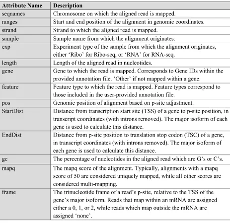
Table 2.1. Description of alignment attributes. Each attribute is contained within a separate metadata column in the GRanges object.
Attribute Name Description
seqnames Chromosome on which the aligned read is mapped.
ranges Start and end position of the alignment in genomic coordinates. strand Strand to which the aligned read is mapped.
sample Sample name from which the alignment originates.
exp Experiment type of the sample from which the alignment originates, either ‘Ribo’ for Ribo-seq, or ‘RNA’ for RNA-seq.
length Length of the aligned read in nucleotides.
gene Gene to which the read is mapped. Corresponds to Gene IDs within the provided annotation file. ‘Other’ if not mapped within a gene.
feature Feature type to which the read is mapped. Feature types correspond to those included in the user-provided annotation file.
pos Genomic position of alignment based on p-site adjustment.
StartDist Distance from transcription start site (TSS) of a gene to p-site position, in transcript coordinates (with introns removed). The major isoform of each gene is used to calculate this distance.
EndDist Distance from p-site position to translation stop codon (TSC) of a gene, in transcript coordinates (with introns removed). The major isoform of each gene is used to calculate this distance.
gc The percentage of nucleotides in the aligned read which are G’s or C’s. mapq The mapq score of the alignment. Typically, alignments with a mapq
score of 50 are considered uniquely mapped, while all other scores are considered multi-mapping.
frame The trinucleotide frame of a read’s p-site, relative to the TSS of the gene’s major isoform. Reads that map within an mRNA are assigned either a 0, 1, or 2, while reads which map outside the mRNA are assigned ‘none’.
and RSamTools for the processing of the alignments;edgeR for inferring differentially translated genes, and ggplot2, cowplot, and grid for producing the wide range of different visualizations (Lawrence et al. 2013, Morgan et al. 2009, Pages et al. 2017, Morgan et al. 2017, Wickham 2009, Robinson et al. 2010). A case study, which demonstrates the use of the platform’s tools within the context of a typical Ribo-seq quality analysis has been provided in Additional file 1.
2.3.2 Graphic customization
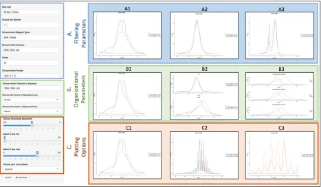
RiboStreamR facilitates graphical customization via various options for plot aesthetic and layout, as well as through dynamic filtering and sub-setting of data. Visualizations are customized through the use of the toolbar, which is located on the left side or bottom of each tool. The toolbar has three different types of adjustable parameters, see Figure 2.4 for examples.
Filtering parameters allow users to select the alignments they want to include in their analysis; alignments can be filtered by any of the attributes listed in Table 2.1. Organizational parameters change how the selected alignments are grouped and positioned within the output.
For example, users may wish to compare the alignments from one sample against alignments from all other samples combined, or they might want to compare subsets across a specific set of attributes, such as feature type, read length, and GC content. Plotting parameters affect the appearance or dimension of the output. Examples include adjustable axis values, axis labels, color schemes, and line types. Through the adjustment of these three types of customization parameters, users are able to create presentation quality graphics which are specific to their exact experimental inquiries. The output from each individual tool is also downloadable as a PDF image.
2.3.3 Reference datasets and anomaly detection
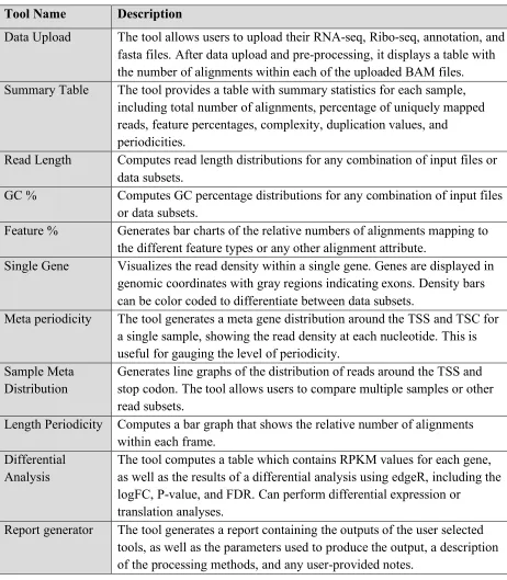
Table 2.2. Description of tools within riboStreamR.
Tool Name Description
Data Upload The tool allows users to upload their RNA-seq, Ribo-seq, annotation, and fasta files. After data upload and pre-processing, it displays a table with the number of alignments within each of the uploaded BAM files. Summary Table The tool provides a table with summary statistics for each sample,
including total number of alignments, percentage of uniquely mapped reads, feature percentages, complexity, duplication values, and
periodicities.
Read Length Computes read length distributions for any combination of input files or data subsets.
GC % Computes GC percentage distributions for any combination of input files or data subsets.
Feature % Generates bar charts of the relative numbers of alignments mapping to the different feature types or any other alignment attribute.
Single Gene Visualizes the read density within a single gene. Genes are displayed in genomic coordinates with gray regions indicating exons. Density bars can be color coded to differentiate between data subsets.
Meta periodicity The tool generates a meta gene distribution around the TSS and TSC for a single sample, showing the read density at each nucleotide. This is useful for gauging the level of periodicity.
Sample Meta Distribution
Generates line graphs of the distribution of reads around the TSS and stop codon. The tool allows users to compare multiple samples or other read subsets.
Length Periodicity Computes a bar graph that shows the relative number of alignments within each frame.
Differential Analysis
The tool computes a table which contains RPKM values for each gene, as well as the results of a differential analysis using edgeR, including the logFC, P-value, and FDR. Can perform differential expression or
translation analyses.
We have implemented four independent anomaly detection strategies with the Summary Table tool:
1. Anomaly detection based on expert defined thresholds for read periodicity, and percent of reads mapped to rRNA, tRNA, and CDS regions;
2. Outlier detection based on interquartile ranges derived from user samples, using Tukey’s fence (Hoaglin 2003).
3. Outlier detection based on user-selected controls. We compute summary QC metrics (including periodicity, feature percentages, and percentage of uniquely mapped reads) from the selected controls, and compare them to the corresponding values derived from the user’s samples. Outliers are defined using percent error calculation, with a threshold of 25% difference (Ambranowitz 1972).
4. Outlier detection based on our supplied reference data sets, using the method describe in item 2 above. We have precomputed a large set of quality metrics for various analysis parameter configurations to facilitate fast and accurate comparisons between the user-samples and our reference data.
Suspicious values found using these methods are flagged and reported in a separate table within the output pane.
2.3.4 Report generation
or analysis steps may also be appended to each graphic. Based on the chosen configuration, the generated reports include important information about the selected analysis parameters, as well as user-provided text as notes, figure summaries, or bibliographies for inclusion in publications. Additionally, individual graphics generated from each tool maybe downloaded independently within each tool for incorporation in publications or research papers.
2.3.5 Performance
Figure 2.4. Examples of riboStreamR’s graphical output customization options. On the left side is a
representation of the toolbar. (A) Filtering parameters are shown in blue, and allow plotting of distinct
subsets of the input data; (A1) Read Length Distribution (RLD) plot where each line represents the
alignments from 3 different samples; (A2) RLD plot where only alignments mapped to the CDS are
included; (A3) RLD where only reads mapping to tRNA or rRNA regions are included. (B)
Organizational parameters, shown in green, allow the user to adjust how the filtered data are grouped
and positioned in the output; (B1) Same as A1; (B2) RLD plots where the two plots separate between
alignments mapped within a CDS and those mapped to any other feature, and the lines separate between
three different samples; (B3) RLD plots where each plot is a separate sample, and the two separate
lines represent reads mapping to different feature types. (C) Examples of different plotting parameters,
shown in orange, which change the aesthetics of the graphical output; (C1) same as A1 and B1; (C2)
reduced bandwidth of line plot to simplify the comparisons between each separate read length; (C3)
reduced range of x-axis range, as well as different color scheme of plots to highlight differences
2.4 Discussion
2.4.1 Need for a web application based Ribo-seq analysis platform
As described above, various tools for streamlining quality control, analysis, and visualization of Ribo-seq data exist. Although extremely useful, the environments, user-interfaces, and infrastructures, of these tools vary considerably, and some of them require the knowledge of command line usage, R, or some other programming language. There exists a need for a platform which provides a consolidated suite of analysis tools that are accessible through a user-friendly GUI. The RiboGalaxy server is a tool that addresses some of these pain points (Michel et al. 2016). However, despite its popularity and usefulness, RiboGalaxy relies on combining processing steps and outputs from various third party tools, and does not focus on consistency and compatibility amongst the different tools. Our riboStreamR application is designed to fill this gap. The platform has the advantage of being an open-ended environment in which it is not a requirement for the user to follow a specific step-by-step procedure to ensure output/input compatibility. RiboStreamR takes advantage of the notable flexibility and functionality that R provides, but in a manner which removes the need for users to have programming experience in R, or another programming language. Of course, there are also certain advantages of alternative software tools over riboStreamR. For example, providing more upstream tools, such as fastq file QC; supporting a wider range of QC metrics, such as codon density analysis; functionality to map footprints to a reference genomes; and supporting integration with command line tools or other NGS software.
2.4.2 Future work
for use in both upstream processing and downstream analysis which are not yet fully implemented. These include additional tools for fastq quality analyses, optimized algorithms to map fastq files of ribosomal footprints to a reference genome, and additional downstream analysis tools to investigate codon usage, and to identify ribosome pause-sites and functional uORFs. Currently our tool has primarily been tested using data from Arabidopsis thaliana, but our goal is to perform extensive validation experiments using data from other species and to include corresponding datasets as references. A further research goal is the development and integration of more robust anomaly detection algorithms tailored for Ribo-seq data. We also plan to expand riboStreamR’s use of automation to simplify user’s analyses. Examples include the automatic generation of textual summaries and the automatic optimization of plotting parameters. Lastly, we plan to further optimize the platform’s underlying R code, for example by compressing sequence attributes to decrease memory consumption.
2.5 Conclusion
CHAPTER 3: Identification of Ribosome Pause Sites Using a Z-Score Based Peak Detection Algorithm
During protein synthesis, the speed of ribosomes as they move along mRNAs can vary based on multiple biological factors. It has been reported that pausing of ribosomes can influence gene expression through multiple mechanisms, such as facilitating protein folding and triggering translational abandonment. However, a deeper understanding of ribosomal pausing and its’ regulation is still missing. Ribosome profiling is a rapidly emerging technique for capturing a global “snap-shot” of ribosome position and activity. The method shows great potential for investigating many aspects of translating ribosomes. In particular, pause sites can be found by identifying peaks in ribosome profiling data. Here we present riboSmoothR, a novel method for identifying such peaks. Our algorithm utilizes the smoothed z-score method to identify anomalous ribosome footprint counts along a transcript. We show that riboSmoothR identifies a larger number of conserved peak sites than other commonly used peak finding methods. Additionally, we describe sequence-based features associated with peak sites identified by our method, such as codon and amino acid biases.
The following work was presented at ICCABS 2018 in Las Vegas, NV, and published in the conference proceeding and in the IEEE Xplore digital library (Perkins and Heber 2018).
3.1 Introduction
resolution within the translatome, and has confirmed the previously asserted notion that the distribution of ribosomes along transcripts is not uniform due to variabilities in the speed of translating ribosomes (Ingolia et al. 2009, Varenne et al. 1984).
The prevalence of sites with abnormally high ribosome occupancies suggest translational pausing. While their biological causes are not yet fully understood, pause sites have been associated with the presence of specific codons, mRNA secondary structures, tRNA availability, and interactions with nascent peptide chains (Woolstenhulme et al. 2015, Pop et al 2014, Gorochowki et al. 2015, Becker et al. 2013). Translational pausing has also been implicated in regulating biological mechanisms, including gene expression, protein folding, and mRNA decay within cells (Burgess Brown et al. 2008, Fluman et al. 2014, Pelechano et al. 2016). The increased resolution provided by modern ribosome profiling techniques offers the opportunity to quantify the global presence of ribosome pause sites in attempts to assess their biological causes and functions.
Potential translation pause sites are commonly identified by searching for significant peaks in ribosome occupancy (Li et al 2012, Gawronski et al. 2018). The distribution of ribosomes is often sporadic throughout individual ORFs, and defining which types of peaks correspond to pause sites can be difficult. Due to pause sites being so loosely defined, the methods which are used to identify them can vary greatly between experiments.
peaks which more accurately represent pause sites, we can attempt to further elucidate the biological context in which they occur.
3.2 Data processing
3.3 Smoothed z-score algorithm 3.3.1 Algorithm Basics
The smoothed z-score algorithm was primarily designed to perform robust and adaptive peak detection for real-time signal processing applications (Catalbas et al. 2017, van Brakel 2018). The algorithm uses a sliding window to scan a set of positions and calculate a moving mean and standard deviation. Significant positions are those which have a z-score higher than some threshold. The generic equation for the z-score is shown in Equation 1.
To increase the robustness of the algorithm, an influence parameter, which varies between 0 and 1, is used to scale the amount which signals effect the moving mean and standard deviation. Therefore, the complete set of parameters required by the algorithm are the lag (l), or size of the moving window, the threshold (thr) constant, which is the minimum z-score of signals, and the
influence (inf). The equations for the components of the smoothed z-score method are shown in
Equations 2, 3, and 4, where xi represents the normalized occupancy value at a single position, and si represents the adjusted value once the influence has been applied.
1
2
3
Figure 3.1. Depiction of how P-site offsets are determined. Meta genes are plotted for each read length, represented by the three different colors. Offsets are determined to by finding distance between the dominant upstream peak and the translation start codon. The 29mers in red have a dominant peak which is out of frame, therefore we search for a secondary peak which retains periodicity.
3.3.2 riboSmoothR
Using the basic properties of the smoothed z-score algorithm described above, we developed a customized algorithm for peak detection in ribosome profiling data. RiboSmoothR takes as input the footprint counts for every position in each coding sequence, divided by the number of seq reads located at the same position. The profiling data are scaled by the RNA-seq levels in order to account for regions which may have increased ribosomal occupancy due to higher transcript levels, as may be the case if multiple isoforms of a transcript are present. Three individual scans of these data-points using variations of the smoothed z-score algorithm are then used to search for peaks:
30 mers
28mers 29 mers
Distance from start codon
N orm al iz ed re ad d en si ty
P-site offset by length
• Scan 1: from 5’ to 3’ using the traditional smoothed z-score method
• Scan 2: from 3’ to 5’ using the traditional smoothed z-score method
• Scan 3: from 5’ to 3’ using a centered smoothed z-score method
The centered z-score scan evaluates a central position relative to the region surrounding Positions which surpass the z-score threshold for all three scans are considered significant peaks and potential pause sites. An example of this process is depicted in Figure 3.2. This combination of three scans is used to specifically identify positions which exhibit a significantly larger occupancy than positions in their local region. Scans 1 and 2 tests for significance relative to upstream and downstream regions, respectively, while scan 3 tests for significance relative to a smaller local region surrounding the position. Each separate scan uses its own unique set of the parameters discussed above (lag, threshold, and influence), allowing it to adjust to subtle differences in ribosome ‘traffic’ upstream and downstream of pause sites, while still ensuring peak intensity relative to neighboring positions.
3.3.3 Peak conservation
Therefore, one way to gauge how effectively a peak finding algorithm detects pause sites is to measure the degree of agreement in the location of peaks between samples. Cohen’s kappa is a popular statistic for measuring agreement between two raters for qualitative features, and can be used to assess the degree of peak conservation between any two samples (Cohen 1960). The
equation for kappa is shown in Equation 5, where is the relative agreement between raters (or
accuracy), and is the probability of agreement by chance.
Figure 3.2. Graphical depiction of the three smoothed z-score scans used by riboSmoothR. The green scan is scan 1, the red scan is scan 2, and the orange scan is scan three. Significant peaks found by each scan are indicated in the bottom three plots, positions in the black boxes are indicated by all three scans, and are therefore deemed significant.
For the calculation of Cohen’s Kappa, we generated an input vector where non-peak positions were coded as a 0s, and peaks were coded as a 1s. Table 3.1 shows that the average kappa value between the replicate samples is higher than between samples with differing treatment/strains. According to these results, samples which share neither a treatment or strain type, as well as samples which share only a treatment type, show much lower peak agreement. These results demonstrate that peaks in ribosome occupancy are conserved between biologically similar samples.
3.4 Algorithm Performance 3.4.1 Parameter Optimization
We can apply the above notion of peak conservation in order to tune the parameters for riboSmoothR by searching for the parameter set which maximizes the peak agreement between samples. First, we assembled a training set of 100 highly expressed and translated genes. A set of possible values for each parameter were then chosen, and the algorithm was run on the set of training genes for the exhaustive list of all possible parameter combinations. This process, also called a grid search, was performed for each of the eight samples. Intraclass correlation, an agreement metric which can be calculated for more than two raters, was employed to measure the agreement between all 8 samples (Shrout and Fleiss 1979). The formula used to calculate intraclass correlation, or ICC, is shown in Equation 6, where nr is the number of samples.
Table 3.1. Average of Cohen’s kappa values between sample pairs. The ‘replicate’ group is the average kappa value between replicate samples; the ‘strain’ group is the average kappa value between all non-replicate samples which share a common strain type, the ‘treatment’ group is the average kappa value between all non-replicate samples which share common treatment type, and the ‘none’ group are non-replicate samples which do not share a common strain or treatment type.
Sample types Kappa value
Replicate 0.211
Strain 0.192
Treatment 0.978
None 0.983
The parameters which achieved the highest conservation between samples are as follows: l1 = 20,
l2 = 20, l3 = 10, thr1 = 4, thr2 = 5, thr3 = 3, inf1 = 0.5, inf2 = 0.5, inf3 = 1, where the numerical subscript
indicates the scan.
3.4.2 Method comparison
which are x (10 and 50, respectively) times larger than the mean (Li et al. 2012, Gawronski et al. 2018). The ICC was used to determine agreement across all eight samples for each method. The methods were run on an identical set of 500 sufficiently expressed and translated genes. As Figure 3.3 shows, peaks identified by riboSmoothR exhibit higher agreement across samples, with lower variability.
3.5 Analysis of peak regions
After running riboSmoothR on the previously mentioned collection of 500 genes using optimized parameters, our goal was to investigate any irregularities in the sequence context of the detected peaks.
The final set of peaks was restricted to those which were found to be significant by riboSmoothR in both samples of a replicate pair. A total of 934 peaks were identified between all four replicate sets, 879 of which were found in exactly one replicate set, 41 in two, 10 in three, and 4 in all four.
3.5.1 Codon and amino acid usage
As can be seen in Figures 3.4 and 3.5, the representation of certain amino acids and codons vary significantly at peak sites when compared to what is hypothetically expected. Interestingly, we found proline sites to be significantly underrepresented at the P-site of peak positions, which contradicts previous studies that have implicated proline as a major factor in the slowing of ribosomes during translation (Woolstenhulme et al. 2015).
The results from Figure 3.5 shows us that this under-representation is due largely to the lack of CCA and CCT codons at peak sites. Arginine is the only significantly overrepresented amino acids at peak sites, with the codons CGC and CGG seemingly being the driving forces of this. Stop codons were also found to be overrepresented in peaks, which is supported by studies which have shown increased ribosome density at translation termination sites (Woolstenhulme et al. 2015).
3.5.2 Strings of amino acids
We also investigated irregularities in the representation of pairs and triplets of amino acids at pause sites. Previous studies have shown that strings of certain amino acids, such as proline, can cause translational pausing. The results from Figure 7 indicates that there are seven pairings that are significantly overrepresented, and one that is underrepresented. The underrepresented pair, SP, contains a proline at the P-site, which is consistent with our previous findings which indicated that prolines are underrepresented at peak sites. Additionally, when looking at the triplets in Figure 7, we find that even though PPP is the most common triplet with a proline is at the P-site, it is still vastly underrepresented compared to what is expected. Although strings of amino acids with proline at the P-site seem common throughout coding regions, our results indicate a lack of these strings at high occupancy locations.
3.5.3 GO enrichment and motif analysis
Figure 3.6. Pairs along the x-axis represent the most common E-P site pairings at peak locations.
A most frequent pair is determined for each different amino acid which occupies the P-site (ex. when A is in the P-site, EA is the most common pair). Blue dots are percent of all peaks which contain each amino acid pair at their E-P sites. Orange dots are the expected percentage based on the global frequency of each pair of amino acids. Blue bars indicate an overrepresentation of the pair compared to what is expected, and orange bars indicate an underrepresentation. Asterisks denote a significant deviation from what is expected, based on a binomial test with an alpha of 0.05.
Table 3.2. GO term and motif analysis. Sequences surrounding peaks from each enriched GO term were submitted for motif analysis.
GO Term Number of genes in term
Number of sequences analyzed
Motifs found at peaks within GO term
response to brassinosteroid; GO:0009741
6 71 CWKMHMMYARMATGWHKDTTDWSTWYMC
AYTGARRTKMYTKMRAWGMBT GATACKGGTTTGTAT
response to organic cyclic compound;
GO:0014070
9 79 ATAGRGWTGHGRWKACWACAA
GRTACKGGTTWGTAT CCWMCCAYCKRDTCYCACGT response to endogenous
stimulus; GO:0009719
16 114 TGCTGATCCCAACATGACGATTAWGKATC
TVYYTTCAAADCYC RGTDSWGTKTGTTGG signal transduction;
GO:0007165
21 120 RAKAGANWNMGGMAKMHSWCMATCRHWYYWYKKGSAAS
R
TNYYTTCAAADCYCD cellular response to organic
substance; GO:0010033
20 90 CAKAGASATSYGAWGASAACA
RYTGARRTTCYTTMRAMGCBT
All peaks 177 934 CWCCRYMWSYHTACWANTCTCCWCCACCA
TAYTCHCCTTCTCCTAAGGTAGASTACAA AGAARAAGRWSAAGR MAAAASMACMAMCAY WBKMATCGCCGCCGCYGWCKDWGAMG GAGDSWMWYGDKGAGATKGYT ATCHABSGGMGGHGAGAKMTC TCCRGAGACTGVTGATCCSAA 3.6 Conclusion