ABSTRACT
POWERS, JASON GANNON. Mapping Replication and Silencing Suppression Elements in the RCNMV Genome. (Under the direction of Steven A. Lommel).
Viruses infect all Kingdoms of life on Earth. Their life cycle represents a constant struggle for survival in hostile cellular environments. To survive the virus must both avoid the host response and replicate in a timely manner. The following dissertation includes
investigations into both of these aspects of viral infection focusing on plant-infecting viruses. When infected by viruses, plants respond by initiating defense pathways. One of these pathways is known as RNA silencing. In this pathway host proteins target double-stranded viral RNA intermediates for cleavage. The product of this cleavage, a short-interfering RNA (siRNA), is incorporated into a protein complex that guides sequence specific cleavage of viral RNA. To cause a productive infection, the virus must devise countermeasures to this targeting. They accomplish this by suppressing the RNA silencing pathway by encoding proteins known as viral suppressors of RNA silencing (VSRs). Identifying these VSRs is critical to the understanding of how any plant virus survives in its host. Commonly used assays for identifying VSRs all use reporters expressed in the nucleus. These nuclear-based DNA reporters are being used to assay for the RNA silencing
use of TCV-sGFP as a reporter for RNA silencing suppression activity TCV-sGFP was used to identify a previously uncharacterized VSR for the plant-infecting virus Red clover necrotic mosaic virus (RCNMV). RCNMV’s replication complex was previously implicated in the suppression of RNA silencing, while the new TCV-sGFP assay allowed us to detect a second suppressor, a protein known to be involved in viral movement, known simply as the
movement protein (MP). Domains of MP were analyzed and it was found that the amino acid residues between positions 122 and 277 were required for suppression activity.
The RNA silencing pathway has two principal components, siRNAs and host proteins. To disrupt the pathway the MP of RCNMV must interfere with one or both of these
components. In this thesis the possibility that the MP suppresses RNA silencing by binding to siRNAs is addressed by employing electrophoretic mobility shift assays to examine MP’s ability to bind to siRNAs. MP was found to have no siRNA binding capability, indicating that it’s mode of action likely relies on binding to or modifiying host proteins.
Finally, initial studies into the temporal regulation of RCNMV replication were undertaken. As mentioned, for a productive infection to take place the virus must go beyond evading the host defense response, it must also properly regulate the timing of expression for its many genes. Using real time PCR, the timing of various replication events in the
RCNMV life cycle were mapped to more fully understand how RCNMV replicates inside the cell. These time course studies have revealed that virion formation has little effect on
Mapping Replication and Silencing Suppression Elements in the RCNMV Genome
by
Jason Gannon Powers
A dissertation submitted to the Graduate Faculty of North Carolina State University
in partial fulfillment of the requirements for the degree of
Doctor of Philosophy
Genetics
Raleigh, North Carolina 2009
APPROVED BY:
_______________________________ ______________________________
Steven A. Lommel Steven Spiker
Committee Chair Committee Co-Chair
________________________________ ______________________________
BIOGRAPHY
ACKNOWLEDGMENTS
First and foremost I would like to thank my family for providing the support I’ve needed to get through these long nearly 6 years of graduate school life. My mother, father, sister, brother, and grandparents have always provided a boost when I needed it. Not to mention a wonderful sanctuary in southwest Florida for when I needed to unwind.
I’d like to thank Steve Lommel, my P.I., not just for direction in my research, but also for being the type of P.I. that understands that to have productive graduate students you must have happy graduate students, and to have happy graduate students there has to be more to their lives than 24/7 lab work. He has always been accommodating when I needed time off, and rarely, if ever, criticized my work ethic. I am also happy to say that we had a good relationship outside of work, I think it is uncommon for P.I.’s to take their graduate students to the Stanley Cup finals, or to a Monday Night Football game, but Steve took me to both, and I appreciate and enjoyed the time we spent outside of the lab.
over the years in the Lommel lab, especially Richard Guenther, who I could always count on to have unique ideas and interpretations of my results.
For a semester of my time here at NC State, I did a study abroad program at Seoul National University in South Korea. While there I worked with Kook-Hyung Kim, who, like Steve, I consider to be both a mentor and a friend. While in Korea members of
Kook-Hyung’s lab helped me to survive in a country I didn’t know and a language I couldn’t understand. In particular Sang Ho, without whom I probably would have hated Korea. I’d like to thank Eva Johannes at the Center for Molecular Imaging Facility for assistance with nearly all the GFP imaging in this thesis, and Alora LaVoy for assistance with the IVIS Lumina imager. I need to thank Clayton Beard, director of research at Global Vacccines, Inc. for agreeing to take me on as a short term intern. He was a great teacher, and I learned a lot in my short time working under his supervision.
The other members of my committee need to be thanked for their guidance, and in particular Steve Spiker for agreeing to be my committee Co-chair. Niki Robertson needs to be thanked for providing some valuable insight into RNA silencing and viruses, as does Robert Franks for agreeing to join my committee despite at the time being a brand new faculty member.
TABLE OF CONTENTS
LIST OF TABLES... ix
LIST OF FIGURES ... x
ABBREVIATIONS ... xii
Chapter 1 A Literature Review of RNA Silencing and Red clover necrotic mosaic virus... 1
Chapter Summary ... 2
Introduction... 2
Innate and Adaptive Immunity ... 3
RNA Silencing... 5
Small RNA silencing-associated RNAs... 8
miRNAs ... 9
piRNAs ... 12
siRNAs... 13
DCLs ... 28
AGOs ... 31
RDRs... 35
RNA silencing targeting plant RNA viruses... 39
VSRs ... 41
Identifying RNA silencing suppressors ... 46
RCNMV... 48
Genome structure, protein function and expression ... 49
Viral replication ... 53
Translation ... 54
Virion packaging... 56
RCNMV-host interactions ... 57
Literature Cited ... 59
Chapter 2 A Versatile Assay for the Identification of RNA Silencing Suppressors Based on Complementation of Viral Movement... 92
Chapter Summary ... 93
Introduction... 93
Results... 96
TCV movement is limited to 3-5 cells in N. benthamiana after CP deletion ... 96
TCV CP delivered in trans complements TCV-sGFP movement... 97
Heterologous viral suppressors complement TCV-sGFP movement ... 99
Co-infiltration of various suppressors results in silencing suppression... 100
Utility of the TCV-sGFP assay for new VSR identifications... 103
A. thaliana vs. N. benthamiana RNA silencing... 108
Materials and Methods... 109
Acknowledgements... 114
Literature Cited ... 114
Chapter 3 The Red clover necrotic mosaic virus RNA-2 Encoded Movement Protein is a Second Suppressor of RNA Silencing... 131
Chapter Summary ... 132
Introduction... 132
Results... 135
RCNMV replication suppresses RNA silencing... 135
RCNMV RNA-2 complements TCV-sGFP... 136
The RCNMV RNA-2 encoded MP is responsible for TCV-sGFP complementation... 137
RCNMV RNA-1 encoded proteins do not complement TCV-sGFP movement.. 138
RCNMV MP complements movement deficient TCV-sGFP... 138
RCNMV MP alanine-scanning mutants resolve 2 distinct roles in viral infection ... 139
Discussion... 141
Suppression of RNA silencing by RCNMV ... 141
Multi-pronged approaches to suppression of RNA silencing... 144
Viral movement and suppression of RNA silencing... 145
Further defining the RCNMV MP functional domains ... 147
Materials and Methods... 149
Acknowledgements... 152
Literature Cited ... 152
Chapter 4 Investigations into siRNA Binding Capabilities of RCNMV MP... 167
Chapter Summary ... 168
Introduction... 168
Results... 171
Expression of p19 and MP... 171
MBP-MP binds to ssRNAs ... 173
MBP-MP cannot bind 21nt siRNAs ... 174
MBP-MP cannot bind 24nt siRNAs ... 174
siRNA methylation does not assist in binding... 175
MBP-MP cannot bind to 21 or 24nt RNA oligos ... 176
Discussion... 176
MBP-MP expression... 177
Silencing suppression by the RCNMV replication complex ... 184
Materials and Methods... 185
Literature Cited ... 189
Chapter 5 Time Course Accumulation Studies of RCNMV... 200
Chapter Summary ... 201
Introduction... 201
Results... 205
RNA-1 and RNA-2 accumulation during a wildtype infection ... 205
RNA-1 minus strand accumulation during a wildtype infection ... 207
The presence of the CP has no effect on RNA-1 accumulation levels ... 208
RNA-1 accumulates to a slightly higher titer during an RNA-1 only infection ... 209
Negative sense northern blots ... 210
Discussion... 211
RNA-1 vs. RNA-2 accumulation... 212
RNA-1 negative and positive sense RNA accumulations... 215
RCNMV coat protein has no effect on viral strand accumulation... 215
Northern blots reveal unexpected RNA bands... 217
Future studies ... 219
Materials and Methods... 221
LIST OF TABLES
Chapter 1
Table 1 – Types of sRNAs and their associations ... 87 Table 2 – DCLs, AGOs, and RDRs of the plant RNA silencing pathways... 88
Chapter 2
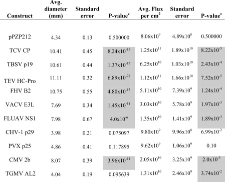
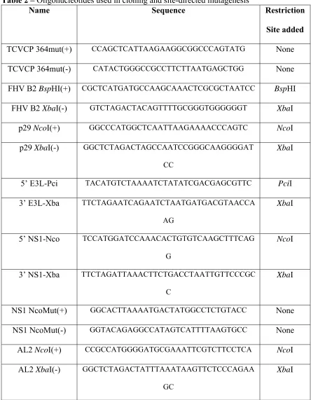
Table 1 –Measurements and P-values for each VSR when analyzed in the TCV-sGFP complementation and PZP-TCV-sGFP co-infiltration assays ... 120 Table 2 –Oligonucleotides used in cloning and site-directed mutagenesis ... 121
Chapter 3
Table 1 –The Functional Domains of RCNMV MP... 158 Table 2 –Oligonucleotides used in Cloning and Site-directed Mutagenesis ... 159
Chapter 5
LIST OF FIGURES
Chapter 1
Figure 1 – Generalized RNA silencing pathway in plants... 89
Figure 2 –RNA silencing targeting RNA viruses in plants ... 90
Figure 3 –Genome organization of RCNMV ... 91
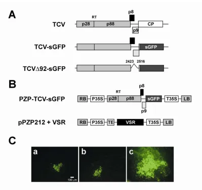
Chapter 2 Figure 1 –Schematic of plasmid constructs and response to transcript inoculation on leaves expressing TCV CP... 122
Figure 2 – TCV-sGFP complementation assay and co-infiltration assay... 123
Figure 3 – TCV CP does not complement movement of p8/p9 deletion mutant... 124
Figure 4 – Complementation of TCV-sGFP movement by TCV CP is not due to virion packaging ... 125
Figure 5 – TCV-sGFP movement can be complemented by a wide range of VSRs.... 126
Figure 6 – PZP-TCV-sGFP co-infiltration assay... 128
Figure 7 – Northern blot of analysis of PZP-TCV-sGFP co-infiltration assays... 130
Chapter 3 Figure 1 –Schematics of viral and Ti-plasmid constructs used ... 160
Figure 2 –RCNMV viral replication suppresses the RNA silencing of a Ti-plasmid driven reporter... 161
Figure 3 –TCV-sGFP complementation assay with full length RCNMV viral RNA Agrobacterium constructs ... 162
Figure 4 – The RCNMV RNA-2 movement protein is responsible for TCV-sGFP complementation... 163
Figure 5 – MP is the only RCNMV protein that can complement TCV-sGFP movement... 164
Figure 6–RCNMV MP facilitates movement of TCV-sGFP p8/p9 deletion mutant . 165 Figure 7 – TCV-sGFP movement complementation by RCNMV MP Alanine-scanning mutants... 166
Chapter 4 Figure 1 –Genome organization of RCNMV ... 196
Figure 2 –Expression and purification of pMAL-MP and p19 constructs ... 197
Figure 3 –Electrophoretic Mobility Shift Assays ... 198
Chapter 5
Figure 1 – Genome Organization and Replication Strategy of RCNMV ... 229
Figure 2 – Viral RNA strand accumulation of RNA-1 and RNA-2 during wildtype infection ... 230
Figure 3 – Negative sense RNA-1 accumulation during wildtype infection... 231
Figure 4 – CP effect on RNA-1 accumulation... 232
Figure 5 – RNA-1 accumulation in the absence of RNA-2... 233
ABBREVIATIONS
ADK Adenosine kinase protein AGO Member of the
ARGONAUTE protein family BiFC Bimolecular fluorescence
complementation CHS Chalcone synthase
CHV-1 Cryphonectria hypovirus 1 EP713
cis-NAT cis-natural antisense transcript
CMV Cucumber mosaic virus
CP Capsid protein CTV Citrus tristeza virus
DCL DICER-LIKE Protein dpi days post
inoculation/infiltration dsRNA Double-stranded RNA DTT Dithiothreitol
endo-siRNA endogenous siRNA FHV Flock house virus
FLUAV Influenza A virus
FS Ribosomal frameshift GFP Green fluorescent protein GUS β-glucuronidase
HCV Hepatitis C virus
HEN1 RNA methyltransferase HIV Human immunodeficiency
virus
HR Hypersensitive response IPTG Isopropyl β
-D-1-thiogalactopyranoside MBP Maltose binding protein miRNA Micro RNA
MP Movement protein
nat-siRNA natural antisense transcript-derived
OAS Origin of assembly sequence Oligos oligonucleotides
ORF Open reading frame
p27 RCNMV replication protein p88 RCNMV replication protein PDS Phytoene Desaturase piRNA PIWI-interacting RNA
PIWI Member of the
ARGONAUTE protein family PolIV RNA polymerase IV
PTGS Post-transcriptional gene silencing
PVX Potato virus X
PVY Potato virus Y
qRT-PCR quantitative real time PCR rasiRNA Repeat-associated siRNA RCNMV Red clover necrotic mosaic
virus
RDR Plant-encoded RdRp RdRp RNA dependent RNA
polymerase R-genes Resistance genes RISC RNA-induced silencing
complex
sGFP synthetic Green Fluorescent Protein
sgRNA Sub-genomic RNA
SGS3 Suppressor of Gene Silencing 3
siRNA Short-interfering RNA sRNA Small RNA
silencing-associated RNA ssRNA Single-stranded RNA TA Trans-activator
TABS TA binding site tasiRNA trans-actingsiRNA
TBSV Tomato bushy stunt virus
TCV Turnip crinkle virus
TEV Tobacco etch virus
TGMV Tomato golden mosaic virus
TGS Transcriptional gene silencing Ti-plasmid Tumor inducing plasmid TMV Tobacco mosaic virus
TRV Tobacco rattle virus
VACV Vaccinia virus
Chapter 1
Chapter Summary
Chapter 1 of this dissertation is a literature review of many of the topics found in the latter chapters of this dissertation, as well as some topics not directly addressed in the
chapters that follow. This wide ranging literature review contains a thorough background on RNA silencing, with a focus on plants. Host proteins involved, along with the various RNA silencing-associated small RNAs are discussed at length. Special emphasis is placed on RNA silencing-associated small RNAs involved in the immune response of the plant against invading pathogens, specifically viruses. Methods of viral counterdefense are discussed, as well as ways to monitor for this counterdefense. Following this review of RNA silencing in plants, the plant-infecting virus Red clover necrotic mosaic virus (RCNMV) is discussed in detail. This literature review is intended to set up the chapters that follow, which focus on RNA silencing targeting viruses in plants, how RCNMV evades the RNA silencing pathway, and examinations into how RCNMV replicates in a cell.
Introduction
life-cycle, far less is known about RCNMV’s interactions with the immune system of its hosts. The work described here centers on an effort to illuminate aspects of these host-virus interactions in an effort to expand the knowledge base of how RCNMV successfully infects and replicates inside its host. Specifically, the interplay of plant viruses with the adaptive immune system in plants that relies on RNA to direct sequence specific anti-viral activities is examined. This activity is generally referred to as RNA silencing and can occur either at the transcriptional level where it is known as transcriptional gene silencing (TGS) or the post-transcriptional level where it is known as post-post-transcriptional gene silencing (PTGS). To survive in the plant host, viruses must circumvent the adaptive immune response of the plant. They do this by encoding viral suppressors of RNA silencing (VSRs), specific viral proteins that disrupt the RNA silencing cascade allowing for a productive viral infection. The study of these proteins in general and RCNMV’s VSRs specifically form the bulk of the work that follows.
Innate and Adaptive Immunity
macrophages can identify lipopolysaccharides found specifically on gram-negative bacterial surfaces through their cell surface ligand called mCD14 (Raetz and Whitfield, 2002; Ulevitch and Tobias, 1994). This identification leads to cytokine production that recruits other host immune system cells to the area and results in phagocytosis of the infectious bacteria (Dobrovolskaia and Vogel, 2002). While most organisms employ the innate immune response, the adaptive immune response in humans is thought to be much more recently evolved (Cannon et al., 2002). Adaptive immunity is triggered by the innate immune
response, but in contrast to innate immunity, adaptive immunity is highly specific. While the innate immune response recognizes generalized pathogen features (i.e. lipopolysaccharide), the adaptive immune response recognizes specific epitopes on specific pathogens, allowing for targeted elimination of the pathogen. This recognition allows not only for specific
targeting of the pathogen in question, but for pathogen “memory” as well. This “memory” of a pathogen is mediated by Memory B and Memory T cells and can lead to long-lasting immunity to the infecting pathogen (Larosa and Orange, 2008).
elicitors) or pathogen-initiated modifications of host proteins often leading to the triggering of the HR (van Ooijen et al., 2007). Adaptive immunity in plants is more widely known as RNA silencing. RNA silencing relies on double-stranded (ds)RNA to trigger an immune system response that eventually leads to sequence-specific targeting of the invading pathogen. As with the human adaptive immune response, this form of plant immunity is highly specific and can lead to resistance to further infection. Viruses are particularly susceptible to this form of targeting by the immune system and have evolved to counteract this defense mechanism by encoding VSRs that can disrupt the RNA silencing cascade (Ding and Voinnet, 2007; Li and Ding, 2006). The RNA silencing cascade, VSRs, and the interaction between the two are discussed in more detail below.
RNA Silencing
implicated in the control of endogenous gene expression (Napoli et al., 1990), repression of transposable elements (Tabara et al., 1999), and heterochromatin formation (Volpe et al., 2002). However, most importantly to this dissertation is the role RNA silencing has been shown to play as a host defense mechanism against pathogens (Brigneti et al., 1998).
A role for RNA specific anti-viral immunity as mechanism of host defense against pathogens was theorized in the early 90’s when a series of papers described the infection of transgenic plants expressing viral coat protein (Fang and Grumet, 1993; Lindbo et al., 1993; Ling et al., 1991). The papers, which focused on Potyviruses, reported that viral infections were severely inhibited in these transgenic plants but were not linked to the CP protein itself. Infected plants typically showed attenuated symptoms and were capable of recovery from infection. At least one group subsequently showed that recovered tissue became resistant to further infection. Protoplasts made from these transgenic plants demonstrated that resistance was functional at the single cell level, as these cells were also resistant to infection. Levels of viral and transgene RNAs were found to be highly diminished in recovered tissues, but somewhat surprisingly, rates of transcription from the transgene were stable. These observations led to the suggestion that the observed viral resistance was likely to be at the RNA stability level (Lindbo et al., 1993). It would be several more years before they would be proved right, as the resistance they described is now known to be due to RNA silencing.
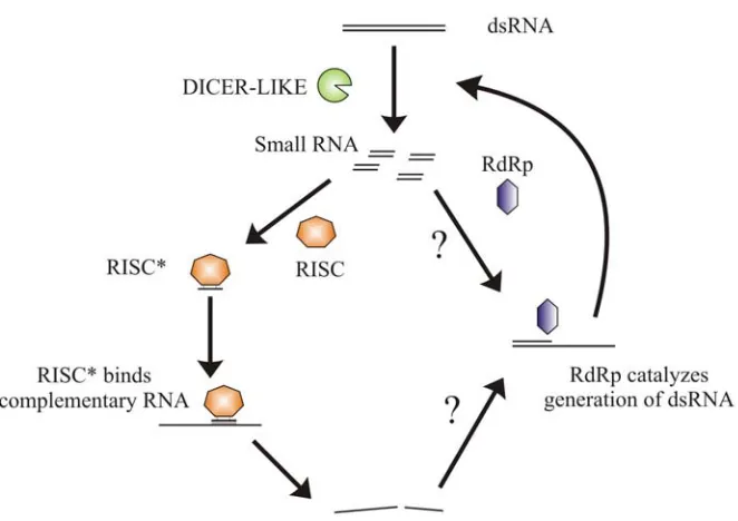
2001). DICER cleaves the dsRNA into small RNA silencing-associated RNAs (sRNAs) that range from 21 to about 30 nt in length (Bernstein et al., 2001). In the next phase, the effector phase, the sRNAs are unwound and one strand is incorporated into the RNA-induced
silencing complex (RISC) where the now single-stranded (ss)RNA is associated with a protein called ARGONAUTE, of which there are two main clades known as AGO and PIWI (Hammond et al., 2000; Liu et al., 2004; Meister et al., 2004; Sasaki et al., 2003). Once part of the RISC complex the sRNA can guide sequence specific targeting of heterologous
Small RNA silencing-associated RNAs
sRNAs can be broadly grouped into three classes, the first of which was identified in Caenorhabditis elegans in 1993, long before it was ever known how sRNAs work to
influence gene expression (Lee et al., 1993). It took many years for researchers to
miRNAs
RISC incorporation is currently unknown (Fang and Spector, 2007; Fujioka et al., 2007; Song et al., 2007). Export from the nucleus could occur before miRNA incorporation into RISC or after. Whenever export from the nucleus occurs, it involves appears to involve HASTY, the Arabidopsis thaliana homolog to the mammalian protein EXPORTIN 5 (Park et al., 2005). In plants the newly generated miRNA in complex with RISC can affect
endogenous gene expression by binding to complementary sequences on target mRNAs and inhibiting translation or – more commonly in plants – directing transcript degradation (Aukerman and Sakai, 2003; Chen, 2004; Llave et al., 2002; Tang et al., 2003).
few examples are known. Notably these examples include Hepatitis C virus (HCV) as well as Human Immunodeficiency virus (HIV), both of which appear to be targeted by host miRNAs (Huang et al., 2007; Pedersen et al., 2007). Experiments in cell culture have shown that miRNAs do have the ability to target and knockdown expression of a virus, but a
widespread role for miRNAs as part of the immune response is still unclear (Lecellier et al., 2005). Logically it would seem unlikely that miRNA targeting of viral genomes would be an effective method of antiviral defense, as viral genomes are known to have high mutation rates. A targeted virus need only mutate a single nucleotide to avoid efficient miRNA targeting, and yet both HCV and HIV seem to be targeted by host miRNAs. Why is this? It has been suggested that HCV and HIV are actually exploiting the host miRNAs for their own life cycle. Both viruses form persistent infections, and these miRNAs might help regulate viral replication to levels that are below the detection threshold of the immune system. In the case of HIV, these miRNAs may help HIV maintain latency, thus allowing it to survive.
HCV appears to use other miRNAs not for negative regulation of replication but instead for the opposite effect, to promote replication. HCV has been shown to utilize the cellular miRNA miR-122 for its replication although the mechanism for this is still unknown (Jopling et al., 2005; Randall et al., 2007). Finally, it is clear that at least some viruses can influence the miRNA profile and/or activity of infected cells (Bazzini et al., 2007; Chen et al., 2004a; Dunoyer et al., 2004). What is currently unknown is whether these changes in
at least some symptomatology in certain plant virus infections is attributable to virally induced changes in miRNA levels, but as with all work involving the interplay between sRNAs and viruses more work is needed (Bazzini et al., 2007; Chen et al., 2004a; Dunoyer et al., 2004).
piRNAs
piRNAs are the newest of the sRNAs to be recognized. As mentioned above the first piRNAs were mistakenly identified as a class of siRNAs, termed repeat-associated
(ra)siRNAs (Aravin et al., 2003). It is now known that in species other than plants rasiRNAs are actually not siRNAs at all, but instead compose a subclass of piRNAs that are derived from areas of the genome that are heavy in repeats (Yin and Lin, 2007). piRNAs can also be found in regions of the genome that do not contain repeats and often map to both the sense and anti-sense regions of transposons (Brennecke et al., 2007).
AGO3 in Drosophila, help to generate the anti-sense piRNAs which preferentially bind to PIWI and AUBERGINE. In turn the anti-sense piRNAs help to generate the sense piRNAs (Brennecke et al., 2007; Gunawardane et al., 2007). Most known sense piRNAs have anti-sense corollaries with 10 bases of complementarity, the 10th base pair appears to be where the PIWI-related protein associated with the piRNAs catalyzes a cleavage event that has been theorized as a possible way to generate the 5’ terminus of the new piRNA (Brennecke et al., 2007; Gunawardane et al., 2007; Saito et al., 2006). This process is thought to be self-reinforcing, so that the newly generated piRNA can now direct the biogenesis of new piRNAs from the other strand.
piRNAs are generally expressed in germline cells and are thought to regulate
transposons and possibly play a role in epigenetic inheritance (Brennecke et al., 2008). The PIWI subclass of ARGONAUTE proteins does not exist in plants, and so plants do not use piRNAs for any of these functions. It is thought that different classes of siRNAs may serve similar roles in these organisms. No known role in viral infection has been found or
theorized for piRNAs. It is for these reasons that piRNAs will not be discussed further in this thesis.
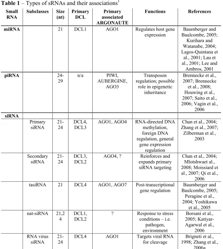
siRNAs
transcript. In plants, endogenous siRNAs can be further subdivided into 4 different subclasses – primary siRNAs, secondary siRNAs, trans-acting (ta)siRNAs, and natural antisense transcript-derived (nat-)siRNAs. Until 2008 no endogenous siRNAs had been characterized in species that lacked an RNA dependent RNA polymerase, such as Drosophila and all mammals. This changed with the discovery of endogenous (endo-)siRNAs in
Drosophila melanogaster as well as mice (Chung et al., 2008; Czech et al., 2008; Ghildiyal et al., 2008; Kawamura et al., 2008; Okamura et al., 2008a; Okamura et al., 2008b; Tam et al., 2008; Watanabe et al., 2008). These endo-siRNAs appear to encompass many of the
endogenous siRNA functions that previously had only been recognized in other organisms such as plants.
Unlike in mammals, endogenous siRNAs have been well characterized in plants and C. elegans. Primary siRNAs come in two broad categories and are the most prototypical of the endogenous siRNAs. Primary siRNAs can be derived from sequences that can self-form double-stranded RNAs, often through inverted repeats that form hairpins. Hairpin-forming inverted repeats are common in the plant genome and contribute to a large population of siRNAs found in plant cells. These dsRNAs are targeted by DCLs for the generation of primary siRNAs (Zhang et al., 2007). These primary siRNAs can then associate with RISC for targeted RNA cleavage. While hairpin-forming repeats are common, in A. thaliana the most common type of primary siRNAs are 24 nt in length and are involved in
Pontier et al., 2005). The exact process of siRNA biogenesis has yet to be fully elucidated, but it is thought that a particular isoform of PolIV, simply designated as PolIVa, transcribes a sequence of DNA which is then made double-stranded by RDR2 (Mosher et al., 2008;
Pontier et al., 2005). The double-stranded form is then cleaved by DCL3 into siRNAs, although DCL2 and DCL4 can functionally replace DCL3 in its absence (Henderson et al., 2006). In conjunction with the DNA methyltransferases DRM and CMT3, a histone methyltransferase (KYP), and AGO4 these siRNAs can then direct sequence specific heterochromatin formation, inhibiting transcription (Cao et al., 2003; Jackson et al., 2002; Zilberman et al., 2003).
Secondary siRNAs are siRNAs that are generated from sequences that have been pre-silenced by primary siRNAs. The role of these secondary siRNAs is simply to amplify and maintain the silencing of these pre-silenced RNAs or loci and to expand the targeted region by a method called transitivity. Secondary siRNAs are particularly important in host defense against exogenous pathogens, but in terms of endogenous RNA silencing it appears as though secondary siRNAs may also play a role in maintaining silenced heterochromatin. While the details have still not been fully worked out, it is thought that the two isoforms of PolIV, the previously mentioned PolIVa and its counterpart PolIVb, somehow work together to generate siRNAs from already methylated DNA sequences. Two competing models have been proposed. The first model, proposed in 2005, suggests that PolIVa synthesizes the primary siRNAs that lead to initial DNA methylation, and that PolIVb recognizes these DNA modifications and is recruited to these sites. Following this recruitment PolIVb, in
methylation, in turn recruiting more PolIVb creating a self reinforcing loop (Pontier et al., 2005). A second model proposed this past year stems largely from the fact that PolIVb has been found to interact directly with the effector complex by way of AGO4 (El-Shami et al., 2007). According to this model instead of synthesizing RNA, PolIVb works with AGO4 to provide some role in DNA or histone methylation, which results in the recruitment of more PolIVa back to initial sites of methylation for further generation of secondary siRNAs (Mosher et al., 2008; Qi et al., 2006).
Another type of endogenous siRNA is tasiRNAs, the synthesis of which is
that there exists another secondary trigger in these transcripts that results in targeting by RDR6 (Axtell et al., 2006). After conversion into the dsRNA intermediate the tasiRNA precursor is further cleaved by DCL4 into 21 nucleotide RNAs (Yoshikawa et al., 2005). These siRNAs are cleaved in a phased manner, meaning that they are cleaved back to back to back, with the original miRNA cleavage site setting the proper phase (Allen et al., 2005). These individual tasiRNAs can then be loaded into RISC alongside either AGO1 or AGO7 and start targeting mRNAs for degradation (Adenot et al., 2006; Baumberger and Baulcombe, 2005; Peragine et al., 2004).
The regulation of tasiRNA production by miRNAs is a novel and interesting
development in the understanding of the role of miRNAs in cells. Up until this point the role of miRNAs seemed to be solely in direct repression of gene expression. The recent work with tasiRNAs shows a different, previously unknown transcript processing function of miRNAs. There is also emerging evidence some tasiRNAs can act as miRNAs, guiding the initial cleavage events that lead to the generation of new tasiRNAs (Chen et al., 2007; Howell et al., 2007).
Despite the existence of large regions of intergenic spaces in plant and animal genomes, examples of pairs of genes that produce overlapping antisense transcripts, cis-natural antisense transcripts (cis-NATs), are abundant. In A. thaliana the number has been put at somewhere between 1000 and 1300 pairs of cis-NATs, while in humans some
form sense–antisense pairs (Chen et al., 2004b). These pairs of cis-NATs could conceivably form dsRNAs that would be ideal targets for cleavage by DICER-related proteins.
The first nat-siRNA was reported in a 2005 article published by Borsani, et al. (Borsani et al., 2005). In that original article the authors were able to show that high salt stress conditions induces A. thaliana expression of the endogenous gene SRO5 which overlaps in an antisense orientation with ∆1-pyrroline-5-carboxylate dehydrogenase (P5CDH), a stress-related gene. Upon this induction a new, 24 nt siRNA appears of the same sense as the SRO5 gene which can target P5CDH for degradation. The biogenesis of this siRNA is dependent on DCL2, as well as RDR6, SGS3, and the large subunit of PolIVa, NRPD1A. In addition, this 24 nt siRNA appears to be capable of directing the synthesis of secondary 21 nt siRNAs, which appear in phase with the original 24 nt siRNA and are dependent upon the same proteins for synthesis as well as DCL1 (Borsani et al., 2005).
It is not yet clear whether nat-siRNAs play a widespread role in defense against invading pathogens. The large numbers of cis-NATs that exist have provided for much speculation as to the possibility for a large role of cis-NATs in gene regulation, but so far only the 2 concrete examples outlined above have been found (Borsani et al., 2005; Katiyar-Agarwal et al., 2006). Still, a recent genome-wide study reported that 646 different cis-NAT pairs in A. thaliana have at least one complementary siRNA (Jin et al., 2008). Unfortunately the biological relevance for this complementarity is unknown, and clearly more research is required. It is possible the nat-siRNAs may play a large role in the innate immune response of plants. The fact that they are endogenously encoded like miRNAs suggests that it is unlikely that they have any direct binding capability to an invading pathogen, but the ability for these nat-siRNAs to downregulate the expression of endogenous genes could prove to be just as useful to a host responding to an invading pathogen, as was shown in the work with P. syringae (Katiyar-Agarwal et al., 2006).
transposons, the function of the rest of the endo-siRNAs is still unclear. Direct RNA target regulation has only been shown for hairpin-derived siRNAs (Czech et al., 2008; Okamura et al., 2008b). No proposals have been made for a role in viral infection for these endo-siRNAs, and like their plant counterparts, it appears that their role, if any, would likely be confined to regulating cellular genes related to pathogen defense rather than directly targeting viral RNAs.
In contrast to endogenous siRNAs, exogenous siRNAs have been shown to play a critical role in host defense against invading pathogens and exogenous nucleic acids in a number of organisms. Plants, fungi, and insects all have demonstrable exogenous siRNA pathways as part of their adaptive immune response, but while endo-siRNAs are now recognized in mammals, no evidence of exogenous mammalian siRNA production has been found. The following will focus on exogenous siRNAs found in plants, which can come from two different sources: exogenous DNA that has been introduced into the genome of the plant (i.e. transgenes, DNA viral genomes), and exogenous RNA that has been directly introduced into the cell, usually through an RNA virus.
now know that this drop off in CHS mRNA was related to RNA silencing of the transgene responsible for CHS transcription. Transgene silencing can be triggered in a number of different ways, and in some ways can be considered to be similar in nature to how
endogenous siRNAs are regulated in plants. If two copies of a transgene are inserted in a both orientations on the chromosome, the transcripts derived from these transgenes are capable of forming RNA duplexes that are strong elicitors of the silencing pathway, reminiscent of trans-NATs (Waterhouse et al., 1998). In another manner similar to
endogenous siRNAs it is also possible for transgenes to be targeted by the silencing pathway if they have inverted repeats which can code for RNAs that form long hairpins.
ß-glucuronidase (GUS) transgene constructs to demonstrate that aberrant mRNA accumulated in the absence of RDR6 and was correlated to RNA silencing. The first construct consisted of direct repeats that lead to premature transcription termination, the second construct lacked a polyadenylation signal leading to improperly terminated mRNA, while the third construct contained dual Cauliflower mosaic virus 35S terminator sequences, ensuring proper
termination. The authors showed that that transgenes that resulted in aberrant mRNA elicited silencing in RDR6 expressing plants much more effectively than the dual terminator
transgene, suggesting that the aberrant mRNA expressed from these loci were effective triggers of RNA silencing (Luo and Chen, 2007).
In addition to primary siRNA generation, it is also clear that transgenes are subject to secondary siRNA generation – a phenomenon often referred to as transitivity – and that this action also requires RDR6 (Vaistij et al., 2002). This transitivity has been found to be dependent on both DCL4 and DCL2. Depending on the source of dsRNA, primary siRNA generation is dependent on DCL4, DCL2, or some combination of the two. Meanwhile secondary siRNA biogenesis related to transitivity appears to be solely dependent on DCL2 (Mlotshwa et al., 2008; Moissiard et al., 2007).
Plant-infecting DNA viruses are also subject to targeting by the RNA silencing machinery of plants. The Geminiviridae are perhaps the best studied of the 3 families of plant DNA viruses in terms of their interactions with host RNA silencing. Geminiviruses are single-stranded circular DNA viruses that replicate in the nucleus. The genomes of
has been shown to require both AGO4 and PolIV (Bian et al., 2006; Dogar, 2006; Raja et al., 2008). Geminiviruses also appear to be targets of PTGS. The 4 DCLs in plants have been found to be partially redundant for silencing of Geminiviruses, but at least one report suggests that DCL4 plays the largest role (Blevins et al., 2006). In addition, RDR6 and SGS3 have been shown to be required for efficient silencing to occur, and siRNAs ranging from 21 to 24 nt in size have been isolated in Geminivirus infections (Akbergenov et al., 2006; Lucioli et al., 2003; Muangsan et al., 2004; Vanitharani et al., 2003). The nuclear localization of Geminivirus genomes has raised the question as to the source of dsRNA for these siRNAs. Three alternative hypotheses have been put forth. With evidence that
supports all three hypotheses, it appears that effective PTGS likely requires a combination of multiple pathways. Some regions of Geminivirus genomes have bidirectional transcription, raising the possibility that at least some targeting of the viral genome may come from cis-NATs. An alternative hypothesis is that siRNAs may be derived from viral mRNA
secondary structure. A third hypothesis is built upon the requirements of RDR6 for effective silencing and suggests that RDR6 converts viral mRNA transcripts into dsRNA that can be cleaved by a DCL (Chellappan et al., 2004; Muangsan et al., 2004; Vanitharani et al., 2005). Evidence for cis-NAT-like targeting is based on observations that viral siRNAs can originate from overlapping transcripts and that these siRNAs are derived from both strands in equal amounts, exactly what one would expect if the siRNAs were derived from dsRNA
were some regions from which siRNAs were preferentially derived, and these siRNAs mapped largely to one strand which suggests that RNA secondary structure might play a role for targeting of the viral genome (Chellappan et al., 2004; Ribeiro et al., 2007). As of now, it is unclear if any of these sources of siRNAs are more important than the others in PTGS, not to mention the relative importance of TGS vs. PTGS in combating a Geminivirus infection in general. Much work is still required to resolve these questions.
The targeting of genomes is more straightforward for RNA viruses than for DNA viruses. Virally-derived siRNAs can come from two sources, two independent
complementary RNAs or RNA secondary structure. In order for a virus to be effectively targeted by the RNA silencing host defense system, the host must be able to recognize one of these two double-stranded forms and process them into siRNAs. While for DNA viruses discussed above this targeting can be somewhat complicated, for RNA viruses the source of these potential target RNAs is relatively simple, as double-stranded intermediates are necessary for viral replication, viral genomes are potential templates for host RDR activity, and viral RNA genomes are known to have a high degree of secondary structure.
siRNAs. While the original hypothesis would predict that virally derived siRNAs would be evenly distributed throughout the genome with a plus to minus sense stoichiometry of approximately 1:1, it has been found that this is not necessarily the case (Ho et al., 2006; Molnar et al., 2005; Pantaleo et al., 2007; Zhang et al., 2008). This argues for specific secondary structure targeting by DCL proteins, resulting in regions from which a large number of siRNAs are derived, known as “hot spots.”
with siRNA accumulation for that specific region nor does the region seem to have any distinct secondary structure (Pantaleo et al., 2007). The lack of distinct secondary structure may actually play a role in the targeting of this region, as it is thought that RNA target
structure contributes to the ability of RISC to access and cleave RNA (Overhoff et al., 2005). At this point it is unclear just how general this phenomenon is, and further work with other viruses will be required.
Slightly after the original work with CymRSV was published, another group out of the United Kingdom published work on siRNAs generated by Turnip mosaic virus (TuMV) and Turnip crinkle virus (TCV). In this work the authors showed that TCV siRNAs were almost exclusively of the plus sense (97.6%), but that TuMV siRNAs were more evenly distributed, with plus sense accounting for about 64.1% of the total population. Although TuMV minus sense siRNAs contributed significantly to the total population, the authors did determine that TuMV siRNA accumulation occurs in viral genome hotspots (TCV siRNA distribution was not analyzed in this study). This work provided even more evidence for viral genome siRNA hot spots (Ho et al., 2006).
Outside of viral RNA secondary structure and viral replicative intermediates, a potential role for RDRs has been suggested in the generation of dsRNA. According to this model host RDRs recognize viral RNAs, possibly due to their lack of a 5’ cap or 3’
polyadenylation, and can use them as templates for RNA polymerization. The dsRNAs that result could then be targeted by the DCLs for cleavage and eventual incorporation into RISC. To date only indirect evidence for generation of primary siRNAs via RDR1 is available (Diaz-Pendon et al., 2007). However, secondary siRNA accumulation via RDRs is clear (Diaz-Pendon et al., 2007; Donaire et al., 2008). How RDRs recognize viral genomes as templates for dsRNA synthesis is currently unknown, but as mentioned it could be due to the absence of a 5’ cap or 3’ polyadenylation. An alternative hypothesis suggests that it could be similar to tasiRNA generation, where target strand cleavage could provide a signal to the RDR to begin RNA synthesis.
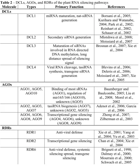
DCLs
The first step in the RNA silencing cascade is the recognition of dsRNA by a host ribonuclease. This protein was first identified in D. melanogaster in 2001 and was termed DICER. DICER proteins have a ribonuclease III domain which allows for the targeting of dsRNA, cleaving it into one of the various sRNAs defined above (Bernstein et al., 2001). In plants these proteins are referred to as DCL proteins, of which there are four (Table 2). The four DCLs are characterized by the types of dsRNA they process and the products of that processing. DCL1 and DCL4 produce siRNAs of 21 nt, DCL2 produces 22 nt siRNAs, and DCL3 produces 24 nt siRNAs (Borsani et al., 2005; Deleris et al., 2006; Gasciolli et al., 2005; Park et al., 2002; Xie et al., 2005; Xie et al., 2004). While each of these DCLs have primary roles in the RNA silencing cascade, knockout mutants have demonstrated that redundancy is a key theme of RNA silencing in plants.
its critical role in plant development, underscoring DCL1’s role in plant viability (Jacobsen et al., 1999; Park et al., 2002; Schauer et al., 2002). In addition to its well defined role in miRNA biogenesis, it is now clear that DCL1 has a role in the generation of other nuclear-derived sRNAs including those nuclear-derived from cis-NATs and tasiRNAs as well as showing some functional redundancy in the targeting of plant viruses (Blevins et al., 2006; Borsani et al., 2005; Henderson et al., 2006; Vazquez et al., 2004). Perhaps more interestingly, DCL1 has also recently been reported to be a negative regulator of anti-viral silencing in A. thaliana (Qu et al., 2008). In this work the plant-infecting virus TCV lacking its suppressor of RNA silencing was used as a reporter for DCL activity. The authors found that in the absence of DCL1 the TCV construct accumulated to a lesser degree than when DCL1 was present. They went on to show that DCL1 down regulates the expression of DCL4 and, to a lesser degree, DCL3 (Qu et al., 2008). It’s unclear exactly how DCL1 accomplishes this, but it is likely that DCL1 processes an endogenous sRNA that either directly regulates the two DCL proteins or regulates a protein related to DCL expression (i.e. transcription factor). In either case, clearly more work is required to elucidate the mechanisms underlying this regulation.
Two studies have recently defined the role of DCL2 in RNA silencing. Until recently it was thought that DCL2 was little more than a backup system to the other DCLs. DCL2 has been found to be subordinate to DCL4 in the processing of an RNA virus and tasiRNAs while subordinate to DCL3 in RNA directed DNA methylation (Deleris et al., 2006;
siRNA (Mlotshwa et al., 2008; Moissiard et al., 2007). These results suggest that DCL2 may play more than simply a subordinate role in anti-viral immunity. Transitivity is thought to be important in host defense, and work has shown that several VSRs are capable of blocking transitivity which results in secondary siRNA accumulation profiles that are remarkably similar to that of a dcl2 knockout (Mlotshwa et al., 2008; Moissiard et al., 2007). Based on A. thaliana knockouts of DCL2, DCL2 processes dsRNA primarily into 22 nt siRNAs although recent work with cis-NATs indicate that DCL2 might also be capable of processing dsRNA into lengths of 24 nt (Borsani et al., 2005; Deleris et al., 2006; Xie et al., 2005).
DCL3 guides epigenetic modifications of the host genome by cleaving dsRNAs into 24 nt siRNAs (Xie et al., 2004). As discussed earlier, these siRNAs direct heterochromatin modification of complementary sequences by recruiting both histone and DNA
The fourth DCL is also one of the most active. DCL4 has been implicated as the primary DCL involved in processing for at least three distinct RNA silencing pathways – the targeting of invading viruses, tasiRNA synthesis, and the initiation of transgene silencing (Blevins et al., 2006; Deleris et al., 2006; Moissiard et al., 2007; Xie et al., 2005). Like all other DCLs, DCL4 is likely to have secondary and subordinate roles in RNA silencing when other DCLs are inactive, however to date only one such role has been defined when DCL4 was shown to direct DNA methylation in the absence of DCL3 (Henderson et al., 2006). Still, DCL4 is by far the most important DCL in host defense against invading viruses, making its activity quite relevant to this work.
AGOs
When sRNAs are generated they move to an ARGONAUTE protein where the PAZ domain specifically binds to the 2 nt 3’ overhangs that are characteristic of sRNAs and the MID domain binds to the 5’ phosphate of the sRNA (Lingel et al., 2003; Lingel et al., 2004; Ma et al., 2004; Ma et al., 2005; Parker et al., 2005; Song et al., 2003). The catalytic domain of ARGONAUTE proteins is the PIWI domain, which structurally resembles RNase H and can catalyze an endonuclease reaction, providing what is often referred to as “slicer” activity (Parker et al., 2004; Rivas et al., 2005; Song et al., 2004). This PIWI domain cleaves one strand of the double-stranded sRNA, the strand not already bound by the PAZ and MID domains (passenger strand), which allows the ARGONAUTE to be loaded with a single-stranded siRNA to guide recognition of complementary targets. While all the
ARGONAUTE proteins have the PIWI domain, not all PIWI domains have catalytic activity, allowing some flexibility in the action of the various ARGONAUTES (i.e. translational inhibition). This also suggests that in some cases the passenger strand is probably separated from the other bound strand by a helicase rather than cleaved to create an active effector complex (Matranga et al., 2005; Rand et al., 2005).
have a particular affinity for sRNAs with a 5’ terminal uridine, which in A. thaliana is the predominant 5’ base of miRNAs (Mi et al., 2008; Rajagopalan et al., 2006). AGO1 is one of the ARGONAUTE proteins that has catalytic function, which is critical for its ability to regulate target RNAs (Baumberger and Baulcombe, 2005; Qi et al., 2005). AGO10 is closely related to the AGO1 protein, suggesting some functional redundancy. Indeed some
functional overlap has been shown between the two proteins for plant development (Lynn et al., 1999). The only specific role for AGO10 so far defined involves shoot apical meristem maintenance and adaxial-abaxial polarity establishment. AGO10 appears to accomplish this by regulating the accumulation of miR165/166 (Liu et al., 2008). AGO5’s role in cell biology also remains elusive. Like AGO1 it shows specificity for sRNAs with a specific 5’ base. In AGO5’s case this specificity is for cytosine (Mi et al., 2008). AGO5 is thus thought to bind to those miRNAs exhibiting a 5’ cytosine, however AGO5 knockout plants show little in the way of developmental defects, leaving this suggestion uncertain (Katiyar-Agarwal et al., 2007; Takeda et al., 2008).
are involved in the transition from juvenile to adult vegetative stages (Adenot et al., 2006; Garcia et al., 2006). Outside of its role in tasiRNA biogenesis AGO7 has recently been implicated in antiviral defense. In AGO7 mutants viral RNA accumulation was found to be slightly enhanced, although not to the level of AGO1 mutants, the AGO thought to be primarily associated with antiviral defense. The authors thus speculated that like the DCLs, AGOs may contain some ability for redundant action (Qu et al., 2008).
DNA methylation and silencing of the viral genome, an important part of the antiviral response of the host (Raja et al., 2008). AGO4’s role in resistance to the bacterial pathogen P. syringae is particularly interesting, as it does not appear to be related to the traditional TGS pathway. The authors showed that bacterial resistance did not rely on DCL3, RDR2, or a variety of DNA and histone methyltransferases normally associated with the TGS pathway. Still, methylation was found to be a factor in pathogen resistance, suggesting the existence of other host factors and perhaps a unique RNA silencing pathway (Agorio and Vera, 2007).
RDRs
Not all organisms encode endogenous RdRps, but those that do, such as C. elegans and plants, rely heavily on these RdRps in a variety of RNA silencing pathways. Plants encode three RdRp proteins involved in the RNA silencing pathway, termed RDR1, RDR2 and RDR6 in A. thaliana (Table 2). All three are thought to somehow recognize targets of RNA silencing and convert the ssRNA into dsRNA, however the mechanism for recognition of target RNA and the synthesis of dsRNA are poorly understood.
RDR2 is a key component of the TGS pathway (Chan et al., 2004; Xie et al., 2004). When areas of the genome are targeted for heterochromatic methylation RDR2 is required for the generation and the maintenance of siRNA accumulation. As noted earlier, PolIV transcribes the DNA region of interest, and RDR2 then converts the resulting transcript into dsRNA. This dsRNA is cleaved by DCL3 to produce primary siRNAs. These primary siRNAs then lead the generation of secondary siRNAs, a process that is again dependent on the ability of RDR2 to convert PolIV-transcribed RNA into a double-stranded form. Beyond these well established roles in TGS, RDR2 has also been linked to viral siRNA accumulation. In a study of TRV, the authors infected several A. thaliana knockout mutants examining the requirements for RDR1, RDR2, and RDR6 for viral siRNA accumulation. No single or double mutants exhibited reduction in viral siRNA accumulation, however triple mutants were severely impaired for viral siRNA production and showed enhanced levels of viral RNA accumulation indicating functional redundancy in the RDR pathway targeting viruses (Donaire et al., 2008).
Of the three RDRs, RDR6 appears to have the heaviest influence in RNA silencing, as it is involved in a variety of pathways. RDR6 was originally recognized as a key
conclusively linked to the generation of tasiRNAs and nat-siRNAs in addition to transgene silencing and antiviral silencing.
suggest that RDRs are required for the generation of dsRNA targets of the various DCLs but information on the modes and triggers of this dsRNA generation are sorely lacking.
One of the more interesting aspects of antiviral silencing in plants is the phenomenon whereby the silencing signal generated from primary leaf infection can spread via the phloem of the plant to upper, non-inoculated leaves rendering them resistant to viral infection. The nature of this silencing signal is so far unknown, but is thought to be a 24 nt siRNA in complex with proteins. The only concrete role for RDRs in RNA virus silencing revolves around the spread of this silencing signal. Using transgenic N. benthamiana, investigators showed that RDR6 was necessary for the phenomenon to occur. RDR6 is required to be active in cells receiving the silencing signal in order for those cells to become resistant to the incoming virus, but is not required for the generation of the silencing signal itself or its spread throughout the plant. What RDR6 actually does with the incoming silencing signal once it is received is still a question. Working under the assumption that the silencing signal is a short RNA, 24 nt or otherwise, RDR6 may utilize the short RNA as a primer and the incoming viral genomes as templates to polymerize dsRNA that can be targeted by the DCLs. A second theory suggests that the silencing signal in complex with RISC might target
expressed in N. benthamiana and immunopurified was used to examine the biochemical requirements for RDR6 activity. It should be noted that all work in this study was performed in vitro, however the authors were able to show that RDR6 has primer-independent RNA polymerase activity on ssRNA and does not distinguish between templates with or without a 5’ cap or poly A tail (Curaba and Chen, 2008). Finally, another study involving CMV showed that RDR1 was critical for siRNA production not only in systemic leaves but in inoculated leaves as well, indicating that RDRs may play a larger role in viral infection than simply systemic immunity (Diaz-Pendon et al., 2007). The RDRs represent one of the remaining major challenges in the quest to fully describe antiviral RNA silencing. With the work that has been done so far it appears as though RDRs might behave like DCLs in that they likely each have their own primary roles to play, but can also act in a redundant fashion if needed. RDR6’s primary role may be as a systemic RNA silencing factor while RDR1 may play a primary role in inoculated tissue activity, but the fact that TRV and the majority of viruses tested were not hypervirulent in the absence of RDR6 in knockout plants suggests that redundancy may be key (Brigneti et al., 1998; Dalmay et al., 2000; Mourrain et al., 2000; Schwach et al., 2005).
RNA silencing targeting plant RNA viruses
an immune system pathway is initiated often leading to tissue necrosis. RNA silencing comprises the adaptive immune response of plants and is particularly important in stemming infections from RNA viruses.
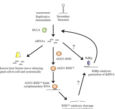
RNA viruses in plants are targeted by the silencing pathway in a very prototypical manner (Figure 2). As discussed earlier, viral dsRNA can come from one of three sources (a replicative intermediate, secondary RNA structure, and endogenous RDR-generated dsRNA), is recognized by the host DCL and cleaved into siRNAs. One strand of these siRNA strands can be incorporated into RISC, leading to targeted degradation of the infecting virus. A silencing signal can also be generated and spread from the initial site of infection cell-to-cell on the infected leaf (short range) and finally via the phloem to other parts of the plant (long range). Although most of the work surrounding the short range spread of RNA silencing has been focused on transgenes, some conclusions can be made about the short distance spread of the silencing signal targeting plant viruses. The spread of the short distance RNA silencing signal occurs over a distance of 10-15 cells, and likely travels through the plasmodesmata (Himber et al., 2003; Voinnet et al., 1998). One class of mutants screened for a defect in the cell-to-cell spread of silencing lost 21 nt siRNA populations but 24 nt siRNA populations were unaffected. This class of mutants was eventually mapped to DCL4, demonstrating a requirement for DCL4-generated 21 nt siRNAs for the spread of silencing (Dunoyer et al., 2005). Other mutants that have been identified as being required for cell-to-cell spread of the silencing signal include PolIVa and RDR2. The fact that these experiments were focused on transgenes and host genes and that PolIVa and RDR2 have been implicated in
but a role for these proteins cannot be ruled out (Dunoyer et al., 2007; Smith et al., 2007). Whatever the mechanisms of this spread, the phenomenon, in combination with DCL cleavage and AGO targeting, results in the targeted degradation of the infecting virus, limiting its ability to survive and cause a productive infection.
Through the process of evolution viruses have come up with their own answer to the RNA silencing pathway. Most, if not all, encode proteins known variously as anti-silencers, VSRs, or simply suppressors. The role of these VSRs is to disrupt – or suppress – the endogenous host silencing pathway to allow the infecting virus a chance to survive and replicate. Without a method of suppressing or avoiding the RNA silencing pathway, an infecting virus has little or no chance of surviving. Work detailed in this thesis has
confirmed this, as a TCV construct lacking its VSR was found to be highly compromised for infectivity, with infections confined to foci of approximately 5 cells (See Chpt. 2; Powers et al., 2008).
VSRs
The VSRs encoded by plant viruses come in a variety of shapes, sizes, and modes of action. Viral genomes are characteristically compact, and the protein sources of suppressor activity reflect this. Many of the known suppressor proteins have other roles in viral
PVX p25, CMV 2b, and Potyvirus HC-Pro (Bayne et al., 2005; Cronin et al., 1995; Ding et al., 1995; Klein et al., 1994).
The first two suppressors identified were CMV 2b and HC-Pro from PVY. Both VSRs were found to have an effect on the silencing of a GFP transgene in N. benthamiana. In these studies the GFP transgene was pre-silenced by the infiltration of an A. tumefaciens culture transformed with a binary vector expressing a GFP expression cassette into 3 week old seedlings. Two weeks later plants were completely silenced for GFP expression with the exception of the extreme meristematic zone, which has been shown to remain non-silenced (Voinnet et al., 1998). The authors found that when silenced plants were infected with CMV or PVY, the effects of silencing were diminished. In the case of CMV, newly emerging leaves were no longer silenced for GFP expression, while PVY-infected plants showed reversal of silencing on leaves already silenced. On the other hand, another virus, PVX, had no effect on GFP silencing. The authors exploited this by using PVX as a viral vector to determine which component of PVY and CMV directed the anti-silencing effects. Eventually the authors determined that PVY HC-Pro and CMV 2b were responsible for suppressing RNA silencing, although exact molecular mechanisms were not determined (Brigneti et al., 1998).
yellows virus p21 (Chapman et al., 2004; Lakatos et al., 2006; Merai et al., 2006; Reed et al., 2003; Silhavy et al., 2002). p19 in particular has been thoroughly characterized, including a crystal structure of p19 bound to a 21 nt siRNA (Vargason et al., 2003). Interestingly, binding of siRNAs by various VSRs has never been shown to be specific, and so the VSR binds to both endogenous and viral sRNAs leading to a massive disruption of plant physiology (Chapman et al., 2004; Dunoyer et al., 2004).
target AGO1 for degradation via their P0 protein which is an F box protein. As part of the SCF ubiquitin ligase complex, host F box proteins can help to mediate ubiquitination of proteins, leading to the degradation of the protein by the proteasome. P0 appears to act in a manner similar to these host proteins, however the degradation appears to be independent of the proteasome, as proteasome inhibitors had little effect on P0’s ability to target AGO1 (Baumberger et al., 2007; Bortolamiol et al., 2007).
While silencing suppression by siRNA binding is well known, there are relatively few demonstrations involving endogenous protein targeting (Baumberger et al., 2007;
All plant viruses studied to date encode at least one VSR. Some viruses have been shown to encode multiple VSRs, while others encode VSRs that act at multiple steps in the RNA silencing pathway. This multi-pronged approach to the suppression of RNA silencing depresses the immune response of the plant even further than a single VSR acting at a single point in the pathway. This ensures that the virus can effectively infect its host, and may be a method by which the virus can account for all the redundancy found in the RNA silencing pathway. As noted earlier, direct inhibition of DCL4 by a viral protein may not be very effective if DCL2 can step in and perform the same dicing function nearly as well.
Disrupting the pathway at multiple points may aid the virus in overcoming the host’s built in backup system.
One of the best known examples of a virus that encodes multiple VSRs is Citrus tristeza virus, which encodes 3 VSRs: CP, which disrupts intercellular silencing, p23, which disrupts intracellular silencing, and p20, which seems to do both (Lu et al., 2004). Another example comes from two Geminiviruses, African cassava mosaic virus and Sri Lankan cassava mosaic virus, both of which encode two suppressors of RNA silencing, AC2 and AC4, although AC2 is significantly less effective for both (Vanitharani et al., 2004).
depress the accumulation of many classes of sRNAs, possibly by blocking their 3’ terminal methylation, a requirement for effective incorporation into the silencing cascade
(Anandalakshmi et al., 2000; Ebhardt et al., 2005; Lakatos et al., 2006; Llave et al., 2000; Mallory et al., 2001; Yu et al., 2006). HC-Pro is not the only protein with multiple methods of silencing suppression as the previously discussed 2b protein can not only bind directly to AGO1, the critical component of the RISC, but can also stem the long distance spread of the silencing signal. A close relative of CMV, Tomato aspermy virus, also encodes a related 2b protein. This protein appears to have some RNA binding capabilities, providing either a third role for 2b, or defining 2b’s mode of action for stemming the long distance spread of the silencing signal (Brigneti et al., 1998; Chen et al., 2008; Zhang et al., 2006a).
Identifying RNA silencing suppressors
The first papers to identify VSRs relied on reporters expressed from transgenes to assay for silencing suppression activity. In these studies the reporter, generally either β-glucuronidase (GUS) or green fluorescent protein (GFP), was silenced, and putative suppressors were then expressed in the transgenic plants to determine if the proteins
the use of a viral vector (Anandalakshmi et al., 1998; Brigneti et al., 1998; Kasschau and Carrington, 1998). Although some of these methods have changed, the same basic formula remains to this day – coexpression of a reporter susceptible to RNA silencing with a putative VSR to determine if the VSR blocks the silencing pathway from targeting the susceptible reporter. The nature of delivery for the reporter, the silencing trigger, and putative VSR has changed over the years, although transgenics still play a key role in the study of VSR
function. The timing of reporter and putative suppressor delivery also now varies. Whereas the original studies relied on a reporter that was presilenced, many current studies look to deliver the putative suppressor into cells prior to the initiation of reporter silencing.
An example of an assay in which the putative suppressor is delivered before silencing initiates involves the co-infiltration of two cultures of A. tumefaciens. The first culture harbors a Ti-plasmid expressing a reporter such as GFP, while the second culture harbors a Ti-plasmid expressing the putative suppressor. These two cultures are mixed and infiltrated onto a leaf, usually via a syringe. Expression of the reporter is subsequently monitored. In this assay the reporter is also the silencing trigger. In the presence of a suppressor, reporter expression can go on for a week or more, while in the absence of a suppressor, reporter expression is minimal three days post infiltration.
Another suppressor assay relies on an endogenous reporter to examine for the
leading to extensive leaf bleaching while in the presence of a VSR this
photo-bleaching is blocked. The putative suppressor can be delivered in any number of ways, by A. tumefaciens, by stable transgenes, or by viral vector and is usually concurrent with delivery of TRV-PDS.
Until recently each of the commonly employed VSR identification assays had
characteristics that made them less than ideal for the study of VSRs from RNA viruses. Each of these assays had reporters and/or silencing triggers whose expression originated in the nucleus. Nuclear expression is subject to a whole host of other silencing related activities, such as TGS, which the cytoplasmic-based RNA virus is not exposed to. This leads to the question as to just how accurate some of these assays are for the identification of VSR activity from plant RNA viruses. To address this question, we generated a new reporter that is viral-based. The known VSR of TCV, its CP, was removed and replaced with sGFP to create a construct termed TCV-sGFP. This construct is susceptible to silencing, is RNA-based, is localized to the cytoplasm, and contains both the reporter and silencing trigger. Details of this work can be found in Chapter 2 (Powers et al., 2008).
RCNMV
RCNMV is a plant-infecting virus classified by the International Committee on Taxonomy of Viruses into the family of positive-sense RNA viruses known as
include Dianthovirus, whose type species is Carnation ringspot virus (Büchen-Osmond, 2006).
Genome structure, protein function and expression
Species in the Dianthovirus family are characterized by bipartite, linear, positive-sense, ssRNA genomes (Büchen-Osmond, 2006). The RCNMV RNAs, simply termed RNA-1 and RNA-2, encode for four proteins – three from RNA-RNA-1 and a single protein from RNA-2 (Fig. 3; Gould et al., 1981; Lommel et al., 1988; Xiong and Lommel, 1989). RNA-1 is 3889 nt in length and encodes three proteins, two from 5’ proximal overlapping open reading frames (ORFs) – a 27 kilodalton (kDa) polypeptide (p27), and an 88 kDa polypeptide (p88) – as well as a 3’ terminal ORF of 37 kDa, known to encode the capsid protein (CP; Bates et al., 1995; Morris-Krsinich et al., 1983; Xiong et al., 1993; Xiong and Lommel, 1989). The 1448 nt RNA-2 encodes for a 35 kDa protein implicated in viral movement, known as the
movement protein (MP; Lommel et al., 1988; Osman and Buck, 1987; Paje-Manalo and Lommel, 1989).
consisted of both p27 and p88 (Bates et al., 1995). p27 is 711 nt in length, has a translation start codon at position 123 and codes for a protein of 236 a.a. p88 is 2304 nt in length and codes for a protein of 767 a.a. The N-terminal sequences of p27 and p88 are identical. Early sequencing of the RNA-1 genome led to the belief that RNA-1 coded for 3 proteins – p27, CP, and a 57 kDa protein called p57, but could not account for an observed in vitro
translation product that was estimated to be 90 kDa in size. This early work led to the postulation that p88 was a critical protein and theorized that ribosomal frameshifting following translation of p27 was required for the production of p88 (Xiong et al., 1993; Xiong and Lommel, 1989). Evidence for ribosomal frameshifting was provided in 1994, when site-directed mutagenesis experiments demonstrated the presence of a heptanucleotide sequence, just upstream of the p27 stop codon, that is required for p88 translation (Kim and Lommel, 1994). This same study reported that p57 was likely not translated in vivo as it was undetectable in infected tissue, and mutations to its putative start codon had no effect on viral infectivity (Kim and Lommel, 1994). By utilizing a GUS reporter, further work