Abstract
Sahana Das Adhikari: The impact of organic acids and pH on the
virulence factor expression of E. coli O157:H7. (Under Direction of Dr.
MaryAnne Drake).
Acidification is used as a hurdle in many minimally processed foods. Decreased pH (pH
5.5) may enhance survival and virulence factor expression of E. coli O157:H7 (EC). The
objective of this research was to determine the effect of different organic acids and pH on
the expression of three virulence factor genes (stx2, hlyA, eaeA) in EC. Gene fusions
containing the lacZ gene inserted into the stx2, eaeA or hlyA genes were created in E. coli
O157:H7 with and without a functional rpoS gene. Overnight cultures were inoculated
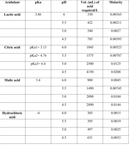
into tryptic soy broth acidified with citric, malic, lactic, or hydrochloric acid at pH 6.0,
5.5, 5.0, or 4.5 or apple juice (pH 3.5). Cell growth characteristics were characterized,
and β-galactosidase activity of stressed or control cells (neutral pH, no acid) was
subsequently determined to follow virulence factor production. Production of all three
virulence factors was increased at pH 5.5 or 5.0 compared to production at neutral pH
(p<0.05). Acid type impacted production of intimin and Shiga toxin, but had no effect on
hemolysin. Production of StxII and HlyA was not detected in apple juice. At pH 5.5, cell
growth was slowest in lactic acid, followed by malic and citric acids then HCl. At pH 5.0,
the slowest growth was observed in citric acid, followed by malic acid, lactic acid and
HCl. At pH 4.5, no growth occurred in citric, malic and lactic acids, and cell numbers
decreased over a period of 5 days. In HCl at pH 4.5, cells grew slowly and increased by 2
logs over a 5-day period. Sublethal acid stress impacts virulence factor expression of E.
coli O157:H7 and these effects are impacted by pH and acid type.
ii
Biography
Sahana Das Adhikari was born on 3rd March 1977 in a small town in India to
parents Ranjana and Sashi Adhikari. Her family also includes her sister Leena and her
husband Prasant Das.
Sahana completed her B.S. degree in Microbiology in October of 1998 and her
M.S. degree in Biotechnology in August of 2000. She then began her M.S. in the
department of Food Science in January of 2003 under the direction of Dr. MaryAnn
iii
Acknowledgements
The author sincerely wishes to express her gratitude for the support and
encouragement provided by her family, friends, laboratory colleagues, and major advisor.
The author expresses her sincere appreciation and gratitude to her major
professor, Dr. MaryAnne Drake for her inspiring guidance, patience, encouragement and
invaluable supervision throughout this program. Thanks are due to Dr. LeeAnn Jaykus
and Dr. Donn Ward for their guidance and encouragement given to her in order to help
her attain this degree.The author is thankful to her friends in the Food Science
Departmentfor all their help and support.
Finally, the author feels privileged to express her deep reverence and gratitude to
her parents, her sister and her dear husband for their support and sacrifice throughout her
iv
Table of contents
List of Tables vi List of figures vii
Introduction 1
Literature Review 4
Types of E. coli 5
Enterohemorrhagic E. coli 9
E. coli O157:H7 10
Disease symptoms 11
Reservoirs of EHEC including E. coli O157:H7 15
Modes of transmission 16
General characteristics of E. coli O157:H7 18
EHEC Pathogenesis 20
Colonization of the gut by epithelial cell adherence 21
Locus of enterocyte effacement 22
Adhesin molecule Intimin 25
Intimin receptor Tir 25
Type III secretion system 27
Shiga toxins in E. coli 27
αααα-Hemolysin and role in pathogenesis 30
pO157 31
Other adherence mechanisms 34
Treatment 35
Detection, enrichment and isolation 36
Genome sequence of enterohemorrhagic E. coli O157:H7 41
Factors influencing the survival and growth of E. coli 41
Temperature 42
pH 42
Sodium chloride 43
Water activity 43
Antimicrobials 43
Adaptive responses to environmental stresses in E. coli 44
Osmotic Stress 47
Acid resistance of EHEC 48
Acid tolerance of EHEC 51
Effect of type of acidulant
55Cold stress 56
Heat shock response 58
Effect of stress on EHEC Shiga-toxin production 61
Conclusion 63 References 64 Manuscript: The impact of organic acids and pH on the virulence factor expression
of E. coli O157:H7 81
v
Materials and Methods 86
Bacterial and culture conditions 86
Creation of lacZ fusions 86
Experimental conditions 89
Preparation of acidified media 90
Experimental protocol 90
Miller assay 91
Statistical analysis 92
Results and Discussion 93
Virulence factor expression- pH and acid effects 93
Growth and survival of E. coli O157:H7 99
Conclusions 105 References 106
Tables and figures 112
Appendix 128
vi
List of tables
Table 1: Some properties and symptoms associated with pathogenic E. coli subgroups 18
Table 2. Significant virulence factors of E. coli O157:H7 21
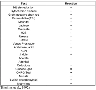
Table 3: Biochemical and physiological behavior of E. coli 40
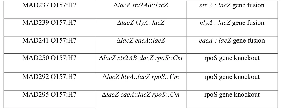
Table 4. Genotypes of strains used in the study 112
Table 5: pH, volume and molarity of each acid used in experiments 113
Table 6. Impact of acid type and pH on production of HlyA. Means are expressed in
Miller units 114
Table 7. Impact of acid type and pH on production of EaeA. Means are expressed in
Miller units 114
Table 8. Impact of acid type and pH on production of StxII. Means are expressed in
vii
List of Figures
Figure 1. Natural history of infection with E. coli O157:H7 and post-diarrheal HUS 15
Figure 2: Isolation of E. coli O157:H7 from foods 39
Figure 3: Flow diagram showing the construction of Shiga toxin, intimin and hemolysin
gene fusions 116
Figure 4: Flow diagram showing the construction of rpoS mutants 117
Figure 5: Mean growth of rpoS+ LacZ gene fusion strains in different acids at pH 6.0 and
pH 7.2 TSB at 25C 118
Figure 6: Mean growth of rpoS+ LacZ gene fusion strains in different acids at pH 5.5 and
pH 7.4 TSB at 25C 119
Figure 7: Mean growth of rpoS+ LacZ gene fusion strains in different acids at pH 5.0 and
pH 7.4 TSB at 25C 120
Figure 8: Mean growth or survival of rpoS+ LacZ gene fusion strains in different acids at
pH 4.5 121
Figure 9: Mean survival of rpoS+ LacZ gene fusion strains in apple juice (pH=3.5) 122
Figure 10: Mean growth of rpoS- LacZ gene fusion strains in different acids at pH 6.0
and pH 7.4 TSB at 25C 123
Figure 11: Mean growth of rpoS- LacZ gene fusion strains in different acids at pH 5.5
and pH 7.4 TSB at 25C 124
Figure 12: Mean growth of rpoS- LacZ gene fusion strains in different acids at pH 5.0
and pH 7.4 TSB at 25C 125
Figure 13: Mean survival/growth of rpoS- LacZ gene fusion strains in different acids at
pH 4.5 at 25C 126
1
Introduction
Escherichia coli O157:H7 is a source of foodborne and waterborne illness of
major public health concern (Buchanan and Klawitter, 1992). It is recognized as a
common bacterial cause of bloody and nonbloody diarrhea in the United States,
accounting for an estimated 20,000 infections each year (Boyce et al., 1995). Three major
virulence genes in E. coli O157:H7 purportedly contribute to its ability to cause disease:
genes encoding Shiga toxin (Stx), intimin (eae), and hemolysin (hlyA) (Law, 2000). The
production of Shiga toxins is one of the defining characteristics of E. coli O157:H7 and
these toxins are thought to be responsible for the principal manifestations of hemorrhagic
colitis (HC) and its complications hemolytic uremic syndrome (HUS) and thrombotic
thrombocytopenic purpura (TTP) (Law, 2000). Two primary classes of Shiga toxins are
Stx1 and Stx2 and these are bacterial lysogens (Muhldorfer et al., 1996; O’Brien and
Holmes, 1987). The eae gene in E. coli O157:H7 codes for intimin, an outer membrane
protein (OMP) required for intimate attachment, allowing the bacteria to adhere to the
intestinal mucosa (Law, 2000). HlyA codes for enterohemolysin, and its precise role in
human infection is unknown (Law, 2000). The gene coding for this protein is present on
the plasmid pO157 (Paton and Paton, 1998).
The involvement of E. coli O157:H7 in foodborne illness associated with the
consumption of acidic foods such as apple cider, fermented sausage, yogurt and
mayonnaise (Morgan et al., 1993; CDC, 1995) has drawn attention to the acid resistance
properties of this pathogen. Many subsequent studies have demonstrated that this
bacterium can survive in a variety of acidic foods (Glass et al., 1992; Miller and Kasper,
2 conditions can further improve the survival of E. coli O157:H7 in foods that are
preserved by low pH and acids (Leyer et al., 1995; Tsai and Ingham, 1997). In addition to
promoting survival in low pH-foods, the development of acid resistance by E. coli
O157:H7 may provide cross-protection against heat, salt, and irradiation preservation of
foods (Buchanan et al., 1998; Leenanon and Drake, 2001). Furthermore, several studies
have shown that acid tolerance of E. coli O157:H7 is enhanced or sustained upon
refrigeration (Clavero and Beuchat, 1996; Lin et al., 1996; Cheng and Kaspar, 1998).
Studies have addressed the expression and production of Shiga toxins. The
influence of temperature on growth and Shiga toxin production by E. coli strains at
various temperatures was investigated by Palumbo et al. (1994). Toxin production was a
function of both time and temperature, and was highest at optimum growth temperatures.
In a similar study, Weeratna and Doyle (1991) reported that the highest titers of Shiga
toxin were produced in both milk and ground beef at optimal growth temperature (37°C)
and that less toxin was produced at 30 or 25°C. On the other hand, Weinstein et al.
(1988) reported that there was no significant effect of temperature on Shiga toxin
production by an E. coli strain that was rendered toxigenic by lysogenization. Elhanafi et
al. (2004) found that the Stx 2 production was highest in early stationary phase. These
results were consistent with the findings of McIngvale et al. (2002) in which stx2 mRNA
expression as determined by RT-PCR reflected the level of mRNA production at the
transcriptional level. Leenanon et al. (2003) demonstrated that oxygen enhanced stx-II
3 Stress may also affect virulence gene expression. Duffy et al. (2000) found that
cells grown at a lower pH (pH 5.6) had lower Shiga toxin production than cells grown at
pH 7.4. Stress conditions such as acid adaptation and starvation enhanced stx-II toxin
mRNA levels but did not enhance subsequent Stx toxin production (Leenanon et al.,
2003). Yuk and Marshall (2003) studied the influence of heat shock and heat adaptation
on intracellular and extracellular Shiga toxin concentration for E. coli O157:H7 with and
without a functional rpoS gene. They found that heat adaptation reduced total Shiga toxin
concentration in both normal and rpoS deficient E. coli O157:H7 cells. However, slightly
lower amounts of Shiga toxin were produced by wild type than by rpoS deficient
mutants, indicating a possible effect of rpoS on Shiga toxin production, a result also
observed in other studies (Leenanon et al., 2003). Elhanafi et al. (2004) found that prior
cold or cold-acid stress had no effect on virulence factor production (Stx 2, eaeA, hlyA)
of E. coli O157:H7. However, growth in acidic media (pH 5.5 media acidified with lactic
acid) enhanced EaeA and HlyA production. An understanding of the stress response of E.
coli O157:H7 and the effect of stress on virulence factor expression is essential. The
objective of this study was to investigate the effect of different acids and pH on the
expression of the Shiga toxin, intimin and hemolysin genes in E. coli O157:H7. Gene
4
Literature Review
Escherichia coli are a group of free-living, Gram negative enteric bacteria. These
are the most intensively studied and best characterized, both genetically and
biochemically, of all bacteria (Record et al., 1998). E. coli was discovered in 1885 by
Theodor Escherich, a German pediatrician, who noted its high prevalence in the intestinal
microflora of healthy individuals as well as its potential to cause disease when directly
inoculated into extra-intestinal sites (Robins-Browne and Hartland, 2002). The key events
that lead to the discovery of E. coli as a primary gut pathogen were reported by Bray and
Bevan in 1948 (Bray and Bevan, 1948; Robins-Browne and Hartland, 2002). They found
that a particular strain of E. coli, which they named Bacterium coli var. neopolitanum,
was significantly associated with infantile diarrhea. A Danish bacteriologist, Fritz
Kauffmann adapted a serotyping scheme that was developed for Salmonella enterica and
later used with E. coli (Kauffmann, 1947). The basis of this scheme is that individual E.
coli isolates can be distinguished from each other from their surface O (somatic) and H
(flagellar) antigens. Currently, it is considered necessary to determine the O and H
antigens to serotype strains of E. coli associated with diarrheal disease. The O antigen
identifies the serogroup of a strain and the H antigen identifies its serotype (Meng et al.,
2001). The current serotyping of E. coli is primarily based on the 171 O antigens and 56
H antigen serotypes (Meng et al., 2001). A phage-typing scheme has also been
successfully developed for E. coli. With its application, over 40 phage types have been
recognized. E. coli O157:H7 strains belong to the phage types 1, 2, 4, and 8 (Ratnam et
5
E. coli is one predominant species of the facultative anaerobic microflora of the
intestinal tracts of warm-blooded animals and is usually harmless to the host (Drasar and
Hill, 1974). Most E. coli strains are harmless, but some are pathogenic and cause
diarrheal diseases (Meng et al., 2001). The main reservoir for this environmentally
ubiquitous organism is the intestinal tract. However, when it is found elsewhere in the
environment, it is considered an indication of fecal contamination and suggests the
possible presence of enteric pathogens (Meng et al., 2001).
Types of
E. coli
Diarrheagenic E. coli isolates are characterized into specific groups based on
virulence properties, mechanisms of pathogenicity, clinical syndromes and distinct O:H
serotypes (Meng et al., 2001). The groups are classified based on their unique virulence
factors and can be identified by these traits. These 6 pathogenic groups include
enterotoxigenic E. coli (ETEC), enteropathogenic E. coli (EPEC), diffuse adhering E. coli
(DAEC), enterohemorrhagic E. coli (EHEC), enteroinvasive E. coli (EIEC),
enteroaggregative E. coli (EAEC), and perhaps others that are not yet well characterized
(Nataro and Kaper, 1998). Each of these pathotypes represent a family of E. coli clones
that share key virulence determinants, which were perhaps acquired by horizontal gene
transfer between E. coli and other bacterial species (Robins-Browne and Hartland, 2002).
EPEC is primarily associated with neonatal and infantile diarrhea in developing
countries (Padhye and Doyle, 1992; Nataro and Kaper, 1998). This is more severe than
most other causes of diarrhea in infants. However, a number of outbreaks of diarrhea
caused by this pathogen have been reported in healthy adults and in adults with
6 with diabetes (Nataro and Kaper, 1998). The diarrhea is watery with mucus, but without
pronounced amounts of blood. EPEC outbreaks have been linked to the consumption of
contaminated drinking water as well as some meat products (Padhye and Doyle, 1992).
The fecal oral route is a major route of transmission with contaminated hands or fomites
serving as vehicles (Padhye and Doyle, 1992). The reservoir for infections by this
organism is believed to be symptomatic or asymptomatic children, or asymptomatic adult
carriers, including mothers and people who handle infants (Nataro and Kaper, 1998).
Volunteer feeding studies have shown that the infectious dose of EPEC in healthy
adult individuals is about 106 organisms. The mode of pathogenesis of EPEC is poorly
understood (Padhye and Doyle, 1992). Pathogenesis of EPEC involves the intimin
protein (encoded by the eae gene) that causes attachment and effacing lesions (Griffin
and Tauxe, 1991). EPEC has been shown to produce intestinal lesions which destroy the
microvilli without any further evidence of invasion. Pathogenesis also involves a
plasmid-encoded protein referred to as the EPEC adherence factor (EAF) that enables
localized adherence of bacteria to intestinal cells (Tobe et al., 1999). Adults acquire
immunity to EPEC and serve as carriers of EPEC without expressing symptoms of illness
(Padhye and Doyle, 1992). EPEC initially targets the M cells of the intestine during its
interaction with the intestinal epithelium. However, it does not penetrate the epithelium
and tends to remain in close contact with the surface of the M cells and erythrocytes. It
gives rise to distinct histopathological changes termed attaching effacing lesions
(Robins-Browne and Hartland, 2002).
EIEC closely resemble Shigella and cause an invasive, dysenteric form of
7 reservoirs; hence the primary source for EIEC appears to be infected humans. EIEC share
a number of key virulence determinants with Shigella, including a large plasmid (140
Mda) that encodes several outer membrane proteins (OMP’s) believed to be responsible
for the invasion of intestinal cells. The pathogenic features of EIEC are virtually identical
to those of Shigellaspp. (Nataro and Kaper, 1998). Although the infective dose of
Shigella is low and in the range of 10 to a few hundred cells, volunteer feeding studies
showed that at least 106 EIEC organisms are required to cause illness in healthy adults
(Padhye and Doyle, 1992). Unlike typical E. coli, EIEC are non-motile, do not
decarboxylate lysine and do not ferment lactose, so they are anaerogenic(do not produce
gas). Pathogenicity of EIEC is primarily due to its ability to invade and destroy colonic
tissue. EIEC can penetrate the intestinal mucosa and can proliferate within the epithelial
cells causing inflammation and mucosal ulceration leading to bloody diarrhea
(Robins-Browne and Hartland, 2002). The colon appears to be the primary site of bacterial
invasion.
ETEC are recognized as the causative agent for two major clinical syndromes:
travelers' diarrhea, and weanling diarrhea among children in developing countries. The
etiology of this cholera-like illness has been recognized forabout 20 years (Mitsuda et al.,
1998). ETEC infections occur commonly in under-developed countries. The
toxin-producing strains of ETEC are responsible for causing mostly endemic disease (Albert et
al., 1995). Traveler's diarrhea is usuallycontracted from contaminated food and water
(Black, 1990; Mattila, 1994). The symptoms of ETEC infection are watery diarrhea,
nausea, abdominal cramps, and low-grade fever. ETEC strains require various adhesive
8 called CFA (colonizing adhesive factors) or PCF (putative colonizing factors). Also, they
secrete two types of enterotoxins known as heat labile and heat stable enterotoxins
through which the ETEC strains cause diarrhea (Robins-Browne and Hartland, 2002;
Sears and Kaper, 1996). Heat-labile toxins can be inactivated at 65°C for 30 min. ETEC
may produce a heat-labile enterotoxin (LT) that is very similar in size (86 kDa),
sequence, antigenicity, and function to the cholera toxin (CT), although its mechanisms
of action is distinct (Sixma et al., 1993; Peterson and Whipp, 1995). Two major
serogroups of LT are LT Ι and II and these do not cross-react immunologically (Nataro
and Kaper, 1998). E. coli strains which express LT I are pathogenic to humans and those
expressiong LT II, which are mostly isolated from animals, have not been linked to
disease in either humans or animals (Nataro and Kaper,1998). The infectious dose of
ETEC is 108 CFU, which induces high attack ratesin volunteers (Levine et al., 1987).
ETEC infections in areas of endemicity tend to be clustered in warm, wet months, when
multiplication of ETEC in foodand water is most efficient (Levine, 1987).
Enteroaggregative E. coli (EAEC), along with ETEC, is also a common
etiological agent of traveler’s diarrhea (Gascon et al., 1998; Vila et al., 2000). EAEC
have a characteristic pattern of adherance to HEp –2 cells known as ‘stacked brick’
(Nataro et al., 1985). The virulence factors that have been best characterized in EAEC are
the plasmid-encoded fimbriae AAF/I and AAF/II. These mediate mucosal adherance
(Savarino et al., 1994; Czeczulin et al., 1997). Some strains of EAEC synthesize a
plasmid encoded toxin or Pet, which has been shown to induce increased mucus release,
exfoliation of cells, and crypt abscess development (Savarino et al., 1994; Eslava et al.,
9 1991). All these virulence factors in EAEC strains are encoded by a ~60-Mda plasmid
common to all EAEC strains (Vila et al.,2000).
Enterohemorrhagic
E. coli
EHEC was recognized as a cause of foodborne diarrhea after a multistate outbreak of
hemorrhagic colitis in 1982, and it has gradually become a significant threat to human
health (Jordan and Davies, 2001). The term enterohemorrhagic E. coli or EHEC was
coined to denote the strains of E. coli that express Stx, produce A/E lesions on epithelial
cells, have a 60-MDa plasmid, and can cause hemorrhagic colitis (HC) (Levine 1987;
Levine and Edelman, 1984; Nataro and Kaper, 1998).There are several serotypes of
Shiga toxin producing E. coli (STEC, E. coli that possess Stx genes), but only the
serotypes clinically associated with hemorrhagic colitis (HC) in humans are designated as
EHEC (Nataro and Kaper, 1998).
The study of EHEC pathogenesis took place along parallel paths of investigation
that resulted in parallel nomenclature systems for the EHEC toxins (Nataro and Kaper,
1998). The term verotoxins or vero cytotoxins (or EHEC toxins) were given to toxins that
have an irreversible cytopathic effect on Vero (African green monkey kidney) cells. The
alternative nomenclature is Shiga toxins (Stx) or Shiga like toxins (SLTs). The previous
two nomenclatures were adopted as one of the cytotoxins produced by EHEC is
genetically and at a protein level identical to the Stx produced by Shigella dysenteriae I.
Again, only Shiga toxin producing strains that can cause human illness in the form of HC
are designated EHEC. EHEC are typified by the production of verotoxins (VT) or Shiga
10 It is now recognized that EHEC strains belonging to a very diverse range of
serotypes are capable of causing very serious diseases in humans. EHEC strains
belonging to 100 different O:H serotypes have been associated with human disease
(Nataro and Kaper, 1998; Law, 2000). Common EHEC serotypes associated with human
pathogenicity include O4:H-, O11:H-, O26: H11, O45:H2, O103:H2, O104:H21,
O111:H8, and O145:H- ( Buchanan and Doyle, 1997; Pradel et al., 2000). The dominant
EHEC serotype associated with both outbreaks and sporadic cases of human disease that
has emerged in United States, Canada and United Kingdom is O157:H7 (Pradel et al.,
2000; Buchanan and Doyle, 1997). This pathogen has been implicated in many food
borne outbreaks (Perna et al., 2001) involving hemorrhagic colitis and hemolytic uremic
syndrome (HUS) (Benjamin and Datta, 1995).
E. coli
O157:H7
E. coli O157:H7 was first isolated in 1975 from a woman having gross bloody
diarrhea (Padhye and Doyle, 1992). It was first recognized as a human pathogen after two
foodborne disease outbreaks in Oregon and Michigan states in 1982 (Riley et al., 1983).
This organism is the prototypic EHEC and is often recognized as a major food-borne
pathogen, implicated in worldwide illness. Close to 75,000 cases of O 157:H7 infections
are now estimated to occur annually in United States (Perna et al., 2001). The recovery
period is sometimes very long, patients can have permanent kidney damage and
approximately 250 people die each year in the United States. The high mortality
associated with E. coli infections differentiates it from other types of E. coli like EPEC,
ETEC and EAggEC (Law, 2000). E. coli O157:H7 mainly affects people of all age
11
Disease symptoms
E. coli O157:H7 illness primarily manifests itself as hemorrhagic colitis (HC) which
is characterized by abdominal cramps and watery diarrhea followed by bleeding from the
large intestine, distinguishing it from other E. coli infections. The main symptoms of HC
are a 3 to 4 day incubation period followed by nonbloody diarrhea and severe abdominal
cramping. Bloody diarrhea follows after 4 to 10 days (Griffin and Tauxe, 1991;
Buchanan and Doyle, 1997). Several life threatening complications can occur in HC
patients, which are as follows:
1) The term hemolytic uremic syndrome (HUS) was introduced by Gasser et al. in
1954 ( as cited in Ruggenenti et al., 2001). It was earlier believed that E. coli serotype
O111:B4 was the causative agent in the majority of the cases (Ruggenenti et al., 2001).
The hallmark of E. coli O157:H7 is hemorrhagic colitis out of which more severe
infections progress to HUS. About 2%-7% of E. coli O157:H7 infections lead to this
complication. In the United States, hemolytic uremic syndrome is the principal cause of
acute kidney failure in children; most cases of hemolytic uremic syndrome are caused by
E. coli O157:H7 (http://www.cdc.gov/ncidod/dbmd/diseaseinfo/escherichiacoli_g.htm),
and it is responsible for about 85-95 % of HUS cases (Buchanan and Doyle, 1997).
Characteristic symptoms of HUS include pallor (pale skin), intra vascular
destruction of red blood cells (microangiopathic hemolytic anemia), depressed platelet
count (thrombocytopenia), decreased or no urine production (oligo-anuria), swelling
(edema), acute renal failure and hemolytic anemia (Ruggenenti et al., 2001; Meng et al.,
2001). Other complications include seizures, coma, stroke, colonic perforation,
12 production of Shiga toxins that damage endothelial cells which trigger the clotting
mechanism. About 0-15% of HC victims develop HUS within a week of developing
symptoms for HC and this disease may lead to permanent loss of kidney function
(http://www.cfsan.fda.gov/~mow/chap15.html; Buchanan and Doyle, 1997).
HUS occurs most commonly in children under 10 years of age. In 5-10% of
children in North America who are infected with E. coli O157:H7, HUS develops soon
after the onset of diarrhea, leading to early development of chronic kidney failure
(Slutsker et al., 1998; Wong et al., 2000). The mortality rate among children with HUS is
3%- 5% (Slutsker et al., 1998; Paton and Paton, 1998). A small number of HUS cases
may recur (Seigler et al., 1993 as cited in Buchanan and Doyle, 1997). Reported but not
confirmed risk factors for the development of HUS includes extremes of age, female
gender, absence or weak presence of P1 antigen expression by red blood cells, bloody
diarrhea, fever, elevated leukocyte count, and treatment with an antimobility or
antimicrobial agent (Besser et al., 1999). Various red blood cell antigens including P1
antigens have been proposed to either play a role in the development of Stx-mediated
HUS or modulate the severity of resulting HUS (Jelacic et al., 2002).
Although diarrhea associated HC is usually self-limiting, patients with HUS
require early and careful management of acute renal failure with fluid and electrolyte
balance. About 50% of the patients diagnosed with HUS require dialysis, and 75% need
blood and platelet transfusion (Buchanan and Doyle, 1997). In the adult form of HUS
(described below), fresh frozen plasma (FFP) is recommended, especially if the patient
has neurologic symptoms (Shah and Rand, 2003). A novel approach to preventing HUS
13
coli O157:H7. However, initial phase II trials have not shown a significant reduction in
the rate of progression to HUS (Besser et al., 1999).
2) Thrombotic thrombocytopenic purpura (TTP) was first described in 1924 by
Dr. Eli Moschcowitz (Shah and Rand, 2003; Chak et al., 2003). It is similar to HUS but
all cases of TTP are associated with adult infections, and it has not been reported in
children (Su and Brandt, 1995; Meng et al., 2001). TTP has an estimated incidence of
3.7 individuals per million population in the United States with less than 10% of the cases
occurring in children (Chak et al., 2003). The mortality rate for this disease without
treatment was 90%, but with effective treatment (plasmaphoresis), the mortality rate has
recently decreased to 10% (Chak et al., 2003).
TTP is a rare microvascular occlusive disorder characterized by a classic pentad
of thrombocytopenia (disorder in which the number of platelets is abnormally low),
microangiopathic hemolytic anemia, neurological and renal abnormalities, and fever
(Chak et al., 2003). It involves the central nervous system and patients may develop
blood clots in the brain (Meng et al., 2001). TTP is also characterized by purplish or
brownish red discoloration easily visible through the epidermis caused by hemorrhage
into the tissues (Ruggenenti et al., 2001; http://www.cfsan.fda.gov/~mow/chap15.html).
TTP patients are treated with a procedure known as plasmaphorsis in which the
patient’s blood is extracted, the damaged plasma portion of the blood is removed and the
blood is returned with additional fresh plasma (fresh frozen plasma or FFP). With daily
treatments, 80-90% of the patients make a full recovery. Other medical treatments
reported in uncontrolled trials include the use of prednisone, vincristine, intravenous
14 also been shown to be effective. However, if the treatment is not successful, removal of
the spleen (splenectomy) and bilateral nephrectomy have been done as a rescue therapy
(Shah and Rand, 2003). However, splenectomy can be very dangerous, and in some
cases, fatal, due to thrombocytopenia and the risk for severe hemorrhage (Shah and Rand,
2003; Chak et al., 2003). Platelet transfusions are done only when there is a
life-threatening bleeding episode since such treatment has been associated with worsening
renal and neurologic status (Shah and Rand, 2003).
Apart from causing hemorrhagic colitis, EHEC also cause bloodless watery
diarrhea. These different clinical manifestations of E. coli O157:H7 may be due to
variations in the infecting strains and the dose of the organism. In some cases, an
outbreak of E. coli O157:H7 infections associated with a single strain have caused a wide
spectrum of disease symptoms, suggesting that host factors are also important in
15 3-4 days
1-2 days
95% 5-7 days 5%
~ 60% resolution
3-5% death
~5% chronic renal ~30% proteinuria failure, stroke and other and other minor major sequelae sequelae
Figure 1. Natural history of infection with E. coli O157:H7 and post-diarrheal HUS (Mead and Griffin, 1998).
Reservoirs of EHEC including
E. coli
O157:H7
Cattle appear to be the main reservoir of EHEC strains as they can carry this
bacterium without showing any symptoms. These strains, along with shiga-toxin
producing E. coli (STEC), are recovered from fecal samples of 10-20% of healthy cattle
in the United States and Europe (Pradel et al., 2000). In a recent Spanish survey, as high
as 37% of cattle fecal samples contained STEC strains (Pradel et al., 2000). E. coli
O157:H7 colonizes the healthy cattle intestine, but also has been isolated from deer,
sheep, goats, horses, birds, and flies and to a lesser degree chickens, cats, and dogs E. coli O157:H7 ingested
Late complications
Resolution HUS
Bloody diarrhea
16 (Griffin and Tauxe, 1991, Meng et al., 2001; Jonge, 2003). It is found in manure, water
troughs, and other places in farms, which may explain the increased risk of infection
observed in people living in rural areas (Griffin and Tauxe, 1991).
Modes of transmission
Most EHEC infections are caused by contaminated food and water (Benjamin
and Datta, 1995), and occasionally through occupational exposure. Most food borne
outbreaks have been attributed to cattle-derived foods, in particular ground beef
(Belongia et al., 1991; CDC, 1993; Bell et al., 1994; Rodrigue et al., 1995); meat patties
(Belongia et al., 1993); high acid foods like unpasteurized apple cider (Besser et al.,
1993; Zhao et al., 1993; Miller and Kasper, 1994; Leyer et al., 1995; CDC 1996, Mc
Carthy, 1996; Tauxe, 1997); fermented sausage (Glass et al., 1992); yogurt (Morgan et
al., 1993; Massa et al., 1997); mayonnaise (Weagant et al., 1994; Zhao and Doyle, 1994;
Erikson et al., 1995; Hathcox et al., 1995); salad dressing (Raglubeer et al., 1995;
Weagant et al., 1994); raw milk (Keene et al., 1997); and fruits and vegetables (Morgan
et al., 1988; Swinbanks, 1996). Contaminated foods from other sources, such as lamb and
jerky, have been involved in some cases. The primary source of cross-contamination is
contact of food with meat or feces contaminated with E. coli O157:H7 (Meng et al.,
2001). Meat probably becomes contaminated at the time of slaughter, and microorganism
is internalized during grinding which may render it more likely to survive cooking.
Fruits and vegetables may also be contaminated as they are often fertilized with cattle
manure, and radish sprouts (Weagant and Bound, 2001), lettuce, and alfalfa sprouts
(CDC 1997) have been implicated in several outbreaks. Radish sprouts were implicated
17 affected more than 6000 schoolchildren (NIHID, 1997; Wantanabe et al., 1996). Another
mode of transmission for E. coli O157:H7 is person-to-person transmission via the
fecal-oral route (Choi et al., 2000). Person-to-person transmission has been reported in daycare
and chronic-care facilities (Belongia et al., 1993; Pavia et al., 1990) and also nursing
homes (Carter et al., 1987). Transmission can also occur through drinking and
recreational water (Vernozy-Rozand, 1997; Pradel et al., 2000; Swerdlow et al., 1992;
Keene et al., 1994). Water-borne outbreaks have occurred as a result of drinking and
swimming in unchlorinated water (Keene et al., 1994; Brewster et al., 1994, CDC, 1996).
EHEC has a low infectious dose (Nataro and Kaper, 1998). Epidemiological data
show that as few as 10 to 100 cells of E. coli O157:H7 per gram of raw ground beef are
sufficient to cause illness (Choi et al., 2000). In one outbreak traced to salami, the mean
infectious dose was estimated to be fewer that 50 organisms (Tilden et al., 1996).
However, there is great variation between populations in the infectious dose of E. coli
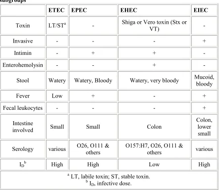
18 Table 1: Some properties and symptoms associated with pathogenic E. coli
subgroups
ETEC EPEC EHEC EIEC
Toxin LT/STa - Shiga or Vero toxin (Stx or
VT) -
Invasive - - - +
Intimin - + + -
Enterohemolysin - - + -
Stool Watery Watery, Bloody Watery, very bloody Mucoid, bloody
Fever Low + - +
Fecal leukocytes - - - +
Intestine
involved Small Small Colon
Colon, lower small
Serology various O26, O111 & others O157:H7, O26, O111 & others various
IDb High High Low High
a LT, labile toxin; ST, stable toxin. b I
D, infective dose.
(http://www.cfsan.fda.gov/~ebam/bam-4a.html#authors).
General characteristics of
E. coli
O157:H7
Evolutionarily, the O157:H7 serotype is a distinct clone that is only distantly
related to other Stx producing enterohemorrhagic E. coli (EHEC). It is most closely
related to an enteropathogenic E. coli (EPEC) clone of serotype O55:H7, a non-Stx
producing strain associated with infantile diarrhea (Whittam et al., 1993). Genetically, E.
coli O157:H7 clones are closely associated with a clone of E. coli O55:H7. It was
19 from the older version 055:H7, which had already developed a mechanism for adherence
to intestinal mucosal cells, when it acquired secondary virulence factors including
Shiga-like cytotoxin production and plasmid-encoded adhesins through horizontal transfer and
recombination (Whittam et al., 1993).
E. coli O157:H7 is different from other E. coli in terms of clinical,
epidemiological, and bacteriological features. E. coli O157:H7 possess biochemical
markers that are significantly different from other E. coli. Virtually all strains have
negative reaction for sorbitol fermentation and positive reaction for raffinose and dulcitol
fermentation (Feng et al., 1998). The clonal nature of E. coli O157:H7 has facilitated its
identification because these organisms, in contrast to approximately 80% of other E. coli
isolates, do not ferment D-sorbitol after overnight incubation and lack β-glucuronidase
(GUD) activity. However, some recent reports indicate that some strains of O157 may
exhibit weak sorbitol fermentation within 24 h-48h. Sorbitol McConkey (SMAC) agar
was developed by substituting the carbohydrate sorbitol for lactose in MacConkey agar,
and SMAC agar has proven to be effective for the isolation of O157 STEC and is the
most widely used medium for this purpose (Feng et al., 1998).
The gene that encodes GUD is intact but non-functional in E. coli O157:H7, and is
nearly identical to the gene in E. coli K-12 with two nucleotide differences (Feng and
Lampel 1994; Feng et al., 1998). It is believed that O157:H7 evolved sequentially from
an O55:H7 ancestor, first by acquiring the stx2 gene and then by diverging into two
branches: one became GUD- SOR-, resulting in the O157:H7 clone that spread worldwide
and the other lost motility leading to the O157:H- clone that is an increasing public health
20 Although O157:H7 is the pre-dominant E. coli serotype incriminated in foodborne
disease, various non-motile and cytotoxigenic O157 variants have also been isolated.
Karch et al. (1993) discovered a novel Stx-producing O157 strain that caused an outbreak
of HUS in Germany. In contrast to typical O157:H7 strains, these non-motile O157
strains ferment sorbitol (SOR+) and have DNA patterns distinct from that of typical
O157:H7 isolates (Karch et al., 1993).
EHEC Pathogenesis
The production of Shiga toxins is one of the defining characteristics of EHEC as these
toxins are thought to be responsible for the principal manifestations of HC and HUS
(Law, 2000). However, the mechanism of pathogenesis of EHEC has not been fully
elucidated (Meng et al., 2001). Pathogenesis of EHEC infection is a multistep process,
involving a complex interaction between a range of bacterial and host interactions. The
main virulence factors and defining characteristics of EHEC include its ability to attach
and efface intestinal mucosal cells and the production of two cytotoxins- Shiga toxin 1
and 2 (Benjamin and Datta, 1995). These toxins show a high degree of homology with
Shiga toxins produced by Shigella dysenteriae. Although Stx I and Stx II are most often
implicated in human illnesses, several variants of Stx II exist. EHEC cells remain in the
intestine and Stx produced in the lumen must be first absorbed by the intestinal
epithelium and translocated into the blood stream. This permits delivery of the toxin to
the specific toxin receptors on target cell surfaces, inducing both local and systemic
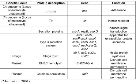
21 Table 2. Significant virulence factors of E. coli O157:H7
Genetic Locus Protein description Gene Function
Chromosome (Locus of enterocyte
effacement) Intimin eae Adherence
Chromosome (Locus of enterocyte
effacement) Tir tir Intimin receptor
Secretion proteins esp A, espB, esp D
Induces signal transduction
Type 3 secretion system
escC, escD, escF,escJ, escN, escR, escS, escT, escU, escV,escQ,
escZ
Apparatus for extracellular protein
secretion
Phage Shiga toxin stx1, stx2, stx2c, stx2d Inhibits protein synthesis
Plasmid EHEC hemolysin EHEC-hly A Disrupts cell membrane
permeability
Plasmid Catalase-peroxidase katP Disrupts cell membrane
permeability (Meng et al., 2001)
It is generally considered that E. coli O157:H7 is more virulent than other
EHEC. The most important virulence factors for E. coli O157:H7 are the production of
Shiga toxin 2 and the adhesin intimin (Law, 2000). Other virulence factors such as
enterohaemolysin, a serine protease (EspP), and a catalase/peroxidase (Katp) may have a
minor role in infection (Law, 2000). The 60-MDa plasmid present in the majority of E.
coli O157:H7 isolates is designated as pO157. This plasmid has been sequenced and
several potential virulence factors have been identified (Law, 2000).
An overview of the steps involved in EHEC pathogenesis is discussed below:
Colonization of the gut by epithelial cell adherence
After surviving the highly acidic pH of the stomach, EHEC cells must establish
22 the cells must compete with other gut microorganisms to establish intestinal colonization
by adhering to the intestinal epithelial cells. The ability of the cells to adhere to the
intestinal epithelial cells and to colonize the intestine is one of the key determinants of
virulence (Paton and Paton, 1998). The processes involved in the establishment and
maintenance of gut colonization by EHEC are poorly understood (Paton and Paton,
1998).
Within the EHEC strains belonging to the serotype O157:H7, there is a great
heterogeneity in adherence, which may reflect different mechanisms. Strains may adhere
in a diffused fashion, or have localized adherence (e.g. form tight clusters or micro
colonies at a limited number of sites on the epithelial surface) or may form a distinct
pattern of adherence called log jam, in which adherence occurs principally at junctions
between cells (Paton and Paton, 1998).
Locus of enterocyte effacement
Some bacterial pathogens possess the ability to produce a characteristic
histological lesion called attaching and effacing (A/E) lesions. The A/E lesion is defined
by the intimate attachment between the bacteria and the epithelial surface and this
phenotype is marked by a loss or effacement of microvilli on the intestinal epithelial cells
at the sites of bacterial attachment (Abe et al., 1998). EPEC is the prototype pathogen
that causes A/E lesions (Goosney et al., 2000). EPEC produce A/E lesions that are
characterized by degeneration and effacement of intestinal epithelial cell microvilli,
intimate adherence of the bacteria to the epithelial cells, and assembly of highly
organized cytoskeleton structures in the cells beneath intimately attached bacteria (Law,
23 One of the important characteristics of EHEC including E. coli O157:H7, is their
ability to produce A/E lesions on a variety of cell types including erythrocytes (Tzipori et
al., 1987; Law, 2000). These lesions involve ultrastructural changes in the erythrocytes,
including loss of erythrocyte microvilli and intimate attachment of the bacterium to the
cell surface. Other members of the A/E family also include a large number of animal
pathogens including those that cause disease in rabbits (REPEC, RDEC-1), pigs
(PEPEC), dogs (DEPEC), and mice (Citrobacter rodentium) (Goosney et al., 2000).
All genes necessary for A/E formation are encoded on a 35 kb chromosomal
pathogenicity island known as the locus of enterocyte effacement (LEE) containing
41 predicted open reading frames (ORFs) inat least 10 operons (Elliot et al., 1999;
McDaniel and Kaper, 1997). The LEE can be divided into three general regions based
upon the specialized functions that contribute to the A/E phenotype. The region to the left
of LEE contains an esc cluster that encodes a type III secretion apparatus which is
responsible for the secretion of Esp (Escherichia coli secreted proteins) proteins.
Mutations in the escV or escN result in abolition of the secretion of the proteins EspA,
EspB, and EspD involved in virulence (Jarvis et al., 1995).
There are two genes in the center of the LEE. The first gene is eae, which encodes
a 94 kDa outer membrane adhesin intimin, required for intimate adherence. The second
gene is tir which encodes the intimin receptor Tir. Upstream of LEE contains at least
three genes espA, espB (Donnenberg et al., 1993),and espD ( Lai et al., 1997) that encode
the secreted proteins, EspA (Kenny et al., 1996), EspB (Donnerberg et al., 1993), and
EspD (Lai et al., 1997), which are secreted by the type III secretion pathway (Perna et al.,
24 within the host cell including the tyrosine phosphorylation of Tir and A/E formation
(McDaniel and Kaper, 1997). The LEE has 23 open reading frames (ORFs) of undefined
function (Elliot et al., 1998; Elliot et al., 1999; McDaniel and Kaper, 1997). Elliot et al.
(1998) reported that the LEE also appears to encode for novel proteins involved in the
type III secretion pathway, new secreted proteins, chaperons, and a regulator/repressor.
All of these genes are conserved in EPEC, EHEC and rabbit EPEC strains (Abe et al.,
1997).
The transfer of the LEE of EPEC is sufficient to confer the ability to form A/E
lesion to non-pathogenic E. coli strains (McDaniel and Kaper, 1997), but the transfer of
EHEC LEE is not sufficient (Elliot et al., 1999). The EHEC LEE was unable to induce
the formation of attaching and effacing (A/E) lesions or to stimulate the secretion of Esp
proteins when it was cloned in E. coli K-12. It is believed that EHEC requires other
non-LEE encoded factors to produce cytoskeleton changes in host cells (Elliot et al., 1999).
These factors have not been identified, but perhaps include regulators and other accessory
factors (Goosney et al., 2000).
In general, the LEE elements in EPEC and EHEC strains are 94% conserved at
the amino acid level and differ by less than 2% in the regions encoding the protein
translocation complex (Elliot et al., 1999). The EHEC O157:H7 LEE also encodes a
cryptic prophage at one end that is not present in the EPEC O127:H6 LEE. Perna et al.
(1998) suggested that the prophage was inserted into the LEE after the island was already
present on the chromosome and it is unlikely that this prophage codes for any known
25 Many EHEC strains involved in severe human disease, including HUS, do not
contain the eae gene or do not express a functional intimin (Pradel et al., 2000). Thus, it
was speculated that attaching and effacing lesions might not be essential for the
development of severe disease and additional factors might be involved (Pradel et al.,
2000).
Adhesin molecule Intimin
The attachment of the bacteria to the epithelial cell surface results in alteration of
the cytoskeleton component beneath the adherent bacteria leading to formation of a
pedestal-like structure that can extend to a pseudopod. The interaction between the
bacterial adhesin molecule, intimin, and its receptor in the host cell membrane, Tir, is
essential for pedestal formation. Intimin is a member of the invasin/intimin-like protein
family. Donenberg and Kaper (1991) showed that intimin mutants could activate
signaling in host cells, inducing tyrosine phosphorylation of Tir and generalized actin
accumulation, but they could not focus actin into pedestal-like structures. Intimin is
essential for the formation of A/E lesions and for complete virulence of the bacteria in
humans and animals (De Grado et al., 1999). The eae gene is a part of the LEE, required
for the formation of the attaching and effacing lesions, initially recognized in
enteropathogenic E. coli strains (Pradel et al., 2000). The EHEC LEE encodes a
homologue for the receptor of intimin, called translocated intimin receptor (Tir
homologue) (Paton and Paton, 1998).
Intimin receptor Tir
The A/E lesions in vitro are mediated by bacteria-host cell interactions including
26 adherence of the bacterial cells to the epithelial cells is mediated by a plasmid- encoded,
bundle-forming pilus (BFP) (Donnenberg et al., 1992), followed by insertion of
translocated intimin receptor, Tir, which is a bacterial protein, into the host plasma
membrane (Kenny et al., 1997). EPEC secretes Tir as a 78 kDa protein using the type III
secretion system (DeGrado et al., 1999). After insertion into the host cell it is
serine/threonine and tyrosine phosphorylated and undergoes an electrophoretic mobility
shift to a molecular weight of 90 kDa. Tir requires secreted proteins EspA and EspB and
the type III secretion apparatus for its translocation.
Tir is generally divergent between EPEC and EHEC, particularly in the C
terminus of the protein. The C terminal tyrosine that is required to be phosphorylated for
pedestal formation in EPEC is not present in EHEC. The A/E lesion formation in EHEC
occurs independently of tyrosine phosphorylation. These differences that allow EHEC to
form pedestals in the absence of Tir tyrosine phosphorylation have not yet been
characterized (Goosney et al., 2000).
Tir has mainly 3 functions that have been identified. It is translocated into the
epithelial cell membrane and serves as a cell surface receptor for intimin. It also nucleates
actin after intimin binding. By focusing actin, it is believed that Tir acts as a bridge
connecting intimin to the host cytoskeleton. Another function of Tir is that it transmits
additional signals to host cells once Tir-intimin interaction occurs. These events trigger
tyrosine phosphorylation of the phospholipase Cγ and other host proteins, resulting in Tir
phosphorylation and other early signaling events (Abe et al., 1998). Tir is a unique
27 it undergoes modifications, acts as an intimin receptor, and induces drastic cellular
changes (DeGrado et al., 1999).
Type III secretion system
The translocation of virulence factors, including Tir, directly into the host cell is
done by the type III secretion systems. The type III secretion systems are multimeric (~
20 protein components) molecular machines that are present exclusively in
Gram-negative bacteria (Goosney et al., 2000). It has been hypothesized that the type III
secretion systems are recent genetic acquisitions to pathogenic genomes although their
origins are unknown. Type III secretion systems mediate translocation and secretion of
critical virulence determinants such as the Ipa proteins of Shigella spp., the Yops of
Yersinia spp., and proteins involved in the cellular invasion of Salmonella spp.
(McNamara and Donnenberg, 1998).
Shiga toxins in
E. coli
The major virulence factor and defining characteristic of EHEC is the production of
Shiga toxins. O’ Brien and Holmes (1987) were the first to report that EHEC produce
Shiga toxins. Members belonging to the Shiga toxin family are bipartite molecules
composed of a single enzymatic A subunit and a multimer of receptor binding B subunits.
(Olsnes et al., 1981). The Stx family contains 2 major, immunologically
non-cross-reactive groups called Stx 1 and Stx 2 (O’ Brien and Holmes, 1987; Nataro and Kaper,
1998). Even though they are immunologically distinct, Stx 1 and Stx 2 share
28 A single EHEC strain may express Stx 1 only, Stx 2 only, or both toxins or multiple
forms of Stx2 (Calderwood et al., 1996). While Stx 1 is homogenous, there are many
variants of Stx2. Many isolates can produce 2 or more forms of Stx 2 (Schmitt et al.,
1991). The different variants of Stx2 are designated as Stx2c (Schmitt et al., 1991),
Stx2v, Stx2vhb, Stx 2d (Pierard et al., 1998), Stx2e (Weinstein et al., 1988) and Stx2f.
The Stx 1 from EHEC is identical to Shiga-toxin from Shigella dysenterae I. Stx1 from
some strains of EHEC may differ from Stx in 1 residue, while Stx from other strains
show no sequence variation (O’Brien et al., 1992). Most (pathogenic) isolates of E. coli
O157:H7 produce Shiga toxin 2 (Stx2) only (O’Brien et al., 1992).
Both toxins are compound toxins composed of a single 32-kDa A subunit and a
pentameric B subunit composed of 7.7 kDa monomers (Olsnes et al., 1981). The A
subunit of the Shiga toxin family is activated by proteolytic processing and it is
proteolytically nicked to yield a 28 kDa peptide A1 and a 4 kDa peptide A2 (O’Brien et
al., 1992). These peptides remain linked by a disulphide bond. The A1 peptide contains
the enzymatic activity while the A2 peptide serves to bind the A subunit to the B subunits.
The B pentamer mediates binding of the toxin to specific glycolipid receptors, known as
globotriaosylceramide or Gb3, which are present on the surface of eukaryotic cell
membranes (O’Brien et al., 1992). While Gb3 is the main receptor for Stx, the receptor
for the Stx2 variant, Stx2e, is Gb4.
The structural genes for Stx 1 and Stx 2 are found on lysogenic lambdoid
bacteriophages (Nataro and Kaper, 1998). Production of Stx is essential for many of the
pathological features and life threatening consequences of EHEC infection (Paton and
29 bacteriophage, presumably directly or indirectly from Shigella (Buchanan and Doyle,
1997; Pradel et al., 2000). The expression of the phage-encoded Shiga toxin is under the
regulatory control of the phage late genes. Induction of the phage lytic cycle is required
for toxin synthesis and release (Shantini et al., 2003). Phage production is increased by
factors such as antibiotic treatment or peroxide released from activated neutrophils, and
these factors can also induce Stx2 production. Non-pathogenic E. coli can produce Stx2 if
infected with the phage encoding the toxin (Shantini et al., 2003).
EHEC strains appear to be unable to invade gut epithelial cells to any significant
extent. Hence, the generation of systemic sequelae must presumably involve translocation
of Stx produced by colonizing bacteria from the gut lumen to underlying tissues and the
blood stream (Paton and Paton, 1998). One possible route for translocation might be
through the lesions in the mucosal barrier caused either by the direct effects of Stx or
other factors such as intimin or perhaps through gaps between adjacent epithelial cells.
An alternative route from the gut lumen to tissues might be through intact epithelial cells
(Paton and Paton, 1998).
After crossing the epithelial barrier and entering the blood stream, Stx targets the
tissues expressing the appropriate glycolipid receptor. The specificity of this interaction
and the distribution of receptors among various cell types have a major impact on the
pathogenesis of the disease (Paton and Paton, 1998). After binding to the receptors, the
toxin molecules are endocytosed by a receptor-mediated endocytic mechanism and are
transported to the golgi apparatus and then to the endoplasmic reticulum (Sandvig and
Van Duers, 1994). The A subunit is translocated to the cytoplasm where the A1 subunit
30 amino acyl tRNA to the 60S ribosomal units, resulting in the inhibition of peptide chain
elongation during protein synthesis and leading to cell death (Brown et al., 1981; Sandvig
and Van Duers, 1994).
Epidemiological evidence indicates that the EHEC isolates producing Stx 2 are more
commonly associated with serious disease than isolates producing Stx 1,or Stx 1 and Stx
2 (Boerlin et al., 1999). The toxicity of Stx2 toward human renal microvascular
endothelial cells (HRMEC) is a 100 fold greater than that of Stx 1. HRMEC’s are the
putative target of the Shiga toxins in the development of HUS (Louise and Obrig, 1991,
1995). Jacewicz et al. (1999) demonstrated that even though Stx 1 had higher binding
affinity for the Gb3 receptor on the human intestinal microvascular endothelial cells
(HIMEC) than Stx 2, these cells were more sensitive to inhibition of protein synthesis by
Stx 2 than Stx1. It was concluded that increased toxicity of Stx 2 to endothelial cells may
be relevant to the higher frequency of Stx 2 producing EHEC strains involved in the
pathogenesis of HUS.
α
-Hemolysin and role in pathogenesis
The hemolytic activity of E. coli was first reported by Kayser in 1903 (cited in
Cavalieri et al., 1984), who found that some E. coli cultures lysed erythrocytes (Cavalieri
et al., 1984). In 1963, Smith was the first to differentiate between cell-bound and cell-free
hemolysin in cultures of E. coli grown in alkaline meat extract broth. Under the same
growth conditions, some hemolytic strains of E. coli can produce cell-free and cell-bound
hemolysin simultaneously. The cell-free hemolytic factor was designated as α-hemolysin,
as it can be obtained free from bacterial cells in culture fluid filtrates and the cell-bound
31 hemolysin which does not hemolyze human or rabbit RBCs, but does hemolyze RBCs of
other species (Cavalieri et al., 1984). Enterohemolysin is another kind of hemolysin,
which is also active in cell free extracts (Buetin et al., 1991).
Several studies have shown that hemolytic E. coli are more frequently isolated
from extra intestinal infections such as urinary tract infections (UTI), bacteremia,
peritonitis, and appendicitis than from the feces of healthy individuals. Some studies have
indicated that colonization before the development of infection may be enhanced by
hemolysin production (Cavalieri et al., 1984). Hemolysin production alone does not
always equate with virulence, but may be a decisive factor in virulence of many
nephropathogenic strains (Cavalieri et al., 1984). The enterohemolysin or
Enterohemorrhagic E. coliHly, a member of the repeat in toxin (RTX) family of pore
forming cytolysins, has been suspected to have a role in pathogenesis. This is because it
has occurred in a majority of the pathogenic EHEC strains tested and is reactive to the
sera of HUS patients (Pradel et el., 2000).
pO157
All strains of O157:H7 contain a highly conserved plasmid, designated pO157
(Schmidt et al., 1994), which varies in size from 93.6 to 104 kb (Schmidt et al., 1996).
This plasmid is also present in O26:H11 strains andis present in most but not all
Stx-producing E. coli strains isolatedfrom humans (Levine et al., 1987). A 3.4-kb fragment of
this plasmid, subsequentlyshown to encode enterohemolysin (Schmidt et al., 1995), was
identified by Levineet al. (1987) as a diagnostic probe for EHEC. In addition to the
enterohemolysin and potential adherence factors described above,this plasmid encodes a
32 suppressionof production of an exopolysaccharide has also been suggested(Fratamico et
al., 1993).
The role of this plasmid in EHEC pathogenesis is unknown. In vivo and in vitro
studies have reportedconflicting results regarding the role of the plasmid in adherence to
epithelial cells. Karch et al. (1987) first reported that pO157was required for the
expression of fimbriae and adhesion to Henle 407, but not Hep-2 cells. Other
investigators have reported that loss of this plasmideither decreased adhesion (Toth et al.,
1990), enhanced adhesion (Junkins and Doyle, 1989), orhad no effect on adhesion
(Fratamico et al., 1993). A study by Hall et al. (1990)reported that for one EHEC strain
of serotype O103:H2, loss ofthis plasmid coincided with reduced adhesion to cultured
epithelialcells while for another EHEC strain of serotype O5: H-, loss ofthis plasmid had
no effect on adhesion. Dytoc et al. (1993) reportedin vivo data supporting the
involvement of this plasmid in intestinaladherence after oral inoculation of adult rabbits.
In this study,E. coli K-12 strain HB101 containing this plasmid adhered to rabbit
intestinal cells whereas HB101 without the plasmid did not adhere.In both rabbit (Li et
al., 1993) and gnotobiotic piglet (Tzipori et al., 1987) models ofdisease, the presence or
absence of this plasmid made no differenceto the amount of diarrhea, the intestinal
histopathology, or theintestinal ion transport. On the other hand, Wadolkowski et al.
(1990) showed that both O157: H7 strain 933, and its plasmid-cured derivative 933cu,
could individually colonize the gut of streptomycin treated mice but that 933cu could not
establish colonization when used together with 933. Even though the same strains were
33 performed. Hence, it is not possible to determine whether the apparent contribution of
pO157 is influenced by host species.
The discordant finding about the role of pO157 may be attributable to differences
in growth and assay conditions, and to differences between O157:H7 strains and to the
fact that the large plasmid itself appears to be heterogeneous even within the serotype
O157:H7 (Barrett et al., 1992). Also, a serious limitation to establishinga role of pO157
in pathogenesis is that there is no suitable animal model that reproducesall aspects of the
EHEC disease, from intestinal inoculation to bloodydiarrhea to renal involvement. The
degree to which either of these animal models reflects colonization mechanisms in
humans is uncertain (Paton and Paton, 1998). Inthese rabbit and piglet studies, the
presence or absence of Stxalso made no difference, further highlighting the limitationsof
animal models. Epidemiologicalevidence suggests a stronger correlation between the
presence of thisplasmid and the development of HUS, rather than diarrhea. As described
above, the enterohemolytic phenotype encoded on this plasmid wasobserved in 16 (88%)
of 18 O111:H- strains isolated from patientswith HUS but in only 4 (22.2%) of
18 O111:H- strains isolatedfrom patients with diarrhea without HUS (Schmidt and
Karch, 1996).
Despite the uncertainty about the significance of plasmid pO157 in disease, it is in
fact widely distributed among human EHECisolates. The initial study by Levine et al.
(1987) on the distributionof this plasmid among human isolates (mostly from North
America)found that 99% of 107 O157:H7 strains possessed the plasmid, asdid 77% of
44 O26:H11 strains. pO157 was also found in 81% of26 Stx-positive strains of serotypes
34 different strain collectionfrom Europe showed similar results, with the plasmid being
presentin 60% of Stx-positive strains of serotypes other than O157:H7and O26:H11
(Willshaw et al., 1992). Another study in Germany found pO157 in ca.90% of all
Stx-producing E. coli isolates from patients. In contrast to the high frequency of the plasmid
in human isolates,only a minority of Stx-positive strains of non-O157: H7 serotypes
isolated from cattle possess this plasmid (Barrett et al., 1992).In addition to the 94- to
104-kb pO157 plasmid, a number of other plasmids ranging in size from 2 to 87 kb have
been foundin strains of E. coli O157:H7 (Willshaw et al., 1992). However, no correlation
has been seen between possession of any of these plasmids and clinicaldisease.
Other adherence mechanisms
There are some factors that affect the adherence of EHEC cells including whole
cells, outer membrane proteins (OMPs) and lipopolysaccharides (LPS) to host epithelial
cells. Studies have shown that antibody to whole cells and outer membrane proteins,
including a 94 kDa OMP and an 8 kDa OMP, but not to H7 flagella, significantly inhibit
the adherence of E. coli O157:H7 to HEp-2 cells.
The O157 LPS (as well as LPS from other bacteria) enhances the cytotoxicity of
Stx on human vascular endothelial cells in vitro,but its effects in vivo are not clear. One
report by Oelschlaeger et al. (1994) stated that E. coli O157:H7 can invade cultured
intestinal cell lines,but a later report (McKee and O’Brien, 1995) disputed these findings,
showing thatO157:H7 strains were no more invasive than E. coli strains fromthe normal
gut flora. Furthermore, there is no in vivo evidence thatinvasion occurs in humans or in
35 Two other putative EHEC virulence factors have been described. These are a
serine protease, EspP, which can cleave human coagulation factor V, and a bifunctional
catalase peroxidase, KatP (Pradel et al., 2000). However, there is no experimental proof
for the role of these factors in the virulence of EHEC (Pradel et al., 2000).
Treatment
Most persons recover from HC without antibiotics or other specific treatment in
5-10 days. There is no evidence that antibiotics improve the course of disease, and it is
thought that treatment with some antibiotics may precipitate kidney complications.
Antidiarrheal agents, such as loperamide (Imodium), should also be avoided
(http://www.cdc.gov/ncidod/dbmd/diseaseinfo/escherichiacoli_g.htm). Treatment of
O157 infection with antibiotics does not affect the E. coli O157:H7 infections, and in
many cases has been associated with worse clinical outcomes (Wong et al., 2000). In
children who are infected with E. coli O157:H7, treatment with antibiotics before the
onset of diarrhea leads to a cessation in the fecal shedding of the organism. However, the
treatment does not prevent hemolytic uremic syndrome (Wong et al., 2000). In vitro
experiments have shown that exposure to various antibiotics cause E. coli to release
Shiga toxin.
The risk of HUS may be increased by antibiotics as the antibiotics cause the
release of Shiga toxin from injured bacteria in the intestine, making the toxin more
available for absorption (Wong et al., 2000). Also, E. coli strains are highly variable in
their antibiotic-induced release of Shiga toxin (Wong et al., 2000). The antibiotics that
show this effect include fluoroquinolones, cephalosporins, and