ABSTRACT
Holtman, Kevin Matthew. An Investigation of the Milled Wood Lignin Isolation Procedure by Solution- and Solid-State NMR Spectroscopy. (Under the direction of Drs. Kadla and
Jameel)
Milled wood lignin (MWL) is only a fraction of the total lignin in wood but is useful
for lignin studies because it is isolated relatively free of carbohydrates. It is isolated from a
finely milled wood powder by solvent extraction and is considered to be the isolated lignin
most representative of native lignin. Because it is only a portion of the total lignin this study presents an extensive evaluation of all isolated lignin portions with the goal of studying the
whole lignin structure intact in wood. In order to achieve this, solution-state NMR experiments
and degradative techniques were performed on soluble lignins to make an estimation of the
interunit linkages. The previously insoluble lignin portions were dissolved in a newly
developed solvent system and examined by solution-state and solid-state NMR.
A systematic study of the MWL and cellulolytic enzyme lignin (CEL) found they were
structurally similar with only minor differences. End groups were somewhat higher in MWL,
especially the oxidized moieties. MWL was lower in β-aryl ether content and had a higher
degree of condensation than CEL. Carbohydrate analyses indicated that MWL may derive
from the middle lamella to a larger extent than CEL. These portions can be combined to create
a higher yield of relatively carbohydrate-free lignin.
Lignin isolated from milled wood and REL are normally insoluble however they were
of structural moieties present. Quantitative 13C NMR however showed that MWL had a β
-O-4’ content lower than that of the wood. Dipolar dephasing solid-state NMR indicated that
MWL had a higher degree of condensation. As a result, REL may be more similar structurally
to the whole lignin in wood than MWL. The milled wood and REL contain a significant
portion of high molecular weight material (~55,000 g/mol) while MWL (~10,000 g/mol) is
lower molecular weight.
A solution-state NMR study of different milling techniques indicated that prolonged
rotary ball milling results in more structural changes than the standard milling technique. It has
been debated whether milling under N2 results in more structural changes than milling in
toluene. This study however clearly shows that there is little difference between these lignins.
The wood milled under the N2 atmosphere is more vigorously milled based on total MWL
yield and contains higher aliphatic and phenolic hydroxyl contents.
The modified DFRC method for analysis of the uncondensed aryl ether structures in
lignin is inefficient because it does not completely cleave all β-O-4’ linkages. DFRC is more
inefficient in higher molecular weight materials than in MWL indicating that molecular weight
plays a role in either accessibility of chemicals or molecular mobility in the reductive cleavage
step. Thioacidolysis completely degrades the β-aryl ether linkages in lignin and is therefore
preferable for analysis of these structures.
Although lignin isolated from finely milled wood can be dissolved using the
the NCSU solid-state NMR facility were incomplete because we did not have the necessary
pulse sequence to perform this last experiment. As a result, a collaboration between our group
and a solid-state NMR group at Iowa State University has been established. Data obtained in
the NCSU facility is presented here while the analysis of the data collected at ISU will be
An Investigation of the Milled Wood Lignin Isolation
Procedure by Solution- and Solid-State NMR Spectroscopy
Kevin M. Holtman
A dissertation submitted to the Graduate Faculty of North Carolina State University
in partial fulfillment of the requirements for the Degree of
Doctor of Philosophy
Wood and Paper Science
I would like to dedicate this dissertation to my wife, Gretchen, and my
family all of whom had a major influence on my life, goals, and
BIOGRAPHY
Kevin M. Holtman was born in Asheville, North Carolina on September 29, 1973.
After graduating from West Florence High School in Florence, SC, in 1991, he entered North
Carolina State University. In the spring of 1996 he received a B.S. degree in Pulp and Paper
Technology and a B.A. degree in Chemistry.
After working full time for the Dept. of Wood and Paper Science for a year, he
entered the graduate program under the guidance of Drs. John F. Kadla, Hou-min Chang, and
Hasan Jameel. During his time in graduate school he interned at Nippon Paper Company in
ACKNOWLEDGEMENTS
The author would like to express his sincere appreciation for the guidance and
support he received from Dr. John F. Kadla and Dr. Hasan Jameel, co-chairs of his advisory
committee. Their constant encouragement, invaluable advice and constructive criticism were
always available when suggestions or stimulating discussions were needed. Their sincere
friendship and guidance have made this study enjoyable and memorable.
Sincere gratitude is also extended to Dr. Hou-min Chang and Dr. Jeff L. White for
their invaluable assistance and guidance. In addition the author would like to thank Dr.
Thomas Joyce for the opportunity to perform research as an undergraduate, an experience
which opened the door to graduate school. Finally, the author would also like to thank Dr.
Ewellyn A. Capanema, Dr. Hanna S. Gracz, Dr. Mikhail Yu. Balakshin, Dr. Tsutomu Ikeda,
and Dr. Satoshi Kubo for their suggestions, stimulating discussions and friendship.
The author expresses his deepest gratitude to Gretchen, for her love, patience,
sacrifice and assistance have made this study an enjoyable experience. Special appreciation
must also be given to the author’s family for their constant support and encouragement
throughout this study.
Finally, the author wishes to thank all those faculty members, colleagues and friends
TABLE OF CONTENTS
Page
List of Figures ix
List of Tables xiii
1. Introduction 1
2. Introduction to Lignin 4
2.1 The Phenylpropane Unit 4
2.2 Lignin Biosynthesis 6
2.2.1 Introduction 6
2.2.2 Shikimic Acid Pathway 8
2.2.3 Biosynthetic Pathway for Primary Lignin Precursors 10 2.2.4 Transport of Monolignol Glucosides into the Lignifying Zone 12
2.3. Polymerization of Monolignols 14
2.3.1 Classification of Lignin 16
2.3.2 The Ultrastructure of Wood 18
2.3.3 Distribution of Components in the Cell Wall 18
2.4. Different Methods of Lignin Isolation 21
2.4.1 Isolation of MWLby the Bjorkman Method 22
2.4.2 Theory on Milling 23
2.4.3 Milling Parameters 23
2.4.4 Extraction and Purification of Milled Wood Lignin 25 2.4.5 Properties of Lignin Extracted with Dioxane 26 2.4.6 Isolation of Lignin (CEL) by Treatment with Cellulolytic Enzymes 26 2.4.7 Mechanochemical Reactions and Changes in Lignin Structure
During Milling 28
2.4.8 Depolymerization of the Lignin Macromolecule
by Ether Bond Cleavage 31
2.4.9 Formation of Carbonyl and Unsaturated Side Chain Structures 32 2.4.10 Possible Formation of Quinoid Structures During Milling 34 2.4.11 Condensation Reactions Occurring During Milling 34 2.5. Characterization of Lignin by Chemical Degradation Methods 36
2.5.1 DFRC 36
2.5.2 DFRC Procedure 36
2.5.3 Discussion of DFRC Method 37
2.6. NMR Spectroscopy of Lignin 53 2.6.1 Introduction to Nuclear Magnetic Spectroscopy 53
2.6.2 The Basic NMR Experiment 57
2.6.3 1H NMR 58
2.6.4 13C NMR Spectroscopy 59
2.6.5 Quantitative 13C NMR Experiments 62
2.6.6 13C DEPT (Distortionless Enhancement by Polarization Transfer) 62 2.6.7 Two-Dimensional Homonuclear Correlation Experiments 63
2.6.7.1 1H-1H COSY (COrrelation SpectroscopY) 63
2.6.7.2 TOCSY (TOtal Correlation SpectroscopY) 64
2.6.7.3 13C-13C INADEQUATE 64
2.6.8 Two-Dimensional Heteronuclear Correlation Experiments 65 2.6.8.1 HMQC (Heteronuclear Multiple Quantum Coherence) 65
2.7 Solid-State NMR 68
2.7.1 Cross Polarization/Magic Angle Spinning (CP/MAS) 68 2.7.2 Interrupted Decoupling (Dipolar Dephasing) 69 2.7.3 Direct Polarization (DPMAS) or Bloch Decay Experiment 70
2.7.4 CP/TOSS (TOtal Sideband Suppression) 71
2.7.5 Efficient CH-group Selection 71
2.7.6 HETeronuclear CORrelation Experiment (HETCOR) 72
3. Methods and Materials 74
3.1 Isolation of Milled Wood Lignin (MWL) and Other Isolatable Lignins 74 3.1.1 Isolation of Lignin via the Bjorkman Isolation Procedure 74
3.1.2 Milled Wood Lignin Isolation 74
3.1.3 Preparation of Cellulolytic Enzyme Lignin (CEL)
and Residual Enzyme Lignin (REL) 76
3.1.4..MWL Isolation Procedures Involving only Rotary-milling of Wood 77 3.2 Derivatization Followed by Reductive Cleavage (DFRC) 79
3.2.1 Standard DFRC Procedure 79
3.2.2 Modified DFRC Method 79
3.2.3 Methylation 80
3.3 Nitrobenzene Oxidation 81
3.4 Thioacidolysis 82
3.5 Gel Permeation Chromatography 83
3.6 1H NMR Analysis 84
3.7 1H-13C 2D Correlation NMR (HMQC) Spectroscopy 84
3.8 Quantitative 13C NMR Spectroscopy 85
3.9 Elemental Analysis 85
3.10 Methoxyl Analysis 85
3.11 Carbohydrate Content 85
3.12 Dissolution of Lignocellulosics in DMSO/N-methylimidazole (NMI) 86
4. Studies on the Effect of Ball Milling on Lignin Structure Using a Modified DFRC
Method 89
4.1 Modified DFRC Method 89
4.2 Effect of MWL Isolation on Lignin Structure 90
4.3 Effect of Ball Milling on Lignin Structure 93
4.4 DFRC Analyses and the Significance of the Results Obtained 97
5. Elucidation of Lignin Structure through Degradative Methods: Comparison of
Modified DFRC and Thioacidolysis 99
5.1 Comparison of Monomer Yields from Thioacidolysis and DFRC Treatment
of Milled Wood and MWL 99
5.2 Effect of Thioacidolysis and DFRC on the Molecular Weight Distribution
of Lignin 102
5.3 1H-13C HMQC NMR Analysis of Thioacidolysis and DFRC Degraded
Lignins 104
6. A Solution-State NMR Study of the Similarities between MWL and CEL 109
6.1 Elemental Composition 109
6.2 Chemical Degradation Analysis of Lignins 110
6.3 Structural Analysis of Lignins via NMR Techniques 111
6.4 Molecular Weight Distributions 122
6.5 Carbohydrate Analysis 123
7. An NMR Comparison of the Whole Lignin from Milled Wood, MWL, and REL
Dissolved by the DMSO/NMI Procedure 125
7.1 Comparison of Lignin Preparations by Degradative Techniques 125
7.2 Comments on Milling 126
7.3 Dissolution of Lignin 127
7.4 1H-13C Two-Dimensional HMQC NMR 127
7.5 Quantitative 13C NMR 131
7.5.1 Oxidized Carbon Region 133
7.5.2 Aliphatic and Phenolic Hydroxyl Content 135
7.5.3 Aliphatic Sidechain Region 136
7.7 Dipolar Dephasing Solid-State 13CP/MAS NMR 138
7.8 Gel Permeation Chromatography 140
8. Quantitative 13C NMR Characterization of Milled Wood Lignins Isolated by
Different Milling Techniques 143
8.1 Modified DFRC 143
9. Solid-State NMR Spectroscopy 157
9.1 Experimental Plan 157
9.1.1 CP/MAS Experiment 157
9.1.2 DP/MAS (Direct Polarization/Magic Angle Spinning) 159 9.1.3 Interrupted Decoupling (Dipolar Dephasing) 160
9.1.4 CH Selection Experiment 162
9.1.5 Comments on Experiments Using Chemagnetics
200 MHz Instrument 164
9.2 Solid-State NMR Analysis of Thermally Treated Kraft Lignins 166 9.3 Quantitative Experiments Performed at Iowa State University 169 9.4 Two-Dimensional HETeronuclear CORrelation experiment (HETCOR) 170
9.5 Final Notes on Solid-State NMR 170
10. Conclusions 171
11. Future Work 175
LIST OF FIGURES
Figure Page
2.1 The primary lignin precursors. 5
2.2 Labelling convention for monolignols, a) wood chemistry terminology
and b) IUPAC nomenclature. 5
2.3 Primary and secondary metabolic pathways leading to the biosynthesis of lignin
and wood components. 7
2.4 Shikimic acid pathway. 8
2.5 Biosynthetic pathway to lignin precursors. 11
2.6 Monolignol glucoside formation. 13
2.7 Dehydrogenative polymerization of lignin monomers. 14
2.8 Radical coupling in lignin polymerization. 15
2.9 Prominent interunit linkages in lignin. 16
2.10 A structural model for softwood. 17
2.11 Structure of the cell wall in wood showing the middle lamella (ML),
the primary wall, the outer (S1), middle (S2), and inner layers (S3). 19
2.12 ESR Spectra of a) cellulose, b) MWL, and c) wood meal, milled with glass beads for three hours in N2 with glass beads. Multiplication number represents
signal intensity. 30
2.13 Free radicals in wood meal milled with steel balls in CO2. 30
2.14 Cleavage of β-O-4 interunit linkage in milling of wood. 31
2.15 ESR of MWL milled in lignin at 77 K. Peak A indicates the presence
2.18 Possible mechanism for the formation of o- and p-quinoid structures during
milling. 34
2.19 Possible mechanism for the formation of a biphenyl linkage during milling. 35
2.20 Schematic of the standard DFRC reaction. 36
2.21 Schematic of the modified DFRC reaction. 38
2.22 Monomeric phenols detected in lignin acidolysis reaction mixtures. 39
2.23 Formation of monomeric phenols from β-O-4’ structures in lignin during
acidolysis. 40
2.24 Acid-catalyzed condensation reaction occurring during acidolysis reaction. 41
2.25 Proposed mechanism of the thioacidolysis reaction with lignin. 42
2.26 Schematic of the modified thioacidolysis reaction. 43
2.27 Major products from nitrobenzene oxidation. 43
2.28 Formation of new phenolic hydroxyl group in the nitrobenzene oxidation.
reaction scheme. 45
2.29 Possible mechanism of nitrobenzene oxidation. 45
2.30 Degradation of β-5’ and β-1’ substructures during nitrobenzene oxidation. 46
2.31 Disproportionation of veratraldehyde to form acid and alcohol. 47
2.32 Major carboxylic acid methyl esters formed during permanganate oxidation
of lignin. 48
2.33 The four-step permanganate oxidation reaction sequence. 49
2.34 Magnetic field generated by positively charged spinning particle. 54
2.35 Precession of spins around the z-axis. 55
2.36 Energy differences between spin states. 56
2.37 Quantitatively edited 13C spectra using the DEPT pulse sequence. 63
2.39 Partial INADEQUATE spectrum showing just the sidechain region, from a 13C-enriched ryegrass isolated lignin that contained substantial polysaccharide
components. 65
2.40 HMQC NMR spectrum of a “methoxy-less” coniferyl alcohol synthetic lignin. 66
2.41 HMBC NMR spectrum of a “methoxy-less” coniferyl alcohol synthetic lignin. 67
3.1 Bjorkman MWL isolation procedure. 75
3.2 Various milling techniques employed in MWL isolation. 78
5.1 Proposed mechanism of lignin thioacidolysis. 100
5.2 Proposed mechanism of DFRC degradation of lignin. 101
5.3 GPC chromatographs of MWL before and after thioacidolysis and
DFRC treatment. 103
5.4 Expansion of the oxygenated aliphatic region of the 1H-13C HMQC spectrum of the A) original MWL, B) acetylated DFRC degraded MWL, and
C) acetylated thioacidolysis degraded MWL. 105
6.1 Quantitative 13C NMR spectra of MWL. Included is an expansion of the
oxygenated aliphatic region of the MWL and acetylated MWL (MWL-Ac). 113
6.2 1H-13C HMQC spectra of the oxygenated aliphatic region of A) MWL and
B) CEL; included are the magnified regions of C) MWL and D) acetylated MWL. 116
6.3 GPC chromatographs of MWL and CEL. 123
7.1 Expansions of the oxygenated aliphatic regions of the HMQC spectra: A) Rotary Porcelain 6 Week Wood; B) Rotary Porcelain 6 Week MWL;
C) Rotary Porcelain 6 Week REL. 129
7.2 Quantitative 13C NMR spectra for A) Rotary Porcelain 6 Week Wood and B) Rotary Porcelain 6 Week Wood, both dissolved and acetylated
using the DMSO/NMI dissolution procedure. 132
8.1 Quantitative 13C NMR spectra of Vibratory MWL. Included is an expansion of the oxygenated aliphatic region of the MWL and acetylated MWL (MWL-Ac). 145 8.2 Commonly found interunit linkages in lignin preparations. 150
9.1 CP/MAS spectrum of Vibratory MWL. 158
9.2 DP/MAS spectrum of Vibratory MWL. 160
9.3 Dipolar dephasing spectrum of Vibratory MWL 161
9.4 Substructures commonly found in the lignin polymer. 163
9.5 CP/MAS spectrum of the Vibratory REL sample showing the effects of
paramagnetic materials on resolution in the NMR experiment. 165
9.6 13C/CPMAS NMR spectra for a hardwood kraft lignin oxidized to different
LIST OF TABLES
Table Page
2.1 Relative frequency of interunit linkages reported per C9 unit in some
softwood lignins. 15
2.2 Frequency of interunit linkages per C9 unit for spruce and birch wood lignins. 18
2.3 Distribution of lignin in spruce tracheids. 20
2.4 Bjorkman’s estimation of the chemical properties of MWL. 26
2.5 Data presented for MWL, CEL-96, and CEL-50. 27
2.6 Relative frequency of carboxylic acid methyl esters from permanganate
oxidation of spruce wood and MWL. 51
2.7 1H NMR chemical shifts for sidechain chemical shifts of common lignin
interunit linkages. 59
2.8 13C NMR chemical shifts for the sidechain carbons of the major interunit
linkages in nonacetylated softwood lignin. 61
2.9 13C NMR chemical shifts for the side chain carbons of the major interunit
linkages in acetylated softwood lignin. 61
4.1 Unit composition and total molar yields for the standard and modified
DFRC methods. 91
4.2 Yields of MWL and CEL from various milling methods. 94
4.3 Unit composition and total molar yields from the modified DFRC
method for preparations of the various milling methods. 95 4.4 Comparison of the total molar yields from the modified DFRC
method and nitrobenzene oxidation. 96
6.3 Assignment of the quantitative 13C NMR spectra. 114
6.4 Quantification of the spectral regions of the 13C NMR spectra. 115
6.5 Estimation of interunit linkages in MWL and CEL via quantitative 13C NMR. 119
6.6 Monosaccharide composition of MWL and CEL. 124
7.1 Comparison of the unit composition and total molar yields of lignin
preparations analyzed by the modified DFRC and thioacidolysis methods. 126
7.2 Peak assignments for quantitative 13C NMR spectra. 133
7.3 Quantitative determination of interunit linkages for RotaryPorcelain 6 Week
MWL and RotaryPorcelain 6 Week MWL by 13C NMR. 134 7.4 Estimations of the degree of condensation for Wiley Wood,
RotaryPorcelain 6 Week MWL, and RotaryPorcelain 6 Week REL,
as determined by the dipolar dephasing solid-state NMR experiment. 140 8.1. Modified DFRC unit composition and total molar yields for the different
MWL preparations. 143
8.2 Assignment of the quantitative 13C NMR spectra. 146
8.3 Quantification of the spectral regions of the 13C NMR spectra. 147
8.4 Estimation of interunit linkages in Vibratory MWL,Vibratory (Dry) MWL,
and RotaryPorcelain 6 Week MWL via quantitative 13C NMR. 149 9.1 Degree of condensation data for some lignins determined by dipolar
1. Introduction
Lignin in the cell wall is an amorphous copolymer of phenylpropanoid units linked
through both ether and carbon-carbon bonds. It provides mechanical support for the plant, as
well as facilitating transport of water and nutrients, and providing defense against attack from
microorganisms. [1-5] Therefore, elucidation of the complete structure and macromolecular
characteristics of the lignin polymer is of interest from the biological perspective. In
addition, lignin must be removed in order to produce high-quality printing and writing paper.
[6] As a result, the structure of lignin is of great importance for optimizing the processes and
understanding the reactions involved in its removal.
For decades, lignin chemists have been attempting to completely isolate lignin from
wood in an unaltered form. However polymerization of monolignols occurs within the cell
wall, embedded in a polysaccharide gel, producing molecular association and possibly
covalent bonds. [4, 7, 8] Therefore quantitative isolation of the complete lignin polymer has
proven impossible.
Milled wood lignin (MWL) is isolated after milling disrupts the crystallinity of the
cellulose in the cell wall and depolymerizes the lignin and polysaccharide polymers to some
extent. [9-12] Solvent can then penetrate the cell wall and extract some low molecular
weight lignin at maximum yields of less than 50 % of theoretical. [13] Due to an inability to
isolate the whole lignin, MWL has been typically considered to be representative of the
impact of milling. As a result, it is essential to perform an in-depth study of different milling
techniques to determine the extent of structural changes that occur during MWL isolation.
While degradative techniques have proven invaluable in the study of the structure of
lignin, they suffer the drawbacks that they supply only partial information about the
macromolecule itself. Additionally, it is not known whether the conditions involved in the
degradation procedure causes changes in the lignin structure resulting in artifacts that
complicate analysis of the data.
The advent of high field nuclear magnetic resonance (NMR) spectrometers and
multidimensional NMR techniques has allowed extensive characterization of lignin structure
in the solution-state. The advantage of NMR spectroscopy is that the entire soluble lignin
polymer can be studied in great detail.
Solution-state NMR spectroscopy has been used to identify several minor
components in lignin. [14-17] It has also shown many of the conclusions made by earlier
wood chemists to be very accurate. Solution-state NMR thus far has suffered a drawback
that all conclusions are based upon only soluble lignins such as MWL, which represents only
the easily isolatable lignin in wood.
There have been many reports on the heterogeneous nature of lignin in wood [18-20]
and therefore anything less than quantitative isolation of lignin cannot be considered
representative. A recently documented technique has been reported which allows for
dissolution of the entire cell wall from a finely milled wood by acetylation in
DMSO/N-methylimidazole in preparation for characterization by solution-state NMR. [21, 22]
Therefore we will attempt to apply this technique to the quantitative estimation of interunit
The impact of milling on the structure of lignin in wood is not precisely known,
limiting the effectiveness of studying even the lignin in a finely milled wood. It is widely
held that Wiley wood meal is not milled extensively enough to alter the structure of the lignin
in wood. As a result, we will attempt to perform an in-depth analysis of the structure of
lignin in Wiley wood meal and later in solid wood by solid-state NMR. Solid-state NMR has
not received a lot of interest in the study of lignin because signals in the spectrum are very
broad and little detailed information about the lignin structure can be obtained. Recently
developed techniques however will enable us to study the structure of lignin in wood meal by
means of quantitative selection of different types of carbons in the NMR spectrum. [23-27]
As a result this document will report on the evaluation of the different milling
techniques and their effects on the lignin structure. Additionally, all isolated lignin fractions
will be evaluated where possible with solution-state NMR. Finally, the progression of
experimental techniques towards the development of a protocol for analysis of the total lignin
2. Introduction to Lignin
The term lignin is derived from the Latin word lignum meaning wood and while it is
not the major component of wood, it nonetheless serves an essential function in the cell wall
of all vascular plants. [1] In 1838, Anselme Payen observed that upon treatment of wood
with nitric acid, a portion disappeared leaving solid, fibrous residue. He explained the
soluble portion, which contained a higher carbon content than the solid residue, as being an
encrusting material. [28] In 1907, Klason proposed that lignin is a polymer consisting of
coniferyl alcohol units joined together by ether linkages. Thus the idea of the lignin
macromolecule had been born and the era of modern lignin chemistry had begun. [29]
Lignin comprises from 15 -36 % of the total weight of wood and is arguably the
second most common biomass component found on Earth. [30] While Payen’s assertion that
the acid-soluble portion is an encrusting material is correct, lignin serves several vital
functions in vascular plants. Lignin provides mechanical strength and structural support,
particularly in the case of trees, to the growing plant. Lignin prevents the permeation of
water across the cell wall, thus facilitating conduction of water and nutrients in the xylem
tissue. Finally, lignin provides the plant a natural defense in the form of a barrier against the
penetration of microorganisms into the tree. [1]
2.1 The Phenylpropane Unit
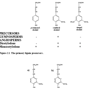
Lignin is formed by polymerization of p-hydroxycinnamyl alcohol units. Figure 2.1
depicts the three primary precursors of lignin, p-coumaryl alcohol, coniferyl alcohol, and
sinapyl alcohol. As a result, lignin is an aromatic biopolymer with propane sidechains which
the labelling of the aromatic and sidechain carbons. Structural information about lignin is
often reported on a C9 basis, indicative of this typical phenylpropanoid structure. While
wood chemists have adopted their own nomenclature, Figure 2.2(a), the more correct IUPAC labelling is shown in Figure 2.2(b). Henceforth in this manuscript the traditional wood chemistry nomenclature will be used to convey structural information. [31]
PRECURSORS
GYMNOSPERMS - + - ANGIOSPERMS
Dicotyledons - + + Monocotyledons + + +
Figure 2.1 The primary lignin precursors.
Figure 2.2 Labelling convention for monolignols, a) wood chemistry terminology and b) IUPAC nomenclature. b) a) OH OCH3 CH CH CH2OH
1 2 3 4 5 6 7 8 9 OH OCH3 CH CH CH2OH
1 2 3 4 5 6 α β γ OH CH p-coumaryl alcohol CH CH2OH
OH OCH3 CH CH CH2OH
OH OCH3 CH CH CH2OH
CH3O coniferyl
alcohol
2.2 Lignin Biosynthesis 2.2.1 Introduction
Vascular plants have the ability to absorb CO2 and water, and with the energy gained
through photosynthesis, convert these basic compounds into simple sugars. The simple
sugars can be then either be used to fuel various biosynthetic processes or stored for later use.
This process is called carbon fixation and is key in the biosynthesis of lignin. [32]
Plants convert a significant portion of the sugars produced by photosynthesis into the
phenylpropanoid precursors in lignin polymerization described in Figure 2.1. Early plants did not produce lignin and there is evidence that the evolution of these pathways was a major
step in plant evolution and played a fundamental role in the adaptation of plants to living on
land. It is likely that lignin initially functioned as an antimicrobial agent based upon the
detection of lignin in ancient algae. [5]
Sugars such as glucose are converted into the lignin precursors, the p-hydroxy
cinnamyl alcohols, by a two-step sequence of reactions known as the shikimic acid pathway
and the lignin precursor pathway. The shikimic acid pathway involves the conversion of
glucose into L-phenylalanine, an aromatic amino acid produced by all higher plants. This is
a primary metabolic process and the L-phenylalanine produced is used in many different
metabolic pathways in plants.
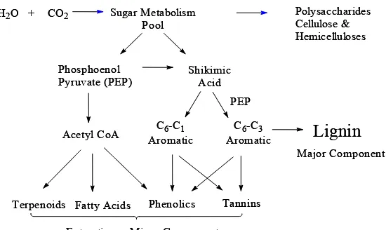
Lignin is a product of secondary metabolism and shares a common precursor,
phenylalanine, with other metabolites including flavonoids, coumarins, stilbenes, and
lignans. (Figure 2.3) At this point the pathways to the production of these different metabolites split. In the case of lignin, the L-phenylalanine is converted to the three p
Figure 2.3 Primary and secondary metabolic pathways leading to the biosynthesis of lignin and wood components.
Recently, lignin and lignification have become very active areas for research of
biological processes. Many of the new studies have involved either identification of new,
alternate pathways in lignin biosynthesis, and/or attempts to genetically manipulate lignin
structures based upon these pathways. [33-37]} New reports indicate that a facile change in
the lignin biosynthetic pathway may have a dramatic effect on lignin structure. [38]
Furthermore, manipulation may cause different monomers to be introduced into the
lignifying zone. As a result, the lignification may be a much more flexible process than
originally believed. [39-41] If this is true, there may be many interesting applications of
genetic modification of lignin structure.
H2O + CO2 Sugar Metabolism Pool
Polysaccharides Cellulose & Hemicelluloses
Phosphoenol
Pyruvate (PEP) ShikimicAcid
Acetyl CoA AromaticC6-C1 AromaticC6-C3 PEP
Terpenoids Fatty Acids Phenolics Tannins
Lignin
Extractives - Minor Components
Major Component H2O + CO2 Sugar Metabolism
Pool
Polysaccharides Cellulose & Hemicelluloses
Phosphoenol
Pyruvate (PEP) ShikimicAcid
Acetyl CoA AromaticC6-C1 AromaticC6-C3 PEP
Terpenoids Fatty Acids Phenolics Tannins
Lignin
Extractives - Minor Components
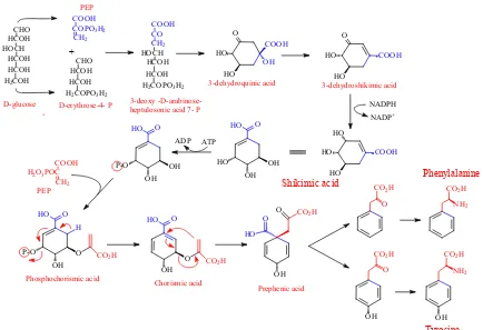
2.2.2 Shikimic Acid Pathway
Figure 2.4 Shikimic Acid Pathway.
Figure 2.4 Shikimic acid pathway.
Figure 2.4 outlines the steps in the shikimic acid pathway. The first step in the shikimic acid pathway is the formation of 3-deoxy-D-arabino-heptulosonate-7-phosphate
(DAHP) via condensation of phosphoenol pyruvate (PEP) and erythrose-4-phosphate
(ery-4-P) mediated by DAHP synthase. PEP and erythrose-4-phosphate are each formed from
glucose via the glycolytic and pentose phosphate pathways, respectively. As a result, even
though the lignin precursors will eventually be formed from glucose, the pathway is not
straightforward but rather the combination of several pathways. The glycolytic and pentose
phosphate pathways will not be discussed in detail, however, all steps of these pathways are
known and can be easily referenced. [32]
Tyrosine Phenylalanine
D-glucose
PEP
3-dehydroquinic acid 3-dehydroshikimic acid
Shikimic acid
COOH H2O3POC
CH2 O HO COOH HO OH
H2COPO3H2
CO CH2 HOCH HCOH HCOH COOH +
H2COPO3H2
CHO HCOH HCOH
COOH COPO3H2
CH2
H2COH
CHO HCOH HOCH HCOH HCOH O HO COOH HO HO HO COOH HO NADPH NADP+ HO OH OH HO O P-O OH OH HO O ATP ADP PEP P-O OH O HO O
CO2H
H
Phosphochorismic acid
OH O
HO O
CO2H
Chorismic acid OH
O
HO
O CO2H
Prephenic acid
O CO2H
OH
O CO2H
OH
NH2
CO2H
NH2
CO2H
DAHP cyclizes and is then reduced by 3-dehydroquinate synthase to 3-dehydroquinic
acid. The next step, formation of 3-dehydroshikimic acid, involves 3-dehydroquinate
dehydratase and creates one of the three double bonds that will eventually be incorporated
into the aromatic ring. Reduction of the ketone by shikimate dehydrogenase to an alcohol
produces shikimic acid. The next three steps serve to form a second double bond and to
introduce the sidechain. A phosphate group is added at the C3 position by shikimate kinase
forming shikimate-3-phosphate and a PEP group at the C5 position to form
5-enolpyruvylshikimate 3-phosphate (EPSP) by the enzyme EPSP synthase. The PEP group
will eventually be modified to become the propane sidechain. Finally, the phospo group is
eliminated by a process mediated by chorismate synthase to create the second double bond,
and the product is known as chorismate. In the field of biochemistry, chorismate is the final
step in the shikimic acid pathway as chorismate is the precursor for other metabolites such as
quinates and folates. [42] However, in the case of wood chemistry, the shikimic acid
pathway is considered to involve production of the aromatic amino acids.
The final three steps convert chorismate to L-phenylalanine. Chorismate mutase
catalyzes a rearrangement with the sidechain carbons migrating from the C5 to C1 is the first
step and produces prephenic acid. A transamination step at the carbon destined to become
Cβ, followed by a reduction involving the loss of CO2 from the C1 position and water
completes the biosynthesis of L-phenylalanine. This is a two-step process with the first step
mentioned above, glucose is not directly incorporated into the shikimic acid pathway, instead
it is incorporated through PEP and erythrose-4-phosphate. Therefore DAHP carries the
radioactive labels at C3 and C7 as the C3 in PEP and C4 in erythrose-4-phosphate are derived
from C1 and C6 in glucose. (Figure 2.4) Cyclization would then dictate that the eventual labels on the aromatic ring will be at the C2 and C6 carbons. In addition, the incorporation of
the additional PEP will introduce another radioactive label at the eventual Cα position on the
phenylpropane sidechain.
2.2.3 Biosynthetic Pathway for Primary Lignin Precursors
The shikimic acid pathway to L-phenylalanine described above is a primary
metabolic pathway. The lignin biosynthetic pathway however is an example of secondary
metabolism. L-phenylalanine is transformed in higher plants to the main lignin precursors
discussed previously through a series of enzyme-mediated reactions. It should also be noted
that some grasses also employ L-tyrosine in the lignin biosynthetic pathway, probably as a
shortcut to p-coumaryl alcohol.
Figure 2.5 outlines in red the classical biosynthetic pathway from L-phenylalanine to the main lignin precursor in gymnosperms, coniferyl alcohol. The first step is deamination
via the enzyme phenylalanine ammonia lyase to produce cinnamic acid. This is followed by a
hydroxylation at the para position on the aromatic ring. This is facilitated by the enzyme
cinnamate-4-hydroxylase (C4H) and results in the creation of p-coumaric acid. This is the
first point at which the lignin precursor pathway can branch. A series of alternating
hydroxylation and methylation steps can convert the p-coumaric acid into the mono- and
the C3 position by p-coumarate-3-hydroxylase (C3H) to form caffeic acid followed
transmethylation of the C3 hydroxyl group via O-methyl transferase (OMT) to produce
ferulic acid. This step can be basically repeated by addition of a hydroxyl group at the C5
position followed by transmethylation to produce sinapic acid. These reactions are facilitated
by ferulate-5-hydroxylase (F5H) and OMT, respectively. [5]
Figure 2.5 Biosynthetic pathway to lignin precursors. CO2H
NH2
CO2H CO2H
OH
CO2H
OHOH
COSCoA
OHOH
COSCoA
OHOCH3 CCoAOMT
CCR
CHO
OHOCH3
CH2OH
OHOCH3 CAD
CH2OH
OHOCH3 H3CO
CHO
OHOCH3 HO
CHO
OHOCH3 H3CO
AldOMT
CAD
PAL C4H C3H
Caffeoyl CoA 3 Feruloyl CoA 4 Conifer-aldehyde 5 Coniferyl alcohol 6 4CL CAld5H 5-Hydroxy-coniferaldehyde 7 Sinapyl alcohol 9 Sinap-aldehyde 8
4-Coumarate 1 Caffeate 2
CO2H
OH OCH3
COSCoA
OH
CH2OH
OHOCH3 HO
COSCoA
OHOCH3
CO2H
OHOCH3
HO
HO
COSCoA
OHOCH3
H3CO CO2H
OHOCH3
HO
4-Coumaroyl
CoA 13
?
?
5-Hydroxyferuloyl
CoA 14
Sinapoyl
CoA 15
5-Hydroxyconiferyl alcohol 20 ? CHO OHOH CHO OH
CH2OH
OH
CH2OH
OHOH ? ? ? ? ? ?
Ferulate 10 5-Hydroxyferulate
11 Sinapate 12
4-Coumar-aldehyde 16
Caffe-aldehyde 17
?
4-Coumaryl alcohol 18
catalyze the formation of feruloyl-CoA from ferulic acid. It however shows little specificity
to sinapic acid and the enzyme for formation of sinapoyl-CoA has not been identified. [45]
The recent identification of the enzyme coniferaldehyde 5-hydroxylase, which has been
shown to catalyze the hydroxylation coniferaldehyde, indicates that the lignin biosynthetic
pathway instead prefers to follow the pathway highlighted in blue in Figure 2.5 to produce sinapyl alcohol. [35-37] After hydroxylation, COMT catalyzes the methylation to form
sinapaldehyde.
The final two steps in the lignin biosynthetic pathway involve the reduction of the
various coenzyme thioesters to their cinnamyl alcohol counterparts. Cinnamoyl-CoA
reductase (CCR) catalyzes the reduction of the thioesters to the corresponding aldehydes, p
-coumaraldehyde, coniferaldehyde, and sinapaldehyde. CCR seems to show relatively the
same specificity for all hydroxycinnamyl thioesters. The last step involves the reduction of
the aldehyde to the final lignin precursors. This is achieved by cinnamyl alcohol
dehydrogenase (CAD). CAD in gymnosperms is generally more responsive to
coniferaldehyde whereas those in angiosperms react equally to coniferaldehyde and
sinapaldehyde. [46] Chiang, et al., however have recently demonstrated that sinapaldehyde
is reduced by the enzyme sinapyl alcohol dehydrogenase (SAD) in angiosperms. [37]
2.2.4 Transport of monolignol glucosides into the lignifying zone
Monolignols are not found in abundance in the lignifying zone but are found as monolignol
glucoside is cleaved in the lignifying zone by a β-glucosidase to free the phenolic hydroxyl
allowing for dehydrogenative polymerization. [47]
Figure 2.6 Monolignol glucoside formation.
OCH3
OH CH2OH
UDP HO
HO
OH O OH
HO HO
OH O OH
O
CH2OH
OCH3
+
coniferyl alcohol glucosyl transferase
Coniferin Coniferyl alcohol
2.3. Polymerization of Monolignols
In 1939, Erdtmann proposed that the lignin macromolecule is formed by a coupling
reaction of phenoxy radicals of coniferyl alcohol formed by enzymatic dehydrogenation. [48]
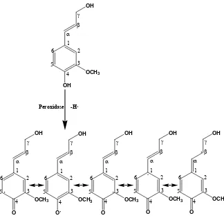
Freudenberg et al., confirmed this hypothesis and demonstrated that a system of laccase/O2
or peroxidase/H2O2 can abstract an electron from the phenolic hydroxyl of coniferyl alcohol
to form a phenoxy radical. (Figure 2.7) [49]
Figure 2.7 Dehydrogenative polymerization of lignin monomers.
This phenoxy radical has five resonance-stabilized mesomeric forms, depicted in
linkages that can be formed based upon the p-hydroxycinnamyl alcohol mesomers and the
probability of formation of these linkages depends upon sterics, electronic effects, and
solvation. [50] Radical coupling therefore occurs based upon the scheme shown in Figure 2.8. Coupling of two monomers results in the formation of a dimer, which can be further polymerized by addition of another monomer to form a trimer or another dimer to form a
tetramer. This process continues to form oligomers and ultimately the three-dimensional
lignin polymer. The most prominent of the interunit linkages in lignin are shown in Figure 2.9.
Figure 2.8 Radical Coupling in Lignin Polymerization.
Table 2.1 Relative frequency of interunit linkages reported per C9 unit in some softwood lignins.
Linkage Type Freudenberg [51]
Adler[198] Sakakibara [52]
Glasser and Glasser[53]
β-O-4’ 35 48 43
α-O-4’ 20 6-8 11
55
β-5’ 15 9-12 14 16
CH CH CH2OH
O
OCH3
CH CH CH2OH
O
OCH3
CH CH CH2OH
O
OCH3
OCH3 O
H2COH
HC CH
CH CH CH2OH
O
OCH3
OCH3 OH H2COH
HC CHOR ROH ROH = acids phenols alcohols
The relative frequencies of the most common interunit linkages in some softwood
lignins are shown in Table 2.1. Discrepancies between estimations may result from differences in species or techniques used to quantify these linkages.
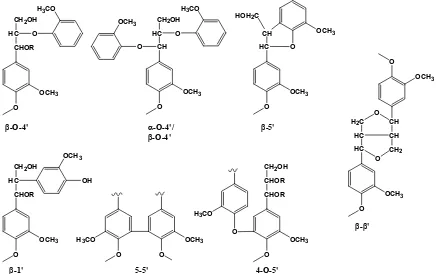
O
OCH3
CHOR HC
CH2OH
O H3CO
O
OCH3
CH HC
CH2OH
O H3CO
O OCH3 O OCH3 HC CH HOH2C
OCH3 O O OCH3 CHOR HC
CH2OH
OH OCH3
O
OCH3
O H3CO
O
OCH3
CHOR CHOR CH2OH
O
H3CO O
OCH3
HC HC H2C
O OCH3 CH CH CH2 O O
β-O-4' α-O-4'/
β-O-4'
β-5'
β-1' 5-5' 4-O-5'
β-β'
Figure 2.9 Prominent interunit linkages in lignin.
2.3.1 Classification of Lignin
As discussed in the previous section, the lignin polymer is formed from
polymerization of the three lignin precursors depicted in Figure 2.1, further contributing to the complexity of the lignin polymer. The types of lignin are classified based upon their
monolignol origin, i.e., lignin arising from primarily p-coumaryl alcohol are referred to as p
-hydroxyphenyl (H) units, guaiacyl (G) units from coniferyl alcohol, and syringyl (S) units
obvious; H units contain zero methoxyl groups, G units contain one methoxyl at the C3
position, and S units 2 methoxyl groups at C3 and C5. While these differences may seem
minor, they provide a dramatic difference in lignin polymer structure based on probability of
interunit linkages.
Figure 2.1 also reveals some of the differences in the heterogeneity in lignins from different plant sources. Gymnosperms (softwoods) are comprised of primarily guaiacyl-type
lignin with only small amounts of H and S units. A typical structural model for a typical
softwood is given in Figure2.10.
small amounts of p-hydroxyphenyl lignin. Table 2.2 shows the relative differences in distribution of interunit linkages between lignins from spruce (softwood) and birch
(hardwood). As can be seen there are higher levels of uncondensed, etherified structures
such as β-O-4’ linkages and lower levels of condensed structures, i.e., 5-5’ and β-5’, in
hardwoods. This is directly attributable to the increased C5 methoxyl units in syringyl units.
During polymerization, this site is not readily available for radical coupling and as a result a
higher incidence of particularly β-O-4’ linkages are observed.
Table 2.2 Frequency of interunit linkages per C9 unit for spruce and birch wood lignins.[31]
β-O-4’ α-O-4’ β-5’ β-1’ 5-5’ 4-O-5’ β-β’
Spruce 48 6-8 9-12 7 9.5-11 3.5-4 2
Birch 60 6-8 6 7 4.5 6.5 3
2.3.2 The Ultrastructure of Wood
Wood consists mainly of cellulose, hemicellulose, and lignin. The cellulose is
composed of microfibrils and the hemicellulose and lignin are deposited in the spaces
between the microfibrils. Cellulose contains both crystalline and amorphous regions while
the hemicellulose and lignin are amorphous.
2.3.3 Distribution of Components in the Cell Wall
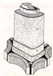
The cell wall consists of several layers which are depicted in the classical
representation in Figure 2.11. The layers of the cell wall from outer to inner are as follows: middle lamella (ML), the primary wall, the secondary wall (divided into the S1, S2, and S3
based on the thickness of the cell wall layer and the microfibril angle. Both of these aspects
will be discussed below.
Figure 2.11 Structure of the cell wall in wood showing the middle lamella (ML), the primary wall, the outer (S1), middle (S2), and inner layers (S3).[29]
As the cell divides it initially forms a cell plate, which is made of primarily pectic
substances such as 1,4-α-polygalacturonic acid. The new cells enclose upon themselves to
form primary walls. Next the cells grow to their final size after which the secondary wall
begins to thicken. As the secondary wall forms, deposition of cellulose and hemicellulose
begins first in the middle lamella and primary wall. While the secondary wall is still
thickening, cellulose and hemicellulose deposition begins in the S1 layer. Lignification also
begins in the cell corners and middle lamella and then proceeding to the primary wall.
Lignification begins in the S1 layer before cellulose deposition is complete in the S3 layer.
result the middle lamella and cell corners contain only 19-28 % of the total lignin in
gymnosperms.
Table 2.3 Distribution of lignin in spruce tracheids. [29, 54]
Morphological
Origin Tissue Volume(%) (% of total) Lignin Lignin Conc. (%)
S 87 72 23
ML 9 16 50
Earlywood
CC 4 12 85
S 94 82 22
ML 4 10 60
Latewood
CC 2 9 100
The primary wall consists of cellulose, hemicellulose, proteins, and pectic substances
embedded in lignin. Although this region of the cell wall contains lignin, it is a very thin
layer, ~0.1-0.2 µm thickness, and therefore contains only a minor portion of the total lignin.
The cellulose microfibrils are randomly oriented in the outer portion of the primary wall but
are nearly perpendicular to the axis of the cell wall in the inner portion.
The majority of the tissue volume is contained in the secondary wall although the
relative concentration of lignin is low. Nevertheless, 72-82 % of the lignin is contained in
the secondary wall. The secondary wall is comprised of three layers: the thick S2 (middle)
layer, which is 1-5 µm in thickness from early to latewood, and the thin S1 (0.2-0.3 µm
thickness) and S3 (~0.1 µm thickness) layers.
Within the cell wall, the cellulose microfibrils are oriented helically around the cell
wall axis. The microfibril angle in the S2 layer is 5 o-10 o, while in the S1 and S3 layers it is
2.4. Different Methods of Lignin Isolation
As discussed in Section 2.3.3, cellulose is deposited first in each layer of the cell wall, followed by hemicellulose, and finally lignin is deposited. As a result the cellulose is
surrounded by a matrix of hemicellulose and lignin, with the latter likely forming some type
of chemical bonds. [4] These components form a tight association in the cell wall, making
their separation difficult. Further enhancing this difficulty is the fact that the major
constituent, cellulose, exists in regions as a highly crystalline material. Due to these
crystalline regions and the high molecular weight of the lignin material, efforts to extract
lignin from whole wood without alteration of structure have met with little success. As a
result it is impossible to completely isolate native lignin as its whole component from wood.
Initially, methods involving acid hydrolysis were performed in an attempt to isolate
lignin from wood. It was soon realized that the acid lignin differed from lignin in wood based
upon its yield from chemical degradation methods such as nitrobenzene oxidation.
Brauns lignin is extracted with 95 % ethanol from wood meal ground to ~100 mesh
and can be isolated in an unaltered form. [55] The yields however are very low, the
preparation contains some carbohydrates and may be contaminated with extractives. The
lignin extracted therefore cannot be considered representative of lignin in wood.
Nord, et al., attempted to isolate lignin by degrading the carbohydrate material with
brown rot fungi. [56, 57] It was determined that the amount of Brauns lignin that could be
isolatable with ethanol. This is evidenced by Brauns admission that even several days of ball
milling would not fragment the lignin polymer to the point that additional lignin could
extracted. [58] Pew proved this point by showing that only 13 % of the total lignin could be
isolated by treatment of a spruce with brown rot even after only 6 % of the carbohydrates
remain. [59]
2.4.1 Isolation of MWL by the Bjorkman Method
Bjorkman first reported in 1954 on “the isolation of lignin from finely divided wood
meal by extraction with neutral solvents”. [60] A significant portion of the lignin could be
extracted and the yield was based upon the extent of milling.
Based upon the previously reported literature which described the isolation of lignin
only by drastic conditions, such as acid or alkaline treatments, Bjorkman hypothesized:
1. Strong chemical bonds exist between lignin and carbohydrates.
2. Lignin has a high molecular weight and forms a three-dimensional network in
wood.
3. Physical phenomena and hydrogen bonding are involved in the retention of lignin
within wood. [61]
Bjorkman suspected that the third possibility played an important role in this problem and
upon this he devised his experimental plan. In reality, it is likely a combination of the three
2.4.2 Theory on Milling
It has been reported that the degree of polymerization (DP) of cellulose decreases
rapidly after only a few hours of milling and that lattice deformation leads rapidly to the
formation of amorphous cellulose. [62, Lai, 1971 #199] Mobility of individual portions of
the macromolecule allows for lattice deformation, whereas immobility of the macromolecule
as a whole leads to degradation. The drop in DP is due to hydrolysis of the glycosidic
linkages rather than oxidative degradation of the cellulose molecule. Additionally, cellulose
and other high molecular weight materials have DP limits upon where no further degradation
can occur. This limit is lower for linear polymers such as cellulose when compared to more
three-dimensional polymers such as lignin. When the particle size is sufficiently small that
molecular movement is no longer restricted, breakage of bonds will cease. [9]
Materials such as lignin will tend to cake with decreasing particle size if milled dry,
therefore it is advisable to utilize wet milling. It is probable that caking limits the minimum
achievable particle size in dry milling by agglomeration of smaller particles. Milling reaches
a “dynamic equilibrium” where no further division of the original material will occur.
Finally, even at this equilibrium point there will always be a “tail” of higher particle size. [9]
2.4.3 Milling Parameters
Bjorkman observed that milled wood would tend to coat the steel balls in the
accumulation of temperature is decreased. Additionally it has been reported that the toluene
will adsorb to the surface of wood particles and act as a radical scavenger thereby reducing
the dramatic impact of the vibratory ball mill. [10, 63] This may help to reduce the amount
of modification occurring in the lignin structure.
In Bjorkman’s original proposal for a standard grinding method, he uses a rotary ball
mill to grind 12 g of wood for 2-3 days and then 6 g for two days in the vibratory ball mill
with yields in the range of 50 %. In fact, he asserts that a two-step milling process seems to
produce an increase in yield. [61] However in the manuscript on the effect of milling he uses
only a vibratory mill and reports that the highest yield (46 %) was obtained with 1 g milled
over the course of two days. [9] An additional 12 days of milling led to only to a 7 %
increase in the yield of MWL.
It is evident that the yield obtained will depend on the milling method, the size and
efficiency of the mill(s), the amount of wood loaded, and the total milling time. As a result,
due to trial and error the milling method and yield can be expected to vary based on
operations at different facilities. For example, a yield of 28 % using only a vibratory ball
mill was reported after 9 days of milling. [64] Glasser et al., reported only a 9.2 % of MWL
after 13 days of rotary ball milling with porcelains balls [65] however Brownell reported
yields similar to Bjorkman after 2-3 weeks of rotary ball milling with flint pebbles. [66, 67]
Furthermore, new orbital mills are being used with more frequency and these mills can
produce a finely milled lignin in a matter hours [68] or even tens of minutes [69]. One
author noted that the mill being used was “more violent than the vibratory ball mill used by
This calls into question the impact of the milling method on the lignin structure.
Therefore in order to quantitatively compare lignins produced by different preparation
methods, it is essential to compare lignins with similar yields.
2.4.4 Extraction and Purification of Milled Wood Lignin
The choice of extraction solvent is not as critical as the milling conditions. Methyl
cellosolve is the best solvent for extraction of lignin from finely divided wood meal,
however, it is not recommended because of the difficulty in completely removing it. A
mixture of 1,2-dichloroethane:ethanol dissolves pure lignin well, but is not suitable for
extraction purposes. Aqueous dioxane was chosen because it extracts low molecular weight
lignin well and it does not extract much carbohydrate.[61]
The dioxane solution containing the dissolved lignin is evaporated under reduced
pressure and 50-60 oC in a heating bath. The concentrated lignin is dispersed in 90 % acetic
acid at a concentration of 50 mg/mL and precipitated into deionized water (1 g/ 250 mL H2O)
to remove impurities such as tannins and some LCCs. The lignin is centrifuged, the
supernatant discarded and the lignin washed with deionized water. Bjorkman evaporated the
remaining water by flowing a rapid air stream over the surface. [61] It is now typical to
freeze-dry the lignin after this step but there have been reports that this changes the solubility
parameters of lignin. [70]
2.4.5 Properties of Lignin Extracted with Dioxane
Table 2.4 lists elemental and functional group analyses as determined by Bjorkman. [71] It was determined that milling in the presence of air resulted in a small increase of
carbonyl content and a decrease in p-hydroxybenzyl alcohol moieties, which represent the α
-hydroxy content. No change in phenolic -hydroxyl content was observed. Weight average
molecular weight (Mw) was determined to be 11,000 and the density of the MWL determined
to be 1.406 g/mL.
Table 2.4 Bjorkman’s estimation of the chemical properties of MWL.[71]
Chemical Formula Phenolic
OH phydroxybenzyl -alcohol
Total
OH C=O Arylalkyl Ether Dialkyl Ether
MWL C9H8.83O2.37(OCH3)0.96 0.30 0.05 0.80 0.18 0.70 0.84
2.4.6 Isolation of Lignin (CEL) by Treatment with Cellulolytic Enzymes
Pew was the first to experiment with enzyme digestion of vibratory ball milled wood.
[59, 72] He observed that solid wood or Wiley-milled wood ground to pass a 20 mesh was
not susceptible to treatment with enzymes. However wood ground for just 10 minutes
allowed for enzyme digestion of up to two-thirds of the carbohydrate in two successive three
day treatments. An additional 14 treatments did not decrease the carbohydrate appreciably.
5 hours of vibratory ball milling would allow for 95 % of the carbohydrate to be removed
and resulted in a lignin containing 12-14 % carbohydrate. 8 hours of vibratory ball milling
allowed for only 96 % of the carbohydrate to be removed. Only limited analysis of these
Chang et al., ball milled spruce in a vibratory ball mill for forty-eight hours in the
presence of toluene. [73] The milled wood was extracted with 96 % (v/v) dioxane to obtain
MWL (17.4 % yield) and then subjected to treatment with a cellulase mixture. Subsequent
96 % (v/v) dioxane extraction yielded an additional 43.1 % of CEL-96. The insoluble
material was then extracted with 50 % (v/v) dioxane yielding 24.9 % of CEL-50 for a total
yield of 95 %. MWL and CEL-96 were very similar based on elemental analysis. The 50 %
(v/v) dioxane fraction however contained twice as much carbohydrates and was much higher
in molecular weight (Mw). (Table 2.5)
Table 2.5. Data presented for MWL, CEL-96, and CEL-50. [73]
Mw Carbonyl Content
per 100 C9 Units
Phenolic OH per 100 C9 Units
KMnO4
Oxidation (mol %)
Nitrobenzene Oxidation
(wt. %)
MWL 16000 0.11 14.4 11.6 37.3
CEL-96 24000 0.08 13.1 10.1 41.4
CEL-50 35000 0.05 8.9 8.1 38.4
Table 2.5 summarizes the data reported by Chang et al., for MWL, CEL-96, and CEL-50. α-carbonyl content was estimated by UV at 305 nm and decreased from MWL to
CEL-96 to CEL-50. Phenolic hydroxyl content followed the same trend as carbonyl content
consistent with the changes in Mw.
Permanganate oxidation yields mirrored phenolic hydroxyl content consistent with
oxidation for CEL-96 was obtained supporting the theory that MWL is more condensed than
CEL-96. Both yields were lower than those for the milled wood, however.
2.4.7 Mechanochemical Reactions and Changes in Lignin Structure During Milling
In order to isolate lignin from wood by solvent extraction, the crystallinity of the
cellulose must be interrupted and the DP decreased. Pew isolated lignin by milling 1 and 5
hours and treating with cellulase to achieve comparable yields. [59] The lignin milled only 1
hour was insoluble in the same solvents in which the lignin milled 5 hours was soluble. This
proves that adequate depolymerization of the lignin macromolecule is necessary to achieve
solubility.
It has been shown that the energy available in the vibratory ball-milling of cellulose is
sufficient to rupture not only glycosidic bonds but also covalent bonds. [10-12] Furthermore,
most organic substances can be decomposed by processes initiated by mechanical and/or
thermal means involving radical processes. [74] It therefore follows that lignin with a rigid
structure containing many ether bonds and a highly conjugated system able to stabilize
radicals once formed, will likely undergo radical decomposition. In addition it is possible
that benzaldehyde-type structures found in MWL may be artifacts from the milling process.
While the existence of free radicals in solid wood is in dispute [12, 74, 75], the fact
they are formed a short time after milling has commenced is indisputable. By electron spin
resonance spectroscopy (ESR), the concentration of free radicals has been shown to increase
20-fold shortly after commencement of milling [76] with concentration increasing with
smaller particle size. [74] All isolated lignin samples will contain free radicals, even after
Milling of cellulose by itself results in only a very weak signal by ESR, indicating
that either radicals are not formed in the milling of cellulose or more likely they dissipate
through secondary reactions rapidly. [77] Lignin or wood meal, on the other hand, exhibit a
signal at least 50 times that of cellulose. (Figure 2.12) This is not surprising based upon the lignin monomer structure and its ability to stabilize a radical through resonance
delocalization. As a result, it is obvious that the free radicals measured originate from the
lignin component of the milled wood. [10]
Data indicates that the formation of free radicals increases rapidly during the initial phases of
milling, reaches a maximum, and then levels off. (Figure 2.13) Presumably the increase of radicals is indicative of ether bond cleavage. The unstable radicals will react leaving the
stable radicals which are pronounced after equilibrium is reached. It is possible that the
stable radicals are predominately phenoxy radicals since they are able to delocalize the
radical throughout the aromatic ring. It is also possible that these radicals are stabilized in
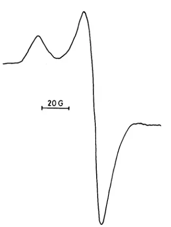
structures with limited mobility and hence have little opportunity to be involved in other
Figure 2.12 ESR spectra of a) cellulose, b) MWL, and c) wood meal, milled with glass beads for three hours in N2 with glass beads. Multiplication number represents signal intensity.[10]
Figure 2.13 Free radicals in wood meal milled with steel balls in CO2. [10]
Additionally, at day 6 when the free radicals level comes to steady-state may coincide
wood, etc., so that this is only an example of the accumulation of free radicals which will
vary from operation to operation.
2.4.8 Depolymerization of the Lignin Macromolecule by Ether Bond Cleavage
As a result, isolation of lignin involves depolymerization which lowers molecular
weight and in turn increases phenolic hydroxyl content. [73] It is evident that the cleavage of
β-O-4’ bonds (Figure 2.14) is the predominant reaction involved in the depolymerization reactions that occur during milling. According to this reaction, the decrease in molecular
weight and subsequent formation of a new phenolic hydroxyl group is achieved. Although
there is also enough energy present to rupture covalent bonds, depolymerization by this
mechanism is probably minimal. While there is probably formation and decay of free radicals
occurring throughout the milling process, it is likely that formation of radicals involves β
-O-4’ cleavage while decay involves processes such as condensation reactions, generation of
unsaturated lignin structures, and possibly the formation of o- and p-quinoid structures.
2.4.9 Formation of Carbonyl and Unsaturated Side Chain Structures
Hon has reported that peroxy radicals will form in toluene even in the absence of
oxygen. [10] He states that oxygen will likely be present as a contaminant from one of the
components used in the milling. In wood milled in toluene at 77 K these peroxy radicals are
readily detected (Figure 2.15) but at room temperature they will react very quickly.
Molecular oxygen can react directly with a benzyl radical to form a peroxy radical as
shown in Figure 2.16. The peroxy radical is unstable at the temperatures involved in ambient ball milling, and hence will decompose to the α-carbonyl structure. The formation
of coniferaldehyde is less likely to be involved in this type of process as it would involve an
unstable primary alkyl radical intermediate. Therefore it is likely that coniferaldehyde is
incorporated into the macromolecule a monomeric precursor in the dehydrogenative
polymerization process.
Figure 2.16 Formation of the α-carbonyl structure during milling.
Likewise coniferyl alcohol can be created by a similar process involving the
secondary alkyl radical formed from β-aryl ether cleavage. (Figure 2.17) Although this is a secondary alkyl radical, it can be somewhat stabilized due to delocalization. Even though β
-O-4’ cleavage is the predominant reaction occurring in the milling process, the relative
proportion of coniferyl alcohol moieties in MWL is rather small. It is likely that
delocalization of the secondary radical results in the ultimate stabilization of the radical as
the phenoxy radical.
Figure 2.17 Formation of coniferyl alcohol during milling of wood.
C C R HO OH OCH3 O2 C C R HO OH OCH3 OO C C R OH OCH3 O ∆ C C R HO OH OCH3 O2 C C R HO OH OCH3 CH CH R OH OCH3 ∆ OO
2.4.10 Possible Formation of Quinoid Structures During Milling
Hydroxy radicals have been shown to react with lignin aromatic moieties to form o-
and p-quinoid structures.[78] Possible mechanisms for these reactions are shown in Figure 2.18. Zhang et al., recently identified a number of quinoid structures present in MWL preparations according to two-dimensional HMQC NMR. [79] Although these moieties are
minor components, they may be indicative of structural changes occurring during milling.
Figure 2.18 Possible mechanism for the formation of o- and p-quinoid structures during milling.
2.4.11 Condensation Reactions Occurring During Milling
Crosslinking reactions would be most likely to occur as particle size decreases and
group achieve greater mobility. This is most likely to coincide with the decrease from the
maximum of free radicals in Figure 2.13 and as milling proceeds. Condensation reactions are believed to most likely follow the mechanism shown in Figure 2.19.
R
O
OCH3
R
O
OCH3 R
O
OCH3 HO
OH
R
O O
o-quinoid
R
O
OCH3
R
O
OCH3 O
OCH3 HO
O
O
OCH3
p-quinoid
![Figure 2.10 A structural model for softwood. [52]](https://thumb-us.123doks.com/thumbv2/123dok_us/1534479.1188152/34.612.165.435.324.609/figure-structural-model-softwood.webp)
![Figure 2.13 Free radicals in wood meal milled with steel balls in CO2. [10]](https://thumb-us.123doks.com/thumbv2/123dok_us/1534479.1188152/47.612.232.393.69.302/figure-free-radicals-wood-meal-milled-steel-balls.webp)