ABSTRACT
CARLIN, KEVIN BRIAN. Engineering Multivalent Protein Affinity Ligands using the
Sso7d Scaffold. (Under the direction of Balaji M. Rao).
Protein affinity ligands have applications in diagnostics, therapeutics and as research tools for probing biological systems. By specifically binding a given target a ligand is useful for detecting that target, blocking a given interaction or useful as non-perturbing biosensors which can monitor the state of a protein in a given system. There are established methods for generating affinity ligands which are continually being refined and improved through various means. However, systematically generating bivalency or multivalency for generating high affinity binding proteins is a potentially very powerful but underutilized approach.
We show that a combinatorial library constructed by random pairwise assembly of low affinity binders can efficiently generate binders with increased affinity. Such a library, from a pool of low affinity binders based on the Sso7d scaffold, contained putative high affinity clones for a model target (lysozyme) at higher frequency than a library of monovalent mutants with equivalent diversity (~ 107). Increased binding affinity was due to intramolecular avidity generated by linking binders targeting non-overlapping epitopes; individual binders of KD ~ 1.3 M and 250 nM produced a bivalent binder with KD ~ 1.7 nM. Furthermore, the bivalent protein retained thermal stability (TM= 84.5 °C) and high recombinant expression yields in E. coli. Finally, when binders comprising the bivalent protein are fused to two of the three fragments of tripartite split-green fluorescent protein (GFP), target-dependent reconstitution of fluorescence occurs, thereby enabling a “mix-and-read” assay for target quantification.
Engineering Multivalent Protein Affinity Ligands using the Sso7d Scaffold
by
Kevin Brian Carlin
A dissertation submitted to the Graduate Faculty of
North Carolina State University
in partial fulfillment of the
requirements for the degree of
Doctor of Philosophy
Chemical Engineering
Raleigh, North Carolina
2016
APPROVED BY:
_______________________________
_______________________________
Dr. Balaji M. Rao
Dr. Jason M. Haugh
Committee Chair
_______________________________
_______________________________
ii
BIOGRAPHY
iii
ACKNOWLEDGMENTS
iv
TABLE OF CONTENTS
LIST OF FIGURES ... 8
LIST OF TABLES ... 11
BACKGROUND AND MOTIVATION FOR ENGINEERING AFFINITY LIGANDS ... 1
1.1 Introduction ... 2
1.1.1 Protein Engineering ... 5
1.1.2 Engineering High Affinity ligands ... 8
1.1.3 Engineering Multivalent Affinity Ligands ... 10
1.1.4 Bivalent ligands as tools for manipulating biological phenomenona ... 15
1.2 Thesis Overview ... 16
1.3 Figures ... 18
COMBINATORIAL PAIRWISE ASSEMBLY EFFICIENTLY GENERATES HIGH AFFINITY BINDERS AND ENABLES A “MIX-AND-READ” DETECTION SCHEME. ... 23
2.1 Introduction ... 24
2.2 Results ... 26
2.2.1 A bivalent library yields a higher frequency of putative high affinity clones. ... 26
2.2.2 Magnetic sorting and FACS identifies a pool of bivalent lysozyme binders with highest affinity. ... 28
v
2.2.4 Binding of NTL-1 and CTL-1 to non-overlapping epitopes on the target can be
exploited to design a “mix-and-read” assay for target quantification. ... 31
2.3 Materials and Methods ... 32
2.3.1 Library construction. ... 32
2.3.2 Comparison of monovalent and bivalent libraries. ... 33
2.3.3 Selection of the highest affinity bivalent ligands ... 34
2.3.4 Simultaneous Yeast Surface Display of CTL-1 and NTL-1 (Dual Display) ... 35
2.3.5 Recombinant expression of BVL-1. ... 35
2.3.6 Estimation of KD ... 36
2.3.7 Size Exclusion chromatography ... 37
2.3.8 Measurement of the Melting Temperature TM. ... 37
2.3.9 Expression and purification of GFP1-9, NTL-GFP11 and GFP10-CTL-1. ... 37
2.3.10 In vitro “mix and read” fluorescence reconstitution assays. ... 38
2.4 Figures ... 39
FUNCTIONAL DIMERIC SSO7D LIGANDS AND EPITOPE ADDITION AS A MODE OF AFFINITY MATURATION IN BINDING INTRINSICALLY DISORDERED DOMAINS ... 48
3.1 Introduction ... 49
3.2 Results ... 53
vi
3.2.2 The first generation tandem library assembled well and yielded high affinity ligands 54 3.2.3 A diversified polypeptide chain was enriched during selections for -catenin binding.
55
3.2.4 Affinity maturation of T2 yields a diversity of advantageous mutations. ... 57
3.2.5 Selection of functional dimeric ligands ... 58
3.2.6 Measurement of soluble phase KDs ... 62
3.2.7 Differential binding observed for ligands to CP peptide sequence alone and in the context of the C-terminal IDR ... 64
3.2.8 ARM Domain Binding ... 67
3.2.9 No binding to β-catenin N-terminal IDR ... 68
3.2.10 Dimerization of T2 and T3 is required for 23CTL functionality. ... 69
3.3 Discussion ... 70
3.4 Conclusions ... 78
3.5 Materials and Methods ... 79
3.5.1 β-catenin Target Expression ... 79
3.5.2 β-catenin constructs cloning ... 80
3.5.3 GST-TCF Expression ... 80
3.5.4 Generation of the peptide pre-targeted library and selection of the M2 ligand. ... 80
3.5.5 Generation of the Tandem Library ... 82
vii
3.5.7 Assembly of the second generation tandem library ... 83
3.5.8 Ligand cloning into bacterial expression vectors ... 85
3.5.9 Blitz Experiments ... 86
3.5.10 KD Measurements ... 87
3.6 Figures and Tables ... 88
CONCLUSIONS AND FUTURE WORK ... 118
4.1 Figures and Tables ... 127
Appendices ... 132
2. Appendix A1 ... 132
3. Appendix A2 ... 135
4. Appendix A3 ... 144
viii
LIST OF FIGURES
Figure 1.1: Flowchart of protein engineering by yeast surface display. ... 18
Figure 1.2: Chelating recombinant ligands generate bivalency regardless of target context ... 21
Figure 1.3: Estimated enhancement for linking ligands ... 21
Figure 2.1: A bivalent library yields a higher frequency of putative high affinity clones... 39
Figure 2.2: Analysis of nine clones from the highest affinity lysozyme ligands selected. ... 40
Figure 2.3: High affinity of bivalent binder BVL-1 is due to synergistic binding of low affinity NTL-1 and CTL-1 subunits. ... 41
Figure 2.4: Size exclusion chromatograms indicating BVL-1 is monomeric. ... 42
Figure 2.5: SDS page of elution samples of the BVL-1 and lysozyme complex from SEC ... 44
Figure 2.6: Differential scanning fluorimetry for estimation of TM. ... 44
Figure 2.7: Binding of NTL-1 and CTL-1 to non-overlapping epitopes on the target can be exploited to design a “mix-and-read” assay for target quantification. ... 45
Figure 2.8: Comparison of monovalent and bivalent libraries by cytometry. ... 47
Figure 3.1: M1 and M2 differ by an L54W mutation. ... 88
Figure 3.2: Generation of a tandem library with M1.2 as the N-terminal ligand. ... 90
Figure 3.3: Generation of the T2 tandem library ... 91
Figure 3.4: PDB diagram of mutations observed in T3-NTL. ... 93
Figure 3.5: SDS page gels indicating M2 and M1 exist predominantly as disulfide bonded homodimers ... 94
Figure 3.6: SDS page gels demonstrating T2, T3 and T3-NTL also exist as disulfide bonded dimers. ... 95
ix
Figure 3.8: Biolayer interferometry data shows the dimeric form of the ligand is functional. ... 98
Figure 3.9: Calculated dissociation rate koff and KD for reduced (1 mM TCEP) and non-reduced M1 based on data recorded on the BlitzTM. ... 99
Figure 3.10: Titration and competition based KD curves for yeast displayed ligands to full length soluble β-catenin... 101
Figure 3.11: Multimodal binding observed in yeast based KD measuremnts. ... 102
Figure 3.12: KD curves for yeast displayed ligands to soluble βCP. ... 105
Figure 3.13: Cytometry histograms for ligands binding the C-terminal IDR. ... 107
Figure 3.14: KD curves for soluble M2 and T3 to yeast displayed C-terminal IDR (666-781). ... 108
Figure 3.15: Yeast surface display of the ARM domain, expression controls and binding by ligands. ... 109
Figure 3.16: Titration based KD’s curves for soluble ligands binding the yeast displayed ARM domain. ... 112
Figure 3.17: Expression level of N-terminal IDR’s on yeast and binding by soluble ligands. ... 112
Figure 3.18: 23CTL requires avidity ... 113
Figure 3.19: Diagrams depicting how monomeric or dimeric M2 may interact with -catenin immobilized on a Blitz sensor. ... 114
Figure 3.20: Proposed binding mechanisms of the ligands engineered in this work to β-catenin. ... 116
Figure 4.1: A sensor for ligand dimerization in the yeast surface display system. ... 127
Figure 4.2: An alternative co-localization sensor using renilla GFP and PLC1-SH2. ... 129
Figure 4.3: A FRET based sort for heterodimeric self-associating ligands. ... 131
Figure A2.1 Biolayer interferometry data recorded for non-reduced M1. ... 136
x
Figure A2.3 Biolayer interferometry data for reduced M1... 138
Figure A2.4 Analysis of biolayer interferometry data for reduced M1. ... 140
Figure A2.5 Biolayer interferometry data and analysis for non-reduced M2. ... 141
Figure A2.6 biolayer interferometry data and analysis for reduced M2. ... 142
xi
LIST OF TABLES
1
CHAPTER 1
2
1.1
Introduction
Engineering protein affinity ligands is a discipline of generating affinity to a target
protein (P) by another protein, referred to as a ligand (L), forming a complex (C). This is
written as a reversible chemical reaction
𝑃 + 𝐿 ⇋ 𝐶
First order kinetics are observed for both the forward reaction, which is referred to as
the association or on rate and the reverse reaction, referred to as the dissociation or off rate. By
convention the constant K
Ddefines this interaction:
[𝑃]𝑒𝑞[𝐿]𝑒𝑞
[𝐶]𝑒𝑞
=𝑘𝑜𝑓𝑓 𝑘𝑜𝑛
≡ 𝐾𝐷
K
Dis a useful term because it is the concentration at which binding is relevant for a
given system. For instance, many diagnostic applications immobilize a ligand on a surface and
then measure the binding of a target protein to that ligand. There are a great variety of methods
through which this binding can be measured, e.g. surface plasma resonance (SPR),
interferometry, sandwich enzyme-linked immunosorbent assay (ELISA), as well as many
non-commercialized techniques which have been developed in published studies
1–3. For each of
these methods the fractional binding of the target (P
o) onto the surface-loaded ligand can be
estimated in cases where the amount bound is small relative to the amount in solution, by the
following equation:
3
If K
Dis much greater then P
othe fraction of the total possible amount of target that can
be bound by the ligand is low. One method of increasing the signal is to engineer a K
Dto be in
the range of or lower than the concentration of the protein target of interest in the sample
mixture. Decreasing detection limits is therefore one motivation for desiring a low K
D. An
alternative motivation to engineer high affinity is in the context of binding a target of interest
in a specific manner such that it is not readily displaced. Such ligands can act as inhibitors of
protein-protein interactions and used as tools to study biological processes
4.
The specific binding of a ligand to a target is referred to as molecular recognition; it is
characterized by low values of K
Dfor the target-specific interaction, relative to other
interactions with non-target species. In case of protein-protein interactions, molecular
recognition is mediated by interactions between amino acid residues of the ligand and target at
the binding interface, including electrostatic, hydrophobic or Van der Waals interactions. In a
survey of 15 different protease inhibitors and antibody complexes it was found that the binding
interface consisted of 34 ±7 amino acids on the ligand
5. Typically specific, high affinity
interactions require many residues on the ligand to be placed in a very exact manner relative
to the target in order to generate recognition.
4
5
Recent publications have been directed at engineering affinity ligands using the Sso7d
protein as a scaffold
13–15. Sso7d is a DNA binding protein from Sulfolobus solfataricus. It
consists of an incomplete beta-barrel with five beta strands and a C-terminal alpha helix
16. The
protein has 63 residues (~7 kDa), has a very high melting temperature (100
C), does not require
disulfide bond formation and expresses well in
E. coli
16. Sso7d is a highly stable form of an
SH3 domain, a natural peptide binding domain found in nature
15–17. Other homologous of
Sso7d bind metal ions, oligosaccharides, nucleic acids and other proteins
15. Therefore, efforts
have been made to establish techniques and methods for selecting high affinity ligands using
Sso7d or the highly homologous Sac7d domain. Initial studies focused on selecting affinity
ligands from a library comprised of mutation of surface exposed residues on the 3 β-sheet face
of the scaffold
13,15. Moderate to high affinity ligands were selected from the naïve library with
little to no loss of thermostability or stability losses due to pH environment or denaturants
13.
This was observed for each of six ligands studied in detail
13. Alternative libraries have been
designed for Sac7d and were successfully used to generate affinity ligands
18. This thesis builds
on these past studies with Sso7d. Techniques to preserve scaffold stability and generate affinity
more expediently using combinatorial methods to generate bivalency are explored. A study is
also done to bind an intrinsically disordered domain. Observations are made as to how these
regions can be pre-targeted and to study mechanisms of affinity maturation for these targets.
1.1.1 Protein Engineering
6
in the region of those mutations. This DNA is inserted into some type of display cassette which
strongly links the DNA to the translated protein. Five display methods are predominately used
in the literature for selection of affinity ligands: phage, yeast, bacterial, ribosome or the very
similar mRNA display system
19. The first three methods are cell based, they incorporate the
DNA for one library member into one cell, and that one library mutant protein is displayed on
the cell surface. The library is comprised of a large collection of mutant expressing cells 10
8to 10
11library members are possible depending on the display system
19. Ribosome display and
RNA display are cell-free systems; for each method RNA is transcribed and directly linked to
the translated protein. To accomplish this translation machinery from prokaryotic or eukaryotic
cells is extracted and used with DNA coding for the library to create the translated protein
library
20. Diversities of 10
12are possible for cell-free systems
20.
7
the library generation steps for RNA display are more cumbersome. This is an important factor
as for the RNA system the library has to be re-created after each selection; whereas, for the
yeast based system selected clones are simply amplified in selective media at yeasts natural
growth rate.
8
surface display plasmid, referred herein as pCTCON, has a tryptophan marker and is
maintained in yeast using casamino acids. Dextrose can be used to expand the yeast population
and a galactose buffer is used to induce protein expression.
Display technologies provide methods through which extremely large pool of mutants
can be generated; selections for productive binding mutants can be carried out, amplified and
further diversified. Typically, initial selections from a naïve library are carried out to capture
low and high affinity ligands
20,28. Generally after enrichment the selected library is
re-diversified through an error prone PCR step. For yeast surface display and other cell based
display systems the DNA is extracted and purified from the cell and an error prone PCR is
performed (EPCR). EPCR generally incorporates about 1-5 mutations/mutant and thereby
re-diversifies the selected binders. Multiple rounds, often 4-8, consisting of enrichment followed
by re-diversification through ePCR are generally done
20,28. During these early enrichment steps
negative selections against proteins or materials used in the selection process are done as well.
Generally for the yeast display system for instance, streptavidin coated magnetic beads are
used in the positive selections to label the target of interest. Therefore streptavidin would be
used alone in negative selections to remove unwanted streptavidin binders. Figure 1.1 shows
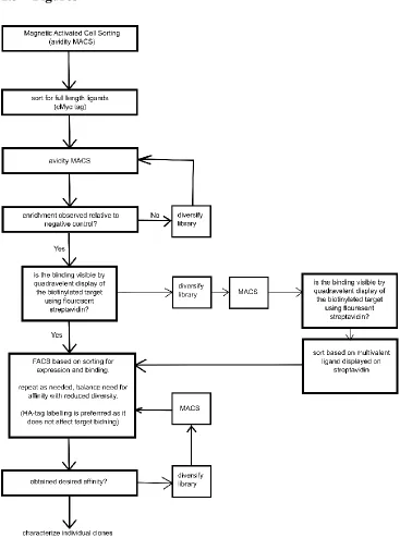
a typical flow chart of how affinity ligands to targets of interest are generated using the yeast
display system
24.
1.1.2 Engineering High Affinity ligands
9
targets that are accessible by a given scaffold and increase the probability of the selection
process yielding ligands that can be easily produced and are not prone to aggregation. An
array of computational methods is being explored toward these ends. One method uses a
scoring algorithm to find a suitable scaffold for a suitable targeted epitope based on structural
data in the Protein Database (PDB)
29. After a scaffold is selected further analysis is done in
silico to select which residues will be mutated to create a library with an optimal probability
for creating successful ligands
29. Alternatively, in silico design can be used to determine a set
of residues that will bind to a site of interest. These residues are then inserted into a scaffold
and screened for binding
4. A third approach uses in silico analysis to generate a focused
library based on target docking studies by a specific ligand
30. The in silico analysis enabled
authors to select which residues to mutate furthermore, at each residue the diversity was
reduced by further by choosing only a subset of amino acids to be evaluated at each positon.
The authors demonstrated that a focused library yielded 30 times greater enrichment over an
alternative naïve library based on saturated mutagenesis at a selected binding interface.
10
highly successful at generating high affinity ligands by panning the naïve library without
affinity maturation
31.
Techniques for improving how the affinity maturation process is carried out have also
been developed for the Fibronectin scaffold. A 0.3 pM lysozyme binding mutant was
selected after nine rounds of mutagenesis
36. Affinity in Fibronectin is generated typically
through three loop regions, in this work the authors showed that varying loop lengths and
interchanging loops during affinity maturation cycles was effective. During rounds of
mutagenesis error prone PCRs were either restricted to just the loop regions or to framework
regions. During each round two libraries were made and then mixed after transformation in
yeast. Interestingly, the loop sequences stopped evolving after the fifth round of affinity
maturation but framework mutations continued to improve the affinity of the ligand. Lastly,
in a final 9th round non-conserved mutations in the best binders were again mutated through
saturated mutagenesis. This step led to an additional threefold improvement in K
D.
1.1.3 Engineering Multivalent Affinity Ligands
11
a similar manner: a particular pair of identical binding domains and targets are present such
that multiple contacts can be made. This is termed intermolecular avidity as two or more copies
of either the target or ligand are required for the generation of affinity.
12
Eight years later a 2003 modelling study endeavored to quantify the magnitude of the
affinity enhancement that could be observed in a chelating system
40. Thermodynamic analysis
reduced the K
Dobserved in a bivalent system to this equation:
𝐾𝐷𝐴𝐵 =
𝐾𝐷𝐴𝐾𝐷𝐵
𝐴𝑒𝑓𝑓
The equation shows that the K
Dfor a bivalent system is equal to the product of the
individual K
D’s for each ligand divided by an affective concentration, A
eff. The effective
concentration is defined by the distance between the epitopes of either ligand and the length of
the linker used to fuse the two ligands together. A linker that is too short will of course not
produce productive binding and a linker overly long will not be optimal. A model was
developed to estimate how the effective concentration varies with the linker length and the
distance between the ligand binding sites
41. The model estimates that the effective
concentration will vary between 0.01 mM and 100 mM for synergistic ligands.
Figure 1.3
shows the bivalent K
Dexpected based on effective concentrations in this range and two ligands
with equal individual K
Ds. The chart shows that even linking the least synergistic pairs, i.e.
even those with low micromolar K
Ds and relatively low effective concentrations that a 10 to
100 fold improvement in affinity should be observed. Substantially compounded affects are
observed as K
Ddecreases and if the effective concentration increases. The first methods for
systematically harnessing avidity were published shortly following this linker study.
13
14
The solution phase ligand binds the target and thus prevents the target from binding the ligand
on the surface. The extent of inhibition is a function of the solution phase ligand concentration
and the solution phase K
D43. Measuring low K
D’s using competition experiments demonstrated
that both chelating ligands were made and that it is possible for the phage display system to be
used to select chelating affinity ligands. This use of phage display for this purpose may not
necessarily have been possible. There are five copies of the library expressed on the surface of
phage in the M13 phage display system, therefore it could have been possible for the target to
only bind the phage displaying the multivalent affinity ligands when binding between separate
phage surface fusions. While the authors did not rigorously show that this was not possible in
the phage display system they do show that their ligands do not require intermolecular avidity
to generate high affinity. Despite the promising results of the avimer scaffold and selection
system few library based multivalent selections have been carried out with avimers or other
scaffolds.
15
functional in the phage system and that the selected bivalent construct gave high affinity in a
competitive binding experiment.
A unique approach to multi-epitope engagement is the affinity clamp, for this construct
two binding domains interface to create a peptide binding site. The original affinity clamp
study demonstrated that a PDZ peptide binding domain could be linked to a fibronectin library
and very specific high affinity peptide ligands could be generated
45. The two affinity ligands
were linked such that the fibronectin binding domain faces and can clamp down on the peptide
and PDZ domain simultaneously. To enable this arrangement the PDZ domain had to be
circularly permutated such that its C-terminus would line up appropriately with the N-terminus
of fibronectin. A later publication adapted this technique to for use with an SH2 domain used
in place of the PDZ domain
46. This demonstrated that the clamp can likely be leveraged to all
SH2 domains and affinity and specificity can be enhanced as desired.
1.1.4 Bivalent ligands as tools for manipulating biological phenomenona
16
inhibitor of the kinases activity. The study by Hackel et al. demonstrates that linking ligands
which bind non-overlapping epitopes on epidermal growth factor receptor (EGFR) can cause
receptor clustering, increase the rate of receptor recycle, resulting in downregulation of the
receptor. In this case a series of high affinity monobodies were selected after several rounds of
affinity maturation and analyzed for the ability to downregulate EGFR without activating
downstream ERK signaling. The selected monobodies did not have the desired effect of
decreasing the presence of EGFR on the cell surface. Ligands were therefore linked together
in pairs as homobivalent or heterobivalent ligands. Pairs which were constituted by
non-competitive binding heterobivalent ligands were found to be most effective at downregulating
the receptor without agonizing signaling.
1.2
Thesis Overview
The focus of this thesis is based around generating intramolecular avidity using
combinatorial approaches. Very few literature examples have pursued this type of approach
despite the demonstration that it is possible and can be very effective. Our motivation for doing
this is to more readily generate high affinity, stable ligands and to establish yeast surface
display as an effective means of doing this.
17
able to bind lysozyme with intermolecular avidity. That is two different surface displayed
ligands are not orientated such that they can synergistically bind lysozyme. We go on to show
that the individual ligands in the bivalent construct can be adapted to the tripartite split GFP
system and used for quantitative detection of lysozyme in an impure mixture.
18
1.3
Figures
Figure 1.1: Flowchart of protein engineering by yeast surface display.
19
21
Figure 1.2: Chelating recombinant ligands generate bivalency regardless of target context
The left panels depicts how an antibody’s ability to generate a high affinity bivalent interaction is dependent upon how the target is displayed. The right panel depicts how the chelating recombinant antibody is designed to generate high affinity regardless of the context of the target. This figure is adapted from44.
Figure 1.3: Estimated enhancement for linking ligands
22
bivalent construct has for its target once the alternate ligand in the bivalent construct binds to its specific epitope on the target. The effective concentration is determined by how far part the individual epitopes for each ligand are from each other and the linker length used. This figure is based on
23
CHAPTER 2
COMBINATORIAL PAIRWISE ASSEMBLY EFFICIENTLY
GENERATES HIGH AFFINITY BINDERS AND ENABLES A
“MIX-AND-READ” DETECTION SCHEME.
24
2.1
Introduction
Binding proteins derived from small non-immunoglobulin scaffolds have gained increasing prominence as affinity reagents and in therapeutic applications. Generation of high affinity binders often involves multiple rounds of random mutagenesis followed by combinatorial screening of the resultant libraries. However, high rates of mutation may result in loss of protein stability, particularly in the context of small protein scaffolds36. The use multivalency or intramolecular avidity is a powerful alternative for generating high affinity binding. In nature, proteins binding their cognate target with high affinity are commonly generated by combining multiple protein subunits that bind distinct, non-overlapping epitopes on the target. For instance, the very high affinity of binding of the transcription factor Oct-1 for its target DNA sequence (KD = 71 pM) is a result of combining two subunits that bind with low to moderate affinities (KD = 150 nM and 1.7 mM)49. Bivalency or multivalency has been exploited to engineer high affinity binding proteins. In one approach,
individual ligands that have typically gone through several rounds of mutagenesis and selection, and are identified as binding non-overlapping epitopes, are linked to obtain high affinity binding39,47,50. A trial-and-error approach is used to identify binders where such linking results in increased affinity. In a second approach, an engineered affinity ligand is linked to a randomized scaffold; higher affinity binders are then selected from the resulting combinatorial library. Examples of this strategy include avimer binders 42and affinity clamps45,46. Here we present an alternative strategy for efficiently generating high affinity binders using multivalency. In our approach, high affinity binders are isolated from a combinatorial library that is constructed by random pairwise assembly of low affinity binding proteins using a flexible linker.
25
protein from the hyperthermophilic archaeon Sulfolobus solfataricus is a versatile scaffold that can be used to generate highly stable binding proteins for a wide spectrum of targets13,14. A pool of low affinity binders to lysozyme was subjected to random mutagenesis and individual mutants were randomly combined pairwise using a flexible PG(PT)8 linker to construct a combinatorial library. We show that such a library has a greater frequency of high affinity clones than a library of equivalent diversity generated using random mutagenesis; notably, the latter is the most commonly used strategy for affinity maturation. High affinity of the selected clones using our approach is a consequence of combining low affinity binders targeting non-overlapping epitopes.
Finally, it is important to note that efficient identification of binders with non-overlapping epitopes on the target is implicit to our strategy; this in turn can be exploited in biosensing
applications. In particular, we show that the two subunit proteins that compose the high affinity bivalent binder can be used to design a simple “mix-and-read” format assay for target quantification. In our scheme, binders that compose the bivalent protein are fused to two out of the three fragments of tripartite split GFP51. In the presence of the target, assembly of GFP and fluorescence
26
2.2
Results
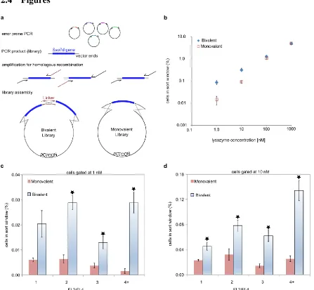
2.2.1 A bivalent library yields a higher frequency of putative high affinity clones.
Affinity maturation of binding proteins is most commonly carried out by multiple rounds of mutagenesis and screening. We hypothesized that the pool of low affinity binders obtained after an initial round of library screening will contain binders to multiple epitopes, and that linking specific pairs of proteins with a flexible linker may generate high affinity binders. Indeed, examples in the literature demonstrate that linking binders with even weak affinities (micromolar to hundreds of nanomolar Kd) can generate high affinity binding41. To evaluate this hypothesis, we used as our starting point a pool of low affinity binders to lysozyme, obtained after magnetic sorting and a single round of screening by fluorescence activated cell sorting (FACS). The selected ligands originated from a yeast surface library of ~ 108 Sso7d mutants; the library was generated by randomization of ten surface residues on the Sso7d scaffold13. DNA from this pool of mutants was subjected to random mutagenesis and pairs of binders were randomly linked using a flexible PG(PT)8P linker52 using homologous recombination in yeast25, to construct a yeast display library of ~ 107 mutants (Figure
27
monovalent and bivalent protein fusion on the yeast surface (Figure 2.5). Figure 2.1b shows that the number of clones that fall in a conservatively drawn sort window is significantly higher for the bivalent library at lower concentrations of lysozyme (1 nM or 10 nM); this suggests a greater
frequency of high affinity clones in the bivalent library. Mutants with higher affinity are characterized by a higher ratio of target binding fluorescence to the fluorescence corresponding to binding of the HA antibody. Indeed, this is the rationale for “sorting along the diagonal” during flow cytometry screening in yeast surface display22. Therefore, this ratio was used as a quantitative metric to further compare the number of putative high affinity clones in each library. Cells were binned into one of four bins based on this ratio. Figure 2.1c and 2.1d show that at lysozyme concentrations of 1 nM and 10 nM, the bivalent library shows a greater frequency of yeast cells with higher values of the ratio across all bins. This result strongly suggests that the bivalent library has a greater frequency of putative high affinity than the monovalent library of similar diversity.
The combinatorial bivalent library was generated with the intent of creating pairs of ligands that bind synergistically when flexibly linked. The comparison of the monovalent and bivalent libraries shown in Figure 2.1B indicates that this mechanism is systemic within the bivalent library. The bivalent library was generated using the identical genetic library as the monovalent library. Therefore, the increase in frequency of selectable ligands as the labeling concentration decreases in
28
2.2.2 Magnetic sorting and FACS identifies a pool of bivalent lysozyme binders with highest affinity.
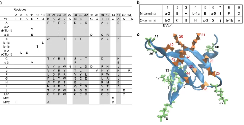
The bivalent library was screened using magnetic sorting and FACS to obtain a pool of binders with highest affinity as described22. DNA sequencing revealed that this pool predominantly contained bivalent binders comprising two distinct domains. 9 out of 14 clones analyzed contained two distinct Sso7d binder subunits. As shown in Figure 2.2a, 16 individual binders from 10 distinct families were identified constituted this pool of bivalent proteins; pairs of binders that form the nine distinct bivalent proteins are shown in Figure 2.2b. In addition to the nine bivalent proteins, four of the clones analyzed corresponded to a set of monovalent proteins derived from the lysozyme binder with highest affinity previously isolated from the naïve Sso7d library of 108 mutants13. The
29
The presence of monovalent and trivalent ligands in the highest affinity pool of ligands selected from the bivalent library motivates analysis on the quality of the bivalent library. This is necessary to determine if constructs with alternative numbers of ligands were enriched for.
Sequencing 18 members of the bivalent library reveals that eleven code for bivalent genes, four code for ligands with a nearly completely truncated N-terminal ligand, one has a significantly truncated C-terminal ligand and two code for monovalent ligands; no trivalent ligands were observed.
N-terminally truncated ligands would not be expressed as they are observed to be terminated by a stop codon prior to expression of the second ligand. This sequencing data indicates that over 10% of the bivalent library is comprised of monovalent ligands which do not contain a linker region. This population is likely highly diversified and therefore the selection of a single monovalent ligand, and mutants of this ligand, indicates that monovalent ligands were not enriched for. The presence of this single monovalent ligand, from this diversified pool, would on the contrary indicate that this ligand is an outlier of the monovalent library.
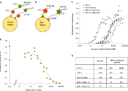
2.2.3 Determination of intermolecular vs intramolecular binding of yeast displayed BVL-1; recombinant expression, stability and binding of lysozyme.
We tested the hypothesis that high affinity binders isolated from the combinatorial bivalent library is largely due to synergistic binding of two low affinity binders separated by a flexible
30
proteins in this study. Nevertheless, KD estimates from yeast surface titrations may be erroneous if the target molecule can simultaneously bind more than one cell surface fusion; in such a case, the
strength of binding affinity is overestimated. In the context of BVL-1, binding of lysozyme to NTL-1 and CTL-1 subunits on two distinct cell surface fusions will result in an increase in apparent affinity. We therefore sought to rule out this possibility of intermolecular binding to lysozyme by distinct yeast surface display fusions. We therefore conducted titrations using a yeast dual-display system where CTL-1 and NTL-1 are expressed as distinct, unlinked cell surface fusions (Figure 2.3b). The apparent KD for the dual-display configuration (160 nM) is ~ 100X higher than the KD for BVL-1, indicating that KD measurements for BVL-1 are not affected by the avidity effect arising from
association of lysozyme with CTL-1 and NTL-1 on distinct cell surface fusions. We further expressed BVL-1 recombinantly and measured the KD of binding with lysozyme using a competitive binding assay53. The KD estimated in this assay (2.7 nM) is consistent with results from yeast surface titrations (Figure 2.3d). These results strongly indicate that the high affinity of BVL-1 for lysozyme arises from synergistic binding of the low affinity NTL-1 and CTL-1 subunits.
The residues selected in BVL-1 tend to be hydrophobic, therefore size exclusion
31
in solution but when bound to lysozyme the complex adopts a more spherical conformation. This evidence supports the conclusion that BVL-1 is a monomeric protein which binds lysozyme in a one to one manner. SEC data can be seen in Figure 2.4. Figure 2.5 is an SDS page gel which confirms that both lysozyme and BVL-1 are observed in the complex elution peak shown in Figure 2.4f. The second peak in Figure 2.4f is shown to correspond to lysozyme alone.
The KD of a clone with highest binding affinity for lysozyme, previously isolated from an Sso7d library of ~ 108 clones, was estimated as 349 nM13. Thus, BVL-1 represents a ~ 200X improvement in binding affinity in a single round of random mutagenesis, library construction and screening. Finally, it is important to note that BVL-1 retains thermal stability. The melting
temperature of BVL-1 was estimated as 84.5 °C in a differential scanning fluorimetry assay (Figure 2.6). Furthermore, BVL-1 could be expressed recombinantly at high yields in E. coli. Yields in not yet optimized shake flask cultures were estimated as 40 mg/L.
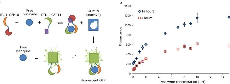
2.2.4 Binding of NTL-1 and CTL-1 to non-overlapping epitopes on the target can be exploited to design a “mix-and-read” assay for target quantification.
NTL-1 and CTL-1 independently bind lysozyme, and when combined through a polypeptide linker yield the high affinity bivalent binder BVL-1; this is consistent with NTL-1 and CTL-1 binding non-overlapping epitopes on lysozyme. Binding proteins that bind non-overlapping epitopes on a target are extensively used in biomolecular detection schemes, e. g. sandwich ELISA. Of particular interest is a mix-and-read assay scheme that eliminates the need for wash steps or protein
32
lysozyme brings GFP10 and GFP11 in close proximity, and allows assembly with the GFP1-9 fragment that is present in solution. The reconstituted complex comprising GFP10, GFP11 and GFP1-9 is fluorescent. As seen in Figure 2.7b, the fluorescence readout is dependent on the concentration of lysozyme over a large concentration range (0-15 M). This result provides direct evidence that NTL-1 and CTL-1 bind non-overlapping epitopes on lysozyme. Interestingly, the fluorescence readout increases as a function of incubation time. This increase may be explained by the irreversible nature of the assembly of the GFP fragments51. Consequently, fluorescence will persist even upon dissociation of lysozyme from the reconstituted complex (Figure 2.7b). The dissociated lysozyme may then further form another GFP complex. Thus lysozyme may act as a catalyst for reconstitution of GFP fluorescence. An alternative explanation for time-dependent increase in the fluorescent readout is the slow kinetics of GFP fluorescence reconstitution in our modified tripartite split-GFP system.
2.3
Materials and Methods
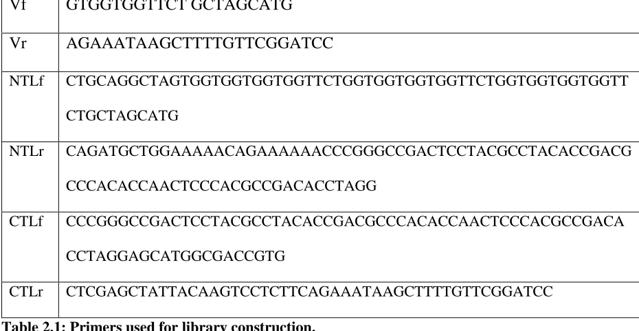
2.3.1 Library construction.
33
were subjected to fluorescence-activated cell sorting (FACS), and all cells exhibiting binding
to both lysozyme and the HA antibody above background were sorted. Plasmid DNA was
extracted from these cells using the ZymoPrep kit (Zymo Research); this was used as template
DNA for library construction.
Linear DNA used for generating both libraries were obtained by error-prone PCR using
the aforementioned template DNA, as described
55. Briefly, 35 cycles of PCR were conducted
with an estimated error rate of ~ 3.8 nucleotides per template, using primers Vf and Vr
,to
obtain DNA product P1. All primers were purchased from Integrated DNA Technologies
(IDT); sequences of all primers used can be found in Table 2.1. To obtain DNA products B1
and B2, coding for the N-terminal and C-terminal portions of the clones in the bivalent library
respectively, P1 was amplified by PCR with two different primer sets. Primers NTLf and NTLr
were used to generate B1; primers CTLf and CTLr were used to generate B2. To obtain DNA
product M1 coding for the clones in the monovalent library, P1 was amplified with primers
NTLf and CTLr. Finally, the bivalent and monovalent libraries were generated by homologous
recombination in yeast (see Figure 2.1). Three transformations were carried out for each
library. 4
g of linearized pCTCON vector and 4
g of M1 were used for each transformation
in case of the monovalent library. For the bivalent library, 4
g of linearized pCTCON vector
and 4
g each of B1 and B2 were used per transformation.
2.3.2 Comparison of monovalent and bivalent libraries.
34
albumin (PBSA), followed by secondary labeling with SA-PE and donkey anti-rabbit IgG DyLight 633 (ImmunoReagents, Raleigh, NC). Labeling volume was chosen to ensure ~ 10-fold excess of lysozyme molecules in solution relative to the yeast-displayed fusions as described22. A BD Accuri C6 cytometer was used to analyze 100,000 cells in all replicates and controls. The number of cells that fall within a conservatively drawn gate (see Figure 2.8) was assessed. For each cell found in this gate, a ratio of the fluorescence signal corresponding to lysozyme binding (FL2 in Figure 2.8) to the signal corresponding to anti-HA antibody binding (FL4 in Figure 2.8) was computed. Gated cells were placed in one of 4 bins (1-2, 2-3, 3-4 and 4+) based on the FL2/FL4 ratio. The fraction of cells in each bin relative to total number of cells analyzed (105) was used to generate histograms (Figure.
2.1c, d).
2.3.3 Selection of the highest affinity bivalent ligands
Library screening was conducted using a combination of magnetic sorting and FACS,
as previously described
22. Briefly, 2x10
8cells expressing cell surface fusions from the bivalent
library were subjected to negative selection by incubation with 50
L of Dynabeads Biotin
Binder beads (Life Technologies) in 2 ml PBSA, at room temperature for 1 hr. Cells that did
not bind the beads were recovered and incubated with 100 nM biotinylated lysozyme in PBSA
(5 ml) for 1 hour at room temperature. Cells were then washed 3 times with PBSA and
incubated with 25 mL of Dynabeads Biotin Binder in 2 mL PBSA, at 4 °C for 1 hr.
Subsequently, bead-bound cells were isolated using a magnet and expanded in culture.
35
labeled with an anti-HA antibody, followed by secondary labeling with SA-PE and G
R633,
and subjected to FACS using a MoFlo cell sorter (Beckman Coulter) to isolate binders with
the highest affinity for lysozyme. Cytometry analysis of cells expanded after FACS showed a
largely homogenous population of cells, as assessed by labeling with 1 nM lysozyme.
Therefore, plasmid DNA was extracted from this pool and sequenced.
2.3.4 Simultaneous Yeast Surface Display of CTL-1 and NTL-1 (Dual Display)
PG(PT)8P-CTL-1 was cloned into the pCT302 yeast display vector containing the Trp selectable marker56. NTL-1 was cloned into a variant of the pCT302 vector containing a Leu selectable marker; this vector was a kind gift from Prof. Eric Boder (University of Tennessee, Knoxville). Additionally, the C-terminal c-myc tag flanking NTL-1 was replaced with a 6xHis tag. Both plasmids were transformed into the EBY100 yeast strain and transformants were selected, and maintained in cell culture, using plates/media lacking Trp and Leu. The presence of both NTL-1 and CTL-1 as distinct cell surface fusions was confirmed by flow cytometry; cells were simultaneously labeled with a chicken-anti-c-myc antibody (Thermo Fisher; 1:250 dilution) and an anti-6xHis antibody (Thermo Fisher; 1:250 dilution), followed by secondary labeling with goat-anti-chicken DyLight 488 (ImmunoReagents; 1:200 dilution) and donkey anti-mouse DyLight 633
(Immunoreagents; 1:200 dilution).
2.3.5 Recombinant expression of BVL-1.
36
protein was then run on a Biorad FPLC using a cation exchange colum ( Biorad High S par # 732-4132). A linear gradient over 25 mls was used to elute the protein using PBS with 1 M NaCl as the elution buffer. To inactivate proteases and further purify BVL-1, product from the chromatography step was heated to 70 °C for 20 minutes, centrifuged to remove precipitates and filtered using a 0.22 m filter. Note that without this heat treatment step, cleavage of the bivalent protein was observed during storage. The protein stock was then dialyzed to PBS.
2.3.6 Estimation of KD
KD values were estimated using yeast surface titrations or using a competition assay with soluble binding protein, as described 24,53. Briefly, for yeast surface titrations, 2.5x10⁶ yeast cells were labeled with varying concentrations of biotinylated lysozyme and anti-HA antibody, followed by secondary labeling and analysis by flow cytometry. Volume of labeling reaction was adjusted to ensure that lysozyme molecules are in ~ 10-fold excess over cell surface fusions. To maintain this ratio at low concentrations fewer cells need to be used therefore, for BVL-1, titration yeast displaying BVL-1 fusions were combined with un-induced yeast cells to facilitate ease of obtaining cell pellets by centrifugation Samples were incubated with nutation at 4 °C for time corresponding to estimates to reach 98% of equilibrium labeling. For estimation of KD by soluble competition yeast cells displaying BVL-1were labeled with lysozyme at a concentration corresponding to ~ 80% of saturating
37
this the free unbound lysozyme fraction was calculated using yeast titration based KD and the fluorescence readout from cytometry.
2.3.7 Size Exclusion chromatography
A GE Superdex 75 10/300 GL column was equilibrated with 50mM HEPES, pH 7.4, 150mM NaCl, 6% Glycerol and 2mM DTT. The protein sample (200ul) was loaded into the column and eluted with 50mM HEPES, pH7.4, 150mM NaCl, 6% Glycerol and 2mM DTT at a flow rate of 0.5 ml/min. UV absorbance at 280 nm was monitored. Eluted 0.5 ml samples were collected using the fraction collector. The purified protein was identified by the resulting absorbance peak and the respective fractions were chosen and pooled. Bio-rad gel filtration standard (catalog# 151-1901) was used to calibrate the column for molecular weight determination.
2.3.8 Measurement of the Melting Temperature TM.
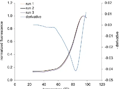
Differential scanning fluorimetry, as described57, was used to determine TM for BVL-1. Samples containing 300 mg/mL BVL-1 and SYPRO Orange according to the manufacturers protocol (Life Technologies) were heated in a real-time PCR machine (Bio-Rad CFX96), at 1C/min from 25C to 99C.
2.3.9 Expression and purification of GFP1-9, NTL-GFP11 and GFP10-CTL-1.
38
restriction sites.
To generate the plasmid vector containing GFP10-CTL1, a synthetic
oligonucleotide containing the sequence of GFP10-linker-K1 was amplified by PCR using
primers PK1f and PK1r, and cloned into pET-28b(+) to generate pET-GFP10; K1 is a coiled
peptide
51. Subsequently, the CTL-1 sequence was amplified by PCR using primers PC1f and
PC1r, and cloned into pET-GFP10 between NdeI and XhoI restriction sites; this step removes
the K1 sequence from pET-GFP10.
GFP1-9 was produced in E. coli Rosetta cells and purified from inclusion bodies as
described51. NTL-1-GFP11 and GFP10-CTL-1 were produced in E. coli Rosetta cells with N- and C-terminal 6xHis-tags, respectively, and purified by immobilized metal affinity chromatography
(IMAC) using a Biorad column (catalog # 7324614). Purified proteins were dialyzed into TNG buffer (100 mM Tris-HCl, 150 mM NaCl, 10% glycerol), pH 7.4. Protein concentrations were quantified by BCA assay.
2.3.10 In vitro “mix and read” fluorescence reconstitution assays.
39
2.4
Figures
Figure 2.1: A bivalent library yields a higher frequency of putative high affinity clones.
40
(FL4) was computed, and cells were placed in one of 5 bins (1-2, 2-3, 3-4, and 4+) based on the FL2/FL4 ratio. The largest fraction of either library was in the 0-1 bin and is not shown, this bin essentially accounts for those members of the library that do not bind lysozyme. The fraction of cells in each bin relative to total cells analyzed is shown for (c) 1 nM and (d) 10 nM lysozyme concentration. All error bars indicate standard error of the mean from triplicate experiments. * indicates p<0.05 based on a two-tailed t-test comparing results from monovalent and bivalent libraries.
Figure 2.2: Analysis of nine clones from the highest affinity lysozyme ligands selected.
41
and outside the 10 residues randomized in the original Sso7d library (green). Residues 2 and 43 cannot be seen in the figure as (2) is surface exposed and faces into the page and (43) faces towards the hydrophobic core.
Figure 2.3: High affinity of bivalent binder BVL-1 is due to synergistic binding of low affinity NTL-1 and CTL-1 subunits.
(a) Schematic for yeast surface display of BVL-1. (b) Schematic for simultaneous yeast surface display of NTL-1 and CTL-1. (c) Yeast surface titrations for estimating KD for BVL-1, NTL-1, and CTL-1, and the apparent KD for cells simultaneously displaying NTL-1 and CTL-1 as distinct fusions. Data from two independent experiments is presented for each construct. Fluorescence data is normalized by the maximum fluorescence value for each construct, obtained from a global least squares fit. (d)
42
experiments is presented. (e) Estimates of KD from data in (c) and (d) using a global least squares fit, assuming a 1:1 binding model.
43
44
Figure 2.5: SDS page of elution samples of the BVL-1 and lysozyme complex from SEC
Non-reducing NuPAGE™ Novex™ 4-12% Bis-Tris Protein Gel stained with Coomassie Brilliant Blue R-250 for elution samples taken from the lysozyme and BVL-1 SEC run shown in Figure 2.4f. (a) Is the Bio-Rad Precision Plus Protein Dual Color Standards molecular weight marker. (b) Lane 1 is the molecular weight standard. Lanes 2-4 were taken during the elution of the first peak and protein bands for both lysozyme and BVL-1 are observed. Lanes 5-7 were taken during the second peak elution peak and only lysozyme is observed.
Figure 2.6: Differential scanning fluorimetry for estimation of TM.
45
Figure 2.7: Binding of NTL-1 and CTL-1 to non-overlapping epitopes on the target can be exploited to design a “mix-and-read” assay for target quantification.
47
Figure 2.8: Comparison of monovalent and bivalent libraries by cytometry.
Yeast cells were labeled with biotinylated lysozyme and an anti-HA antibody, followed by secondary labeling with SA-PE (FL2) and DyLight 633 conjugated secondary antibody to detect the anti-HA antibody (FL4). (a) forward scatter and side scatter gating (b) gating to select singlet cells (c)-(e)
bivalent library labeled at 100 nM, 10 nM and 1 nM lysozyme (f)-(h) monovalent library labeled at 100 nM, 10 nM and 1 nM lysozyme (i) no lysozyme control for bivalent library (j) no lysozyme control for monovalent library.
Vf
GTGGTGGTTCT GCTAGCATG
Vr
AGAAATAAGCTTTTGTTCGGATCC
NTLf CTGCAGGCTAGTGGTGGTGGTGGTTCTGGTGGTGGTGGTTCTGGTGGTGGTGGTT
CTGCTAGCATG
NTLr CAGATGCTGGAAAAACAGAAAAAACCCGGGCCGACTCCTACGCCTACACCGACG
CCCACACCAACTCCCACGCCGACACCTAGG
CTLf CCCGGGCCGACTCCTACGCCTACACCGACGCCCACACCAACTCCCACGCCGACA
CCTAGGAGCATGGCGACCGTG
CTLr CTCGAGCTATTACAAGTCCTCTTCAGAAATAAGCTTTTGTTCGGATCC
Table 2.1: Primers used for library construction.
48
CHAPTER 3
FUNCTIONAL DIMERIC SSO7D LIGANDS AND EPITOPE
ADDITION AS A MODE OF AFFINITY MATURATION IN
49
3.1
Introduction
Intrinsically disordered proteins (IDPs) and intrinsically disordered regions of proteins (IDRs) that compose over 40% of the human proteome. These proteins play important roles in cell signaling and transcription and their importance is highlighted by the fact they make up 80% of the proteins which have been identified as being responsible for neurodegenerative diseases and cancers58,59. They are defined by regions of 30 or more residues that do not form a stable tertiary structure. Their importance has led to the continued improvement in methods for identifying their presence in the proteome, modelling and measuring their dynamic behavior, as well as in searching for drugs to alter their behavior 58–60. Targeting oncogenic transcription factors containing IDPs is an active area for small molecule drug discovery. One candidate that shows promise is a c-Myc inhibitor that prevents it from binding Max, a partner which makes c-Myc active61. The mechanism by which the drug works is to prevent the IDR of c-Myc from undergoing a change in conformation which is necessary to bind Max. Small molecules readily cross the cell membrane and therefore, in a therapeutic context, intracellular delivery does not represent the challenge that it does for larger engineered antibodies or protein affinity ligands. However, in the context of research tools for obtaining important biological information engineered protein affinity ligands have been successful47,62,63. The work herein expands the literature base on targeting a peptide sequence within an IDR using the non-antibody Sso7d scaffold. We make observations on the ability to pre-target a specific epitope within an IDR; and report findings made when affinity maturing pre-targeted ligands to a protein containing an IDR.
50
canonical Wnt signaling pathway64. Wnt signaling is critical to many cellular processes including proliferation, migration and stem cell differentiation65,66. In the absence of Wnt signaling, -catenin binds E-cadherin and -catenin near the cellular membrane, on the cytosolic side, and is involved in making cell to cell connections67. Cytoplasmic levels of β-catenin are maintained at very low levels because of a β-catenin destruction system68. However this changes when extracellular Wnt ligand is present. Secreted Wnt ligands bind to and co-localize the Frizzled and LRP5 or LPRP6 extracellular receptors. This co-localization results in the sequestration of the -catenin destruction complex69. Β-catenin then builds up in the cytoplasm and through a not well-understood mechanism translocates to the nucleus where it binds to DNA binding proteins and other transactivators to transcribe Wnt responsive genes69. Β-catenin is composed of a 151 residue, intrinsically disordered N-terminus, a folded ARM domain composed of twelve alpha-helical repeat units (residues 151-666) and an intrinsically disordered C-terminus (666-781) 70. The disordered C-terminus is a transactivation domain that binds to both transcriptional elements and chromatin modifying elements66.
51
A second approach would be to use a peptide itself as a targeted region in an IDR. This is a strategy used to make vaccines for targeted antibody generation by animals73. However, the resulting antibodies then need to be screened for functional binding to the target in the context applicable for the antibody. Affinity ligands engineered ex vivo have also been generated towards peptides which represent sequences of interest that appear in an IDR or IDP46. Importantly, in this example the peptide is the target, the ligand is never selected to bind the actual IDR of interest. Additionally, binding of the ligand to the actual IDR is typically not measured quantitatively, in its place selectivity measurements are typically done46. However, as the targeted peptide epitope may take on different behavior in the context of the entire IDR sequence, this could be an important issue in certain cases and could be a source of unexpected results.
52
expressed from E. coli through an avid interaction. The selection was carried out using biotin binder magnetic beads immobilized with biotinylated β-catenin protein. Subsequently, this pool of ligands was subject to a mutagenic PCR which covered the entire Sso7d gene and from this a second
generation yeast surface display library was generated. The highest affinity members of this library to -catenin were then selected using the avidity MACS technique and subsequent rounds of FACS (fluorescence activated cell sorting). One ligand from the selected highest affinity population, termed M2, was studied in detail. We refer to M2 as being pre-targeted to an epitope within an IDR and affinity matured, through mutagenic PCR, and subsequently selected for binding to the target protein containing the IDR, -catenin.
53
are studies left for future work. Here, many important observations about the ligands generated through these methodologies have been made through in vitro assays and these are the focus of this chapter.
The highest affinity ligand chosen for further analysis using the tandem approach is termed T2, this is shorthand for tandem affinity ligand from a second generation library. As an affinity matured bivalent ligand to an IDR has not yet been generated to this author’s knowledge, a T3 ligand was also generated. The name indicates that is a tandem ligand and generated after three different libraries have been generated. While profoundly high affinity was expected from T3 and observed to some extent, we were able to make other important observations into the nature of how a ligand with an IDR can bind a target with an IDR.
3.2
Results
3.2.1 L54W is a conserved mutation in the highest affinity ligands selected from the affinity matured monovalent library pre-targeted to the β-catenin C-terminal IDR.
54
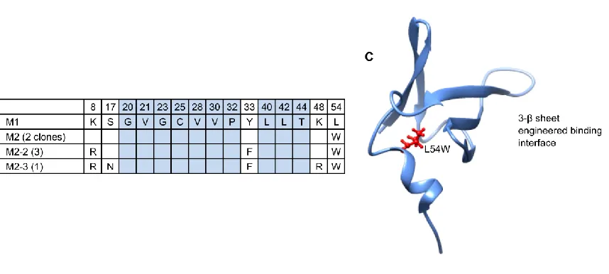
beads and the library was incubated with the -catenin displaying beads. After this selection four of five sequenced clones were found to be mutants of the M1 ligand. The highest affinity ligands were selected after several rounds of FACS, each selection was carried out at higher dilutions of biotinylated -catenin. Six ligands were sequenced from the selected pool of ligands and they were all found to be mutants of M1. All six had a conserved L54W mutation; 2 had only this mutation; 2 others also contained K8R and Y33F mutations; and two others had the same 8R and 33F mutations as well as S17N and K48R. For this work we chose to study the single L54W mutant, referred to from here on as M2. Figure 3.1 shows (A) the M1 sequence; (B) the mutations present in the pool of highest affinity ligands sequenced which differ from M1; (C) a PDB diagram display the location of the 54th residue position in Sso7d. The tryptophan in wild type Sso7d faces internally and is on the side opposite the original engineered binding interface on Sso7d. This interface is the location of the ten residues mutated in the naïve library13. These mutations are all on the 3 -sheet surface which is also identified in Figure
3.1C.
3.2.2 The first generation tandem library assembled well and yielded high affinity ligands
55
possible codons. Also for this library, the 5’ end of the PCR product has homology to the linker region and not the yeast surface display vector. To enable precise recombination the codons used to code for the (PT)15P were highly diversified. Figure 3.2 shows how the tandem library was assembled.
The transformed tandem library was estimated to have a diversity of 2x10⁸. Nine random library mutants sequenced contained the (PT)15P linker in frame and without mutations. Four of those nine mutants contained mutations only at the library positions of the C-terminal ligand; the remaining five mutants had stop codons in library positions or just following a library position. Sequence analysis also identified inadvertent mutations in each of the N-terminal M1 ligands: a K63 deletion and a K62Q mutation. These mutations are at the C-terminus of M1, K63 is the terminal residue in Sso7d. This mutant will be referred to as M1.2. It is shown that these mutations did not affect the ability of a tandem ligand selected in this library to bind the βCP sequence.
3.2.3 A diversified polypeptide chain was enriched during selections for -catenin binding.
This first generation tandem library was initially sorted at 100 nM using biotinylated β-catenin. Cells which bound β-catenin at this concentration were collected after washing using magnetic biotin binding beads. This method is based on a previously published protocol76. The advantage of this magnetic bead capture step relative to FACS is the extreme high throughput and low cost. 2x10⁹ cells would take an unreasonable amount of time to sort whereas MACS can be readily done after a few hours or overnight incubation for a fraction of the cost and time. Typically an initial selection would be carried out using an avidity-based MACS wherein β-catenin is immobilized on magnetic beads prior to the selection. However, in this case each ligand already bears a β-catenin binding domain so an avidity based selection would be ineffective.
56
biotinylated β-catenin and HA-tag expression was carried out. The HA tag was labelled with a primary rabbit antibody and then a goat anti rabbit conjugated with Dylight 633 secondary antibody; biotinylated -catenin was labelled with streptavidin Phycoerythrin (PE). Sequencing nine members of the library at this point showed that only four were the expected M1.2 ligand, linker and a second Sso7d protein. The other five consisted of M1.2 but were truncated prior to the second ligand and contained a conserved peptide sequence. The unexpected peptide sequences seen in the linker region are shown in
Table 3.1. The randomized polypeptide sequences likely arose from errors in the manner in which the yeast homologous recombination system combined the yeast display vector and library. The presence of the enriched sequence of ligand plus polypeptide linker would suggest that this would likely be a successful alternative approach for creating bivalent ligands. This alternative approach would use a short linker and a randomized peptide sequence, consisting of 8-10 sequential degenerate DNA codons. This randomized peptide would take the place of the Sso7d library that is used here.
57
been termed T2 and was chosen for further characterization. Table 3.2 shows the linker mutations observed as well as the library mutations selected in the C-terminal ligand for the six tandem ligands sequenced.
3.2.4 Affinity maturation of T2 yields a diversity of advantageous mutations.
T2 was affinity matured by randomly mutating the DNA for the N-terminal ligand (NTL) and linker sequence in a first PCR and in a second PCR the linker and C-terminal ligand (CTL) DNA sequence was mutated. The two libraries were generated in this case using a mutagenic PCR protocol based on Taq polymerase and an optimized buffer55. A T2 tandem library was assembled by the yeast homologous recombination system when the two libraries were transformed into electrocompetent yeast with the yeast display pCTCON vector. Figure 3.3 depicts how this second generation tandem library was generated. The library diversity was estimated to be to be 1x10⁸. Library sequencing showed that 7 of 10 sequences contained two Sso7d mutants on either side of the linker with 0-3 amino acid mutations in either ligand; two library sequences had an additional linker segment in-between the two ligands and the other library member sequenced was a single Sso7d clone which was a mutant of the C-terminal ligand in the T2 construct.
58
The C-terminal ligand in both T2 and T3 are identical and therefore this ligand is referred to as 23CTL throughout the remainder of this work. Table 3.4 shows the nomenclature for each of the mutants selected and characterized in this work, summarizes how they were engineered and the individual ligands which make up the T2 and T3 tandem ligands.
3.2.5 Selection of functional dimeric ligands