1071-412X/97/$04.00
1
0
Copyright
q
1997, American Society for Microbiology
Relation of Impaired Lymphocyte Proliferative Function to
Other Major Human Immunodeficiency Virus Type 1-Induced
Immunological Changes
HONG Z. BASS,
1JOHN L. FAHEY,
1* PARUNAG NISHANIAN,
1ROGER DETELS,
2,3WILLIAM CUMBERLAND,
2MARGARET KEMENY,
4ANDSUSAN PLAEGER
5Center for Interdisciplinary Research in Immunology and Disease and the Jonsson Comprehensive Cancer Center,
Department of Microbiology and Immunology,
1Department of Psychiatry,
4and Department of Pediatrics,
5UCLA
School of Medicine, UCLA School of Public Health
2and Los Angeles Center, Multicenter AIDS Cohort Study,
3Los
Angeles, California 90095
Received 7 March 1996/Returned for modification 2 May 1996/Accepted 16 September 1996
Human immunodeficiency virus (HIV) type 1 (HIV-1) induces impairment of immune function reflected in
reduced lymphocyte proliferative responses. Many other immune changes are induced by HIV-1, but their
relationship to lymphocyte functional defects is not known. The present study was designed to correlate
functional defects with other HIV disease parameters. Cryopreserved samples from 118 HIV-1-positive subjects
and 40 seronegative individuals were examined. The main findings were that impaired proliferative responses
to mitogens correlated with (i) decreased cell surface expression of the interleukin-2 receptor (CD25), (ii)
increased expression of HLA-DR antigens on CD4 cells, (iii) reduced CD4 and increased CD8 cell numbers,
and (iv) increased levels of serum immune complex dissociated p24 antigen. However, impaired function was
not associated with increased serum neopterin,
b
2-microglobulin, or soluble interleukin-2 receptor or with
CD38 antigen expression on lymphocytes. In summary, proliferative functional impairment correlated with
some, but not all, immunological changes associated with HIV-1 infection. Most of the phenotypic markers that
correlated with altered function are cell surface molecules with significant roles in lymphocyte proliferation
and were associated primarily with CD4 cells, compatible with the view that dysregulation of CD4 cells is
responsible for impaired function.
Human immunodeficiency virus (HIV) type 1 (HIV-1)
causes a progressive breakdown of immunity that can be
quan-tified by several different immunological assays. The selective
depletion of CD4 lymphocytes has been well described (7, 23).
The number of peripheral blood CD4 T cells is commonly used
to determine the stage of disease and to predict disease
pro-gression (19, 24). Impairment of T-cell function is a separate
measurement and occurs early in HIV-1 infection, when CD4
T-cell numbers are still in the normal range. T-cell function
decreases further with disease progression (5, 20). In addition
to the response to phytohemagglutinin (PHA) (12, 13),
prog-nostic assessments have indicated that the proliferative
re-sponse to pokeweed mitogen (PWM) is a sensitive prognostic
marker for AIDS occurrence.
Extensive activation of the immune system is another major
effect of HIV-1 infection. The activation occurs early and
in-creases throughout the course of the infection. HIV-1-induced
activation results in the production of serum immune
activa-tion products, such as neopterin,
b
2-microglobulin (
b
2M),
sol-uble interleukin-2 (IL-2) receptor (sIL-2R), and solsol-uble CD8
(8, 14, 22, 27). Changes in these markers have also been shown
to predict HIV-1 disease progression and to be largely
inde-pendent of CD4 T-cell numbers (25). The increase in serum
immune activation markers may reflect specific
cytokine-me-diated processes. For example, serum neopterin reflects
pri-marily an increased production of gamma interferon, whereas
b
2M reflects increases in other cytokines (1, 15). sIL-2R does
not correlate closely with the changes of serum neopterin and
b
2M (16) and presumably reflects a different aspect of HIV-1
immunopathogenesis.
Changed expression of lymphocyte surface antigens also
re-flects the dynamic interaction between the human immune
system and HIV-1. HLA-DR (major histocompatibility
com-plex class II molecule) CD38 (a cell immaturity and activation
marker), and CD71 (transferrin receptor) are significantly
in-creased while CD25 (IL-2 receptor alpha chain) is significantly
decreased in HIV-infected subjects (2, 10, 11, 16, 21).
Multipa-rameter phenotypic analyses offer opportunities to examine
changes within specific lymphocyte subpopulations, e.g., CD4
T cells, CD8 T cells, or B cells. Because CD4 T cells are central
to proliferative function and are a major target for HIV-1
infection, the phenotypic changes of these cells are of special
interest. Finally, the virologic marker of serum p24 antigen,
both free and complexed with antibody, also is known to relate
to disease prognosis (26). This marker reflects active, ongoing
viral replication.
Thus, studies of AIDS pathogenesis have identified
numer-ous potential surrogate markers for prognosis and therapy in
HIV-1 infection. With many different measurements emerging,
little work has been done to relate these markers to each other.
A goal of the current study was to determine whether the
functional changes in lymphocytes indicated by decreased
pro-liferative responses to mitogens (PHA and/or PWM)
corre-lated with other HIV-1-associated immune changes. These
pa-rameters include (i) the levels of four major lymphoid cell
subsets (CD4, CD8, B, and NK cells), (ii) expression of four
lymphoid activation markers (HLA-DR, CD38, CD71, and
CD25) in the total lymphocyte population as well as in the
major lymphocyte subsets, (iii) levels of free and total serum
p24 antigen, and (iv) three soluble immune activation markers
* Corresponding author. Mailing address: Department of
Microbi-ology and ImmunMicrobi-ology, UCLA School of Medicine, Los Angeles, CA
90095-1747. Phone: (310) 825-6568. Fax: (310) 206-1318.
64
on August 17, 2020 by guest
http://cvi.asm.org/
(serum neopterin,
b
2M, and sIL-2R). A total of 118
HIV-1-seropositive individuals were compared to 40 seronegative
ho-mosexual men. The data indicate that reduced proliferative
function correlated with several, but not all, immune
alter-ations in HIV-1 infection.
MATERIALS AND METHODS
Subjects.Cryopreserved peripheral blood mononuclear cells (PBMC) from 118 mildly symptomatic and asymptomatic HIV-1-seropositive homosexual and 40 HIV-1-seronegative homosexual individuals were selected from the Multi-center AIDS Cohort Study repository at Los Angeles, Calif. All subjects had been monitored approximately every 6 months since 1985 for presence of HIV-1 antibody, major lymphocyte subsets, antiretroviral therapy, and health status. Details of the recruitment procedures and the characteristics of the cohort at baseline have been reported previously (6, 18). Samples obtained in 1988 were obtained from the frozen repository. Of 118 HIV-1-seropositive subjects without any antiviral treatment, 60 had a baseline CD4 number of,500/mm3, with a
mean of 364/mm3. The remaining 58 subjects had a baseline CD4 number of
.500/mm3, with a mean of 728/mm3.
Preparation of PBMC.PBMC were originally isolated from fresh peripheral blood by centrifugation on a Histopaque density gradient (Sigma) and suspended to 103106/ml in complete RPMI 1640 medium (GIBCO, Grand Island, N.Y.)
containing 20% human serum and 20% dimethyl sulfoxide (Sigma). One-milli-liter cell aliquots were dispensed into 2-ml cryovials (Nalgene) for programmed-rate freezing (Cryomed, New Baltimore, Mich.) to2808C and stored in liquid nitrogen. The vials of cryopreserved cells were placed in a 378C water bath until just thawed, and 5 ml of RPMI 1640 medium with 10% human serum was added dropwise. The cells were washed twice, and viability was determined by exclusion of 0.2% trypan blue dye. The viability of all thawed samples was over 90%.
Proliferative assay.One-hundred-microliter aliquots of PBMC (2.53105
lymphocytes/ml) in RPMI 1640 medium containing 10% human AB serum were pipetted into triplicate wells of a 96-well round-bottom microtiter plate. Mito-gens at a concentration of 360mg/ml for PHA (Wellcome Diagnostics, Research Triangle Park, N.C.) or a dilution of 1:250 for PWM (GIBCO) were added at 100 ml per well. These concentrations had been previously determined as optimal by titration using cells from at least three healthy subjects. The microtiter plates were incubated at 378C in a humidified atmosphere of 5% CO2in air. After 72 h,
the PHA-stimulated plates were pulsed with 10ml of [3H]thymidine prepared as
a fresh stock at 200mCi/ml. After 5 days, the PWM-stimulated plates were pulsed with [3H]thymidine. The plates were incubated further for 18 h, and then cells
were harvested with an automated cell harvester (PHD, Cambridge, Mass.). The incorporated radioactivity was measured with a liquid scintillation counter (Beckman LS-1800). The results (in counts per minute) were given as medians of triplicate determinations.
Measurement of serum immune activation markers.Serum was stored at 2208C prior to testing. Serum neopterin levels were quantitated by a commercial radioimmunoassay (Neopterin, RIAcid; Henning, Berlin, Germany). Serum b2M levels were measured by automated microparticle enzyme immunoassay (IMX; Abbott Diagnostics, Abbott Park, Ill.). The details of the methods have been described elsewhere (14, 22). Serum sIL-2R was assayed by using an enzyme-linked immunosorbent assay kit from T Cell Diagnostics (Cambridge, Mass.) according to the manufacturer’s instructions.
Lymphocyte immunophenotype analysis.PBMC were distributed among 12-by 75-mm plastic tubes (53105cells per tube) containing the following
mono-clonal antibody combinations: CD3-CD56116-CD8, HLA-DR–CD38-CD8, CD71-CD25-CD8, CD57-CD56-CD8, HLA-DR–CD38-CD4, CD71-CD25-CD4, and CD71-CD25-CD19 (all from Becton Dickinson Immunocytometry Systems, San Jose, Calif.). In each case, the first monoclonal antibody was labeled with fluorescein isothiocyanate, the second was labeled with phycoerythrin, and the third was labeled with peridinin chlorophyll protein (PerCP). The tubes were incubated for 15 min at room temperature, washed, and resuspended in 0.25 ml of fixation buffer (1% paraformaldehyde in 13phosphate-buffered saline). Cells were analyzed by using a FACScan flow cytometer (Becton Dickinson). Lym-phocytes were identified by gating on forward (low-angle) and 908C (wide-angle) light scatter parameters, and anti-CD45 (pan-leukocyte) and anti-CD14 (mono-cyte) served as markers for validating lymphocyte scatter gating. The fluores-cence negatives were defined by using isotype control antibodies, and compen-sations were adjusted by using the CD3-CD56116-CD8 tube. List mode data were collected for 10,000 events in the total gate. The absolute number of cells within a subset was determined by multiplying the absolute lymphocyte count by the percent cells positive for that marker(s). The absolute lymphocyte count was obtained from the leukocyte count and differential, which were determined for fresh whole blood prior to cell separation and cryopreservation.
Free and total serum p24 antigen measurement.Free serum p24 antigen (not bound into immune complexes) was measured by an enzyme immunoassay using the Coulter (Hialeah, Fla.) HIV p24 antigen assay kit according to the manu-facturer’s procedure (26). Total serum p24 antigen (free plus immune complexes dissociated) was measured after acid pretreatment (26). A 100-ml volume of serum from study subjects or standards were mixed with 50ml of 0.5 N HCl, incubated for 1 h at 378C, and then neutralized with approximately 50ml of 0.5
N NaOH to pH 6.5 to 7.5. Thereafter, the assay was performed as for free p24 antigen, with the exception of an overnight incubation at 48C instead of 1 h at 378C as the first step.
Statistical analysis.Correlations were calculated by using Spearman’s rank correlation coefficient. P values are two-sided alternatives, representing the prob-ability of a value as large as (or larger than) that observed if there were no association between the variable measures. For group comparisons and the stratified analysis, comparisons were made with the Wilcoxon rank sum test. In the stratified analysis, groups were obtained from the HIV-seropositive subjects on the basis of absolute CD4 count (above or below 500/mm3).
RESULTS
Lymphocyte proliferative responses to mitogens.
Results of
proliferative responses to PHA and to PWM in 40
HIV-1-seronegative controls and 118 seropositive individuals are
shown in Fig. 1. Seropositive individuals were divided into
those with CD4 T-cell levels of greater or less than 500/mm
3.
There was no difference in the response to PHA between
HIV-1-seronegative and -seropositive subjects with CD4
num-bers of
.
500/mm
3. A significant difference, however, was
found between the responses of the seronegative control
sub-jects and HIV-1-seropositive subsub-jects with CD4 counts of
,
500/mm
3. PWM responses, on the other hand, were
signifi-cantly different between seronegative controls and both groups
of seropositive subjects (Fig. 1). Furthermore, the seropositive
group with low numbers of CD4 cells had a significantly poorer
response to PWM (P
5
0.0015) than did the seropositive group
with higher CD4 cell levels.
Similar results were obtained when proliferative responses
FIG. 1. Proliferative responses of PBMC to PHA and PWM. Means and standard deviations are shown. HIV(2), HIV negative; HIV(1), HIV positive. CD4 counts per microliter are indicated.on August 17, 2020 by guest
http://cvi.asm.org/
of the HIV-infected subjects were calculated as a percentage of
the mean value of the seronegative controls measured in the
same assay or when the proliferation results were expressed as
stimulation indices (data not shown). There was a good
corre-lation (P
,
0.0001) between the proliferative responses to
PHA and PWM in seropositive individuals.
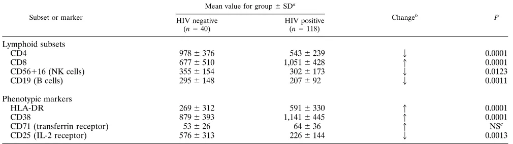
Lymphocyte phenotype changes and correlation with
prolif-erative response.
The mean levels of the major lymphocyte
subsets and cells expressing activation antigens in the
seropos-itive men were compared with those of HIV-1-seronegative
controls measured concurrently (Table 1). The absolute
num-bers of CD4 T cells, NK, B cells, and cells expressing CD25
were significantly lower in HIV-1 infection. In contrast, the
numbers of CD8 T cells and cells expressing HLA-DR and
CD38 were significantly higher.
In the HIV-1-seropositive group, impaired lymphocyte
pro-liferative responses showed a clear positive correlation with
lowered numbers of CD4 cells as well as with decreased total
lymphocyte expression of CD25 (Fig. 2). Decreased CD25
expression on CD4 and on B-lymphoid subpopulations also
correlated with the proliferation impairment. Increased
ex-pression of HLA-DR or CD71 on CD4 cells correlated
in-versely with proliferative responses (Table 2). There was no
significant correlation between lymphocyte proliferative
re-sponses and phenotypic markers in the HIV-1-seronegative
group (data not shown).
Correlation of proliferative responses and serum p24 levels
in HIV-1 infection.
Total serum p24 antigen was detected in 62
subjects, and the levels correlated inversely with reduced
pro-liferative response (P
5
0.0273) (Table 3). Only 8 of the 118
seropositive subjects had detectable levels of free p24 (immune
complex dissociated) antigen at the time of the study, and the
levels inversely correlated with impaired proliferative response
(P
5
0.0165).
Lack of correlation of proliferative response with
pheno-typic marker changes on CD8 T cells or with serum immune
activation markers.
CD8 cell expression of CD38 and
HLA-DR was significantly increased and expression of CD25
was decreased (data not shown). However, these changes did
not correlate with the proliferative response impairment. Two
serum immune activation markers, neopterin and
b
2M, were
significantly increased in seropositive subjects (P
,
0.0001). In
comparison with a mean
6
standard deviation of 6.6
6
1.9
nmol/liter for 40 HIV-1-seronegative individuals, the mean
neopterin level for seropositive subjects with CD4 counts of
.
500/mm
3was 11.9
6
3.7 nmol/liter. This further increased in
subjects with CD4 counts of
#
500/mm
3(14.25
6
3.5 nmol/
liter). Similarly, the mean value (
6
standard deviation) for
b
2M in the seronegative group was 1.3
6
0.3 mg/liter, whereas
the levels increased to 2.2
6
0.6 mg/liter in seropositive
sub-jects with CD4 counts of
.
500/mm
3and 2.6
6
0.6 mg/liter in
the seropositive group with CD4 counts of
.
500/mm
3. sIL-2R
was tested only in the HIV-1-seropositive subjects with CD4
,
500/mm
3and was also significantly increased. However,
these three serum immune activation parameters appeared to
be independent of cell proliferation and did not correlate with
either PHA or PWM proliferative responses (Table 3). The
correlations of proliferative functional impairment with other
immunological changes in HIV infection are also summarized
in Table 3.
DISCUSSION
Clinical parameters, such as the onset of opportunistic
in-fection, certain neoplasms, or death, according to the clinical
definition of AIDS, are long-term endpoints for evaluation of
HIV-1 infection or its treatment. Thus, effective and reliable
surrogate markers are needed for more-expedient evaluation
of disease progression and therapeutic efficacy. Whereas many
immune changes during HIV-1 infection have been identified,
few studies have examined their interrelationships. In a
previ-ous study, several serum immune activation and lymphoid
phe-notypic markers were assessed, and it was shown that some
serum markers (neopterin,
b
2M, and soluble CD8) and
lym-phoid phenotypic markers (CD38, CD45RA, and 1-selectin)
reflect related aspects of immune dysregulation associated with
HIV-1 infection (3). Prince et al. reported similar results (28).
The relationship of serum and lymphoid phenotypic markers
to lymphocyte function, however, has not been determined
previously.
The impairment of proliferative function in HIV-1 infection
is particularly interesting because this change can be seen
be-fore CD4 lymphocytes are substantially reduced in number (5,
20) and because such changes are reported to be related to
disease progression (13). Furthermore, proliferative responses
return toward normal levels transiently after initiation of
zidovudine therapy (29), indicating that this change is induced
by HIV-1 infection. Proliferative evaluations, however, are
rarely used in HIV-1 disease stage determination and
thera-peutic evaluation, in part because the assay is more laborious
TABLE 1. Summary of lymphocyte subset and phenotypic changes in HIV-1 infection
Subset or marker
Mean value for group6SDa
Changeb P HIV negative
(n540)
HIV positive (n5118)
Lymphoid subsets
CD4
978
6
376
543
6
239
2
0.0001
CD8
677
6
510
1,051
6
428
1
0.0001
CD56
1
16 (NK cells)
355
6
154
302
6
173
2
0.0123
CD19 (B cells)
295
6
148
207
6
92
2
0.0011
Phenotypic markers
HLA-DR
269
6
312
591
6
330
1
0.0001
CD38
879
6
393
1,141
6
445
1
0.0001
CD71 (transferrin receptor)
53
6
26
64
6
36
1
NS
cCD25 (IL-2 receptor)
576
6
313
226
6
144
2
0.0013
a
Mean number of lymphocytes per cubic millimeter (for lymphoid subsets) or mean number of lymphocytes expressing the phenotypic antigen (for phenotypic markers).
b
Direction of change in HIV-1-seropositive subjects compared to seronegative controls. c
NS, not significant (P.0.05).
on August 17, 2020 by guest
http://cvi.asm.org/
than serologic or flow cytometric assays and usually must be
done with freshly obtained blood. Cyropreserved cells were
used in the current study because batch testing allowed for
better standardization. We included cryopreserved cells from
the same healthy subject as an interexperimental control for
each batch, and the tests were performed in a limited period by
the same technician using a single lot of reagents. The
coeffi-cients of variation for the interexperimental control were 32%
for PHA and 24% for PWM.
Previously, PWM was found to be a more sensitive stimulus
FIG. 2. Correlations between impaired proliferative responses to PHA (A and B) and PWM (C and D) and the levels of CD25 expression (reductions) on lymphocytes and the levels of HLA-DR expression on CD4 T cells. Correlation coefficients and P values are indicated for each correlation.TABLE 2. Significant correlations of proliferative responses with changes in lymphocyte phenotype
Parameter Changea Proliferative response b
PHA PWM
% of total lymphoid cells expressing phenotypic marker
CD4 cells
2
0.22 (0.0157)
0.38 (0.0001)
CD8 cells
1
2
0.20 (0.0284)
2
0.35 (0.0001)
CD71 cells
1
2
0.20 (0.0375)
NS
cCD25 cells
2
0.26 (0.0061)
0.25 (0.0089)
% of lymphoid subset expressing phenotypic marker
CD4 cells expressing:
HLA-DR
1
2
0.28 (0.0026)
2
0.24 (0.00109)
CD71
1
2
0.35 (0.0001)
NS
CD25
2
0.19 (0.0436)
0.18 (0.0489)
CD19 B cells expressing CD25
2
0.29 (0.0016)
0.20 (0.0384)
aSee Table 1, footnote b.
bSpearman’s rank correlation coefficients are given, with P values in parentheses. cNS, not significant.
on August 17, 2020 by guest
http://cvi.asm.org/
than PHA for detecting the impairment of proliferative
re-sponse associated with HIV-1 infection (13). Rere-sponses to
PHA were reduced only in more-advanced HIV-1-seropositive
subjects, while the response to PWM was reduced in most
HIV-1-seropositive asymptomatic men (17). Our results also
showed that PWM responses can distinguish between
seropos-itive subject groups with CD4 numbers of greater or less than
500/mm
3. This may be explained, at least partially, because
PHA acts via both the CD2-dependent and the T3/Ti pathway
while PWM involves primarily the T3/Ti-induced responses.
The proliferative defect associated with HIV-1 infection alters
principally the T3/Ti-induced responses (17).
Previously, it was reported that IL-2 receptor expression
(CD25) on the major lymphoid subsets is significantly reduced
in HIV-1 infection (16). Since IL-2 production and IL-2
recep-tor expression are essential steps in lymphocyte proliferation, it
was expected that reduced CD25 expression would correlate
with the functional defect in HIV-1 infection. This was
con-firmed for CD4 T cells in the present study. CD4 lymphocytes
are known to be central to the proliferative responses and are
responsible for producing IL-2 and other cytokines necessary
for cell proliferation. However, it has been shown that IL-2
cytokine mRNA is not increased in CD4 cells in HIV-1
infec-tion (9), and IL-2 is known to be a stimulus for IL-2 receptor
(CD25) expression. Thus, impaired IL-2 receptor expression
and IL-2 gene expression may both contribute to the
prolifer-ation impairment in HIV infection.
Interestingly, CD4 T cells have been shown to have
in-creased expression of gamma interferon mRNA in HIV
infec-tion (9). This may relate to the changes of other cytokines
which contribute to HIV-associated increases in HLA-DR and
CD71 on CD4 T cells (Table 3). Altered proliferative function
may reflect aspects of HIV-1 pathogenesis distinct from those
reflected by other immune changes. This possibility is
sup-ported by the fact that the kinetics of zidovudine-induced
changes in immune activation markers, lymphoid phenotypes,
and proliferative response differ markedly. In HIV-1-infected
subjects treated with zidovudine, elevated levels of serum
ac-tivation markers and lymphoid phenotypic markers are most
reduced (closest to normal) by 1 to 4 weeks of therapy (2, 4).
In contrast, the recovery of proliferative response and increase
of CD4 T cells after initiation of zidovudine treatment peak
later at about 2 to 3 months (29). Whether zidovudine-induced
reduction in viral activity results in functionally competent
CD4 T cells or whether other factors are responsible for this
transient functional improvement remains to be determined.
Unfortunately, wide variability in proliferative response to
mitogens is a major problem inherent in the test system. A
standard deviation of 50% is a common finding with this type
of assay. This introduces uncertainties in establishing
correla-tions between impairment of proliferation with other
measure-ments of immunological change in HIV-1 infection. Although
we batch-tested samples to minimize variation, the possibility
of missing some existing correlation between proliferative
re-sponses and other measurements cannot be completely ruled
out.
The complexity of the immune changes induced by HIV-1
infection is emphasized by our findings. The results show that
measurements of lymphocyte functional change reflected by
proliferative impairment provide additional information not
obtained by measuring the changes in serum immune
activa-tion markers or several lymphoid phenotypic markers on CD8
T cells.
ACKNOWLEDGMENTS
We are indebted to all the subjects participating in the Multicenter
AIDS Cohort Study for their contribution to the study; to John
Thomas for assistance with flow cytometry; to Daisy Wang for
assis-tance with proliferation testing; to Diana Liao, Judy Pinal, and Susan
Stehn for assistance in data management; to Joy King for statistical
analysis; and to James Moore for assistance in manuscript preparation.
This study was supported by NIH grants AI 36086, AI 72631, AI
27660, and CA 09120 and by the State of California as directed by the
Universitywide Task Force on AIDS (90RCC 86LA).
REFERENCES
1. Barak, M., D. Merzbach, and N. Gruener. The effect of immunomodulators on PHA or gamma-IFN induced release of neopterin from purified macro-phages and peripheral blood mononuclear cells. Immunol. Lett. 21:317–322. 2. Bass, H. Z., W. D. Hardy, R. T. Mitsuyasu, J. M. Taylor, Y. X. Wang, M. A.
Fischl, S. A. Spector, D. D. Richman, and J. L. Fahey.1992. The effect of zidovudine treatment on serum neopterin andb2-microglobulin levels in midly symptomatic, HIV type 1 seropositive individuals. J. Acquired Immune Defic. Syndr. 5:215–221.
3. Bass, H. Z., P. Nishanian, W. D. Hardy, R. T. Mitsuyasu, E. Esmail, W.
Cumberland, and J. L. Fahey.1992. Significant correlations and differences in serum markers and lymphoid phenotypic antigens. Clin. Immunol. Immu-nopathol. 64:63–70.
4. Bass, H. Z., W. D. Hardy, R. T. Mitsuyasu, Y. X. Wang, W. Cumberland, and
J. L. Fahey.1992. Eleven lymphoid phenotypic markers in HIV infection: selective changes induced by zidovudine treatment. J. Acquired Immune Defic. Syndr. 5:890–897.
5. Clerici, M., N. I. Stocks, R. A. Zajac, R. N. Boswell, D. R. Lucey, C. S. Via,
and G. M. Shearer.1989. Detection of three distinct patterns of T helper cell dysfunction in asymptomatic, human immunodeficiency virus-seropositive patients. J. Clin. Invest. 84:1892–1899.
6. Detels, R., B. R. Visscher, J. L. Sever, M. Gavell, D. L. Madeen, K. Schwartz,
J. P. Dudley, P. A. English, and H. Powers.1987. Predictors of clinical AIDS in young homosexual men in a high-risk area. Int. J. Epidemiol. 16:271–276. 7. El-Sadr, W., M. Marmor, S. Zolla-Pazner, R. E. Stahl, R. Lyden, D. William,
S. D’Onofrio, S. H. Weiss, and W. C. Saxinger.1987. Four-year prospective study of homosexual men: correlation of immunologic abnormalities, clinical status and serology to human immunodeficiency virus. J. Infect. Dis. 155: 789–793.
8. Fahey, J. L., J. M. G. Taylor, R. Detels, B. Hofmann, R. Melmed, P.
Nisha-nian, and J. V. Giorgi.1990. The prognostic value of cellular and serologic markers in infection with human immunodeficiency virus type 1. N. Engl. J. Med. 322:166–172.
9. Fan, J., H. Z. Bass, and J. L. Fahey. 1993. Elevated IFN-gand decreased IL-2 gene expression are associated with HIV infection. J. Immunol. 151: 5031–5040.
10. Giorgi, J. V., P. G. Nishanian, I. Schmid, L. E. Hultin, H. L. Cheng, and R.
Detels.1987. Selective alterations in immunoregulatory lymphocyte subsets in early HIV (human T lymphotropic virus type III/lymphadenopathy-asso-ciated virus) infection. J. Clin. Immunol. 7:140–150.
11. Giorgi, J. V., and R. Detels. 1989. T-cell subset alterations in HIV-infected homosexual men: NIAID MACS. Clin. Immunol. Immunopathol. 52:10–18. 12. Hofmann, B., B. O. Lindhardt, J. Gerstoft, C. S. Petersen, P. Platz, L. P.
Ryder, N. Odum, E. Dickmeiss, P. B. Nielsen, and S. Ullman.1987. Lym-phocyte transformation response to pokeweed mitogen as a predictive marker for development of AIDS and AIDS related symptoms in
homosex-TABLE 3. Correlations of proliferative capacity with other
immunological changes in HIV-1 infection
aMarker tested Correlationb
Serum p24 antigen levels
Free ...
1
Total...
1
% of CD8 cells expressing:
HLA-DR ... NS
CD38... NS
CD71... NS
CD25... NS
Serum activation marker levels
Neopterin ... NS
b
2M... NS
sIL-2R... NS
aCorrelation results were similar for PHA and PWM stimulations for all HIV-positive subjects.
b1, P,0.05; NS, P.0.05 (not significant).
on August 17, 2020 by guest
http://cvi.asm.org/
ual men with HIV antibodies. Br. Med. J. 295:293–296.
13. Hofmann, B., I. Bygbjerg, E. Dickmeiss, V. Faber, B. Frederiksen, J. Gaub,
J. Gerstoft, B. K. Jakobsen, K. D. Jakobsen, and B. O. Lindhardt.1989. Prognostic value of immunologic abnormalities and HIV antigenemia in asymptomatic HIV-infected individuals: proposal of immunologic staging. Scand. J. Infect. Dis. 21:633–643.
14. Hofmann, B., Y. X. Wang, W. C. Cumberland, R. Detels, M. Bozorgmehri,
and J. L. Fahey.1990. Serumb2M level increases in HIV infection: relation to seroconversion, CD4 T-cell fall and prognosis. AIDS 4:207–214. 15. Hofmann, B., H. Bass, P. Nishanian, M. Faisal, R. A. Figlin, G. P. Sarna,
and J. L. Fahey.1992. Different lymphoid cell populations produce varied levels of neopterin,b2M and sIL-2R when stimulated by IL2, IFN-gamma or TNF-alpha. Clin. Exp. Immunol. 88:548–554.
16. Hofmann, B., P. Nishanian, J. L. Fahey, I. Esmail, A. L. Jackson, R. Detels,
and W. Cumberland.1991. Serum increases and lymphoid cell surface losses of IL-2R CD25 in HIV infection: distinctive parameters of HIV-induced change. Clin. Immunol. Immunopathol. 61:212–224.
17. Hofmann, B., K. D. Jakobsen, N. Odum, E. Dickmeiss, P. Platz, L. P. Ryder,
C. Persersen, L. Mathiesen, I. B. Bygbjerg, and V. Faber.1989. The HIV-induced immunodeficiency: relatively preserved PHA as opposed to de-creased PMM responses may be due to preserved responses via CD2/PHA pathway. J. Immunol. 142:1874–1880.
18. Kaslow, R. A., D. G. Ostrow, R. Detels, J. P. Phair, B. F. Polk, and C. R.
Rinaldo.1987. The multicenter AIDS cohort study: rationale, organization and selected characteristics of the participants. Am. J. Epidemiol. 126:310– 318.
19. Lane, H. C., J. M. Depper, W. C. Geene, G. Whalen, T. A. Waldmann, and
A. S. Fauci.1985. Qualitative analysis of immune function in patients with the acquired immunodeficiency syndrome. Evidence for a selective defect in soluble antigen recognition. N. Engl. J. Med. 313:79–84.
20. Lane, H. C., H. Masur, E. P. Gelmann, D. L. Longo, R. G. Steis, T. Chused,
G. Whalen, L. C. Edgar, and A. S. Fauci.1985. Human lymphoblastoid interferon treatment of Kaposi’s sarcoma in the acquired immune deficiency syndrome. Clinical response and prognostic parameters. Am. J. Med. 78: 737–741.
21. Martinez-Maza, O., E. Crabb, R. T. Mitsuyasu, J. L. Fahey, and J. V. Giorgi. 1987. Infection with the human immunodeficiency virus (HIV) is associated with an in vivo increase in B-lymphocyte activation and immaturity. J. Clin. Immunol. 138:3720–3724.
22. Melmed, R. N., J. M. G. Taylor, R. Detels, M. Bozorgmehri, and J. L. Fahey. 1989. Serum neopterin changes in HIV-infected subjects: indicator of sig-nificant pathology, CD4 T cell changes and the development of AIDS. J. Acquired Immune Defic. Syndr. 2:70–76.
23. Miedema, F., M. Tersmette, and R. A. VanLier. 1990. AIDS pathogenesis: a dynamic interaction between HIV and the immune system. Immunol. Today
11:293–297.
24. Moss, A. R., S. Bacchetti, D. Osmond, W. Krampf, R. E. Chaisson, D. Stites,
J. Wilber, J. P. Allain, and J. Carlson.1988. Seropositivity for HIV and the development of AIDS or AIDS related condition: three year follow up of the San Francisco General Hospital cohort. Br. Med. J. 296:745–750. 25. Nishanian, P., B. Hofmann, Y. X. Wang, A. L. Jackson, R. Detels, and J. L.
Fahey.1991. Serum soluble CD8 molecule is a marker of CD8 T cell acti-vation in HIV-1 disease. AIDS 5:805–812.
26. Nishanian, P., K. R. Huskins, S. Stehn, R. Detels, and J. L. Fahey. 1990. A simple method for improved assay demonstrates that HIV p24 antigen is present as immune complexes in most sera from HIV-infected individuals. J. Infect. Dis. 162:21–28.
27. Prince, H. E., S. H. Kleinman, V. C. Maino, and A. L. Jackson. 1988. In vitro activation of T lymphocytes from human immunodeficiency virus (HIV)-seropositive blood donors: soluble interleukin 2 receptor (IL2R) production parallels cellular IL2R expression and DNA synthesis. J. Clin. Immunol.
8:114–120.
28. Prince, H. E., S. Kleinman, C. Czaplicki, J. John, and A. E. Williams. 1990. Interrelationships between serologic markers of immune activation and T lymphocyte subsets in HIV infection. J. Acquired Immune Defic. Syndr.
3:525–530.
29. Rinaldo, C., X. L. Huang, P. Piazza, J. Armstrong, G. Rappocciolo, G. Pazin,
D. McMahon, P. Gupta, Z. Fan, and Z. Zhang.1991. Augmentation of cellular immune function during the early phase of zidovudine treatment of AIDS patients. J. Infect. Dis. 164:638–645.