THEMED ISSUE – LASERS IN DERMATOLOGY: CURRENT STATUS
Copyright © 2017 Harsh S and Patil SB. This is an Open Access article distributed under the terms of the Creative Commons Attribu-tion-NonCommercial 4.0 International License (http://creativecommons.org/licenses/by-nc/4.0/), permitting all non-commercial use, distribution, and
PERSPECTIVE
Facial laser surgery
Shree Harsh
*, Surendra B. Patil
Department of Plastic and Maxillofacial Surgery, Government Medical College and Hospital, Nagpur, India
Abstract: Lasers have a number of clinical applications on the face, ranging from aesthetic uses such as the rejuvena-tion of ageing face to funcrejuvena-tional ones such as the correcrejuvena-tion of bleeding vascular malformarejuvena-tions. The vast growing uses of lasers on the face emphasises the need to have knowledge of the subject. Though the vast spectrum of lasers is very difficult to compile in an article, the authors give an overview of the application of lasers in the facial region and discuss the most defining treatment of the individual disease processes.
Keywords: Face; laser; surgery
Citation: Harsh S and Patil SB. Facial laser surgery. J Surg Dermatol 2017; 2(T1): 139–147; http://dx.doi.org/10.18 282/jsd.v1. it1.137.
*Correspondence to: Shree Harsh, Department of Plastic Surgery, G.MC Nagpur, India, [email protected].
Received: 20th February2017; Accepted: 22nd March 2017; Published Online: 12th April 2017
Introduction
Background and history
Laser is an abbreviation for “light amplification by stim-ulated emission of radiation”. It generates light energy in the photon beam form. The concepts to build laser was postulated in 1917 by Albert Einstein, which was pub-lished in The Quantum Theory of Radiation. Maser, an acronym for microwave amplification by stimulated emission of radiation, was devised by Charles H Townes and Arthur L Schalow in 1958. Townes along with Ale-ksandr Mikhailovich Prokhorov and Nikolay G Basov were awarded Nobel Prize in 1964 for their contribution in quantum electronics which helped in the discovery of Laser and Maser[1].
Helium-neon laser, the first gas laser which produced a continuous beam, was developed by physicists William Bennett and Ali Javan in 1961. Argon laser was thereaf-ter discovered, followed by 10,600-nm carbon dioxide (CO2) laser by engineers Kumar Patel et al. in 1964[2].
Neodymium-doped yttrium aluminium garnet (Nd:YAG) was developed by scientists JE Geusic, HW Marcos and LG Van Uitert in 1964 and was used first for the control of gastrointestinal bleed. Dye lasers were discovered by physicists PP Sorokin and JR Lankard as well as FP Schafer et al., in 1966[3].
Later, excimer lasers, copper vapour lasers and other lasers were discovered and continued to be added to the armamentarium of the treating doctors. The principle of selective photothermolysis was proposed in 1983 by Anderson and Parrish where they explained the process of selective destruction of certain tissues by absorption of a particular wavelength by a chromophore[4]. To standardise the practice of laser, many societies were established such as The Laser Institute of America, In-ternational Society for Laser Medicine and Surgery, American Society for Laser Medicine and Surgery, and The American National Standard Institute for the organi-sation and evolution of lasers.
Facial laser surgery
The use of lasers on the face has a very wide spectrum. They are amongst the most popular options in aesthetic practice for facial rejuvenation. They can also be used for the treatment of vascular and pigmented lesions, remov-ing unwanted hair, treatment of facial scar and for the treatment of some dermatological disorders. We will discuss them in the article.
Safety in laser surgery
med-Facial laser surgery
140
ical lasers on the basis of the ability to cause damage to ocular and cutaneous structures (Table 1).Class I laser does not cause damage. Class II lasers can cause ocular damage only if someone overcomes the natural aversion response towards bright light. Class III (mainly IIIb) lasers can cause ocular damage, and class IV lasers can be harmful to eyes and skin and has potential fire hazard.
Ocular hazard by long wavelength lasers (CO2
and Erbium-doped yttrium aluminium garnet
(Er:YAG)) can cause corneal burns. Holmium: YAG (Ho:YAG) laser can cause injury to the lens and cornea. It spares the retina. Short wavelength lasers can cause retinal damage[5]. Appropriate safety eyewear should be used for the protection of the eyes of all members who are inside the chamber where the session is going on. Fire hazard is associated mostly with the use of high power systems. Fire safety should be a priority inside the chamber. Fire extinguisher should be available near the laser room. The chamber should have restricted entry at the time of the procedure to avoid any accidental ex-posure.
Evacuation systems and ultra–low particulate air filter should be used to evacuate the plume and protective de-vices should be used by all members of the team.
International standards can be followed by observing the guidelines laid by the International Electrotechnical Commission for manufacturers, clinicians and adminis-trators.
Ethics statement
The figures of the patients exhibiting various conditions are from the Department of Plastic and Maxillofacial Surgery, Government Medical College and Hospital, Nagpur, India. Informed consent was taken from the patients (or their parents, in the case of minor patients).
Table 1. Laser safety
Class of Laser Safety profile
I Safe
II Safe but harmful if natural
aversion response to light averted
III Harmful to eyes
IV Harmful to eyes, skin.
Discussion
Laser for rejuvenation of the face
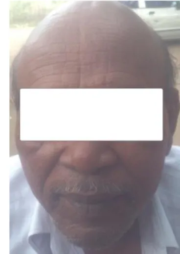
The features of the aging face appear due to a combina-tion of intrinsic and extrinsic factors[6]. These include wrinkles, malar depression, actinic changes, excess of skin, furrows, accumulation of submental fat and the showing of mandibular teeth. Sagging occurs due to the loss of skin elasticity, which decreases from an early age[7]. Wrinkles (Figures 1and2) appear due to the de-crease of procollagens I and III and collagen VII[8]. Fitz-patrick skin phototypes[9] are used to classify the responses to sun exposure by the skin (Table 2).There are many lasers available for skin resurfacing. They can be used in the treatment of skin wrinkles, acne scars, actinic and seborrheic keratosis, photo-aging and lentigines. Table 3 summarises the different types of lasers.
Figure 1. Aging face with wrinkles
Harsh S and Patil SB
141
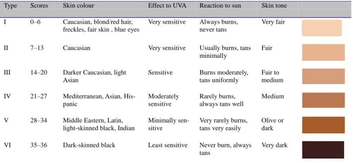
Table 2. Fitzpatrick skin types
Type Scores Skin colour Effect to UVA Reaction to sun Skin tone I 0–6 Caucasian, blond/red hair,
freckles, fair skin , blue eyes
Very sensitive Always burns, never tans
Very fair
II 7–13 Caucasian Very sensitive Usually burns, tans
minimally
Fair
III 14–20 Darker Caucasian, light Asian
Sensitive Burns moderately, tans uniformly
Fair to medium IV 21–27 Mediterranean, Asian,
His-panic
Moderately sensitive
Rarely burns, always tans well
Medium
V 28–34 Middle Eastern, Latin, light-skinned black, Indian
Minimally sen-sitive
Very rarely burns, tans very easily
Olive or dark VI 35–36 Dark-skinned black Least sensitive Never burn, always
tans
Very dark
Table 3. Summary of lasers
Laser type Wavelength (nm) Indications Pulse duration (ms) Complications
CO2 10,600 Skin resurfacing,
wrinkles
50 Edema, erythema, pruritus, contact dermatitis, hyperpigmentation
Erbium:YAG 2940 Mild-to-moderate
rhytids, scars, pig-mentation
.25 Minimal burning
Nd:YAG 1064, 1320,1440 Wrinkles, pigmenta-tion
50 Pain, redness, itching, swelling
IPL 500–1200 Dyspigmentation,
wrinkling, telangiec-tasia, hair removal
1–300 Erythema, discomfort, blisters, pain, hyperpigmentation, crusting, purpura
PDL 585, 595 Vascular lesions,
wrinkling, dyspig-mentation
45 Pain, purpura, swelling
KTP 532 Photo-damaged
red-brown discol-ouration
20–50 Edema, pain, crusting, erythema, telangiectasia
Alexandrite 755 Tattoo, hair removal 3 Pain, redness, swelling, itching
Diode 600–1020 Facial rhytids,
telan-giectasia, hair removal
30 Hyperpigmentation, erythema, edema
Ablative techniques
This is more aggressive as compared to non-ablative lasers. CO2 laser (10,600 nm) vaporises the epidermis
and dermis layer by layer. It rejuvenates the skin by epidermal regeneration and reorganisation, and by the strengthening of collagen bundles. It is more useful in severe facial wrinkles, challenging skin textures, and dyspigmentation[10]. Fractional ablative lasers provide adequate skin resurfacing safely as compared to the unfractionated models used earlier. Non-ablative la- ser spares the epidermis, decreases fine wrinkles, chang-
es the texture and tone of skin and treats dyspigmenta-tion.
The scarring produced in continuous wave (CW) laser is more when compared to the short-pulsed CO2 laser.
Currently, high power pulse (ultrapulse) and CW CO2
Facial laser surgery
142
minimally[12]
Ultrapulse CO2 lasers for midfacial region are treated
with 90 mJ/45W with first-pass density setting of 7, and less for the upper and lower eyelids and even lesser for hairline and jawline. In the second pass, lesser density is required as compared to the first. Some wrinkles may require a third pass.
Postoperative period may be complicated with swell-ing (which subsides in a week), erythema, pruritus and contact dermatitis. Post-inflammatory hyperpigmen-tation, which occurs 2–3 weeks post-therapy, may be decreased with prior treatment of retinoic acid and hy-droquinone[13].
Er:YAG lasers
It has a wavelength of 2,940 nm with water as its chro-mophore. It is used for superficial rhytids, actinic kerato-sis, dyschromia and Favre-Racouchot disease[14]. The penetration and ablation is more superficial compared to CO2. About 10–20 µm of thermal damage is caused by
10– 40 µm of skin impact. Skin tightening is achieved by the second and third pass. Wrinkles are reduced up to 50% in 2–3 passes[14]. Er:YAG has less crusting and erythema compared to CO2 laser[15]. Erbium can be
com-bined with CO2 to give uniformity in the treatment of
different areas of the face.
Fractional photothermolysis
It was developed by Relent technologies. The wave-length is 1,550 nm. It is among the recent options for skin rejuvenation[16]. It treats only a fraction of skin. The thermal side effects are decreased.
Non-ablative techniques
These include plasma skin regeneration, pulse dye laser, Nd:YAG, intense pulsed light (IPL), light-emitting di-ode(LED) devices and photodynamic therapy. It involves the sparing of epidermis, and affects the dermis directly, which helps in early recovery. The mechanism is by tar-geting chromophores such as melanin, haemoglobin and collagen. The wavelength is in the visible to infrared region, targeting the upper and middle part of the dermis. There is the activation of dermal fibroblasts, which helps in healing.
Plasma skin regeneration
Nitrogen plasma delivers energy to the skin. There is no chromophore mediator and the energy is delivered in a uniform and smooth way to the dermis by pulses of plasma. It is a non-ablative method used to treat facial rhytids, benign facial skin lesions and actinic keratosis.
Pulse dye laser
Long pulse dye laser is used for the treatment of facial
lentigines and wrinkles. There is no scarring or changes in skin pigmentation post-operatively[17]. There is an in-crease in dermal collagen with the use of 585-nm pulsed dye laser (PDL) between days 0–90 with maximum in-crease in the periorbital region[18].
Intense pulse light
Dyschromia, ephelides, senile and solar lentigines can be treated with intense pulsed light (IPL)[19]. It is a non-coherent light with a wavelength between 500–1,200 nm. It works on the basis of selective photothermolysis targeting haemoglobin, melanin and water. Skin tighten-ing in IPL occurs due to the contracture of the heat-ed collagen fibers. Photoaging due to telangiectasia and pigmentation shows significant improvement within three treatments[20]. Side effects include mild crust-ing, blistercrust-ing, erythema and purpura, all of which are transient and self-limiting.
Potassium titanyl phosphate
The 532-nm lasers produce energy pulses with small spots. They target oxyhemoglobin and melanin. Patients with Fitzpatrick types I to III are good candidates for these lasers[21]. They are effective in treating pho-to-damaged red and brown discolourations. Side effects of the therapy include erythema and edema.
Light-emitting diode
Light-emitting diode (LED) was invented in 1962. It stimulates collagen synthesis and accelerates fibro-blast-myofibroblast transformation. The wavelengths are 590, 633 and 830 nm. For fine wrinkles, the perior-bital area show more improvement than nasolabial area[22]. LED has shown to decrease erythema, edema, pain and bruising following blepharoplasty and periocu-lar resurfacing by Er:YAG/CO2 laser
[23]
.
Near-infrared laser
Rejuvenation with this laser is produced by long-lasting elastin stimulation. Significant improvement in the skin texture and wrinkles has been observed[24]. The increase in collagen and elastin improves skin texture, although without significant improvement in hyperpigmented le-sions[25].
Photodynamic therapy
It involves use of light with photosensitising substance ( amino levulonic acid). It has an additional advantage of destroying precancerous cells, in addition to the treatment of sun-damaged fine lines and pigmentation.
Facial laser surgery
recovery for these patients. The fractional lasers provide the best combination to give optimal results.
Treatment of pigmentation
Solar lentigines
Though there are many topical therapies available, the treatment of solar lentigines by laser provides al-most complete clearance. The options include Q-switched lasers, long-pulsed lasers and HGM K1 krypton lasers. Long-pulsed dye laser have shown near complete clearance of the lesion and reduced ma-chine-produced index factor[26]. Q-switched Nd:YAG with 532-nm wavelength is a good option for light-skinned patients with lentigines, and 1064nm for darker-skinned patients. Q-switched Nd:YAG is better than fractional CO2 laser for treating solar lentigines[27].
Solar elastosis
CO2, Nd:YAG, diode, IPL and Er:YAG can be used for
solar elastosis. Diode laser has the advantage of preserv-ing the epithelial layer with resurfacpreserv-ing effects, same as that of CO2 laser[28].
Melasma
Treatment options for melasma include:
1. Q-switched ruby laser
2. Erbium:YAG – refractory melasma 3. PDL – recurrent melasma
4. Fractional laser
5. IPL
Lasers should be used in patients of melasma who are refractory to topical therapies.
Use of lasers in facial acne
Potassium titanyl phosphate (KTP) laser may act through selective photothermolysis of the blood vessels or by a photodynamic effect on Propionibacterium acnes[29]. The 585-nm PDL has been used in treatment of acne scar[30]. It also decreases post-acne erythema. 1450-nm diode laser and 1540-nm Erbium glass laser are also used for acne with the latter causing decreased oiliness.
They can be used in the inflammatory phase of acne by acting on haemoglobin and water as chromo-phores and for the management
Laser for treatment of vascular lesions on
the face
Hemangioma
It is the most common type of vascular tumor (Figure 3).
Children should be treated when the hemangioma fails to regress. PDL with wavelength ranging from 585–600 nm with pulse width of about 0.45 ms[21] can be used to treat these lesions. Residual scarring can be treated by CO2 or
Erbium lasers.
Laser photocoagulation is the method of choice to treat port-wine stain. Argon laser can be used for the treatment of hypertrophied nodule of thickened port-wine stain. However, the disadvantage with Argon, CO2 and Nd:YAG is scarring[31]. Best results are with
flashlamp-pulse dye laser[32]. Patients are treated at in-terval of 4–6 weeks or on clinical judgement.
Venous malformation
Nd:YAG can be used for deeper and superficial lesions with 595–1,064-nm laser (Figure 4).
Lymphatic malformation
Laser is useful in treating superficial lymphatic malfor-mation of the head and neck area. 10,600-nm CO2 laser
is of help for treating mucosal lesions.
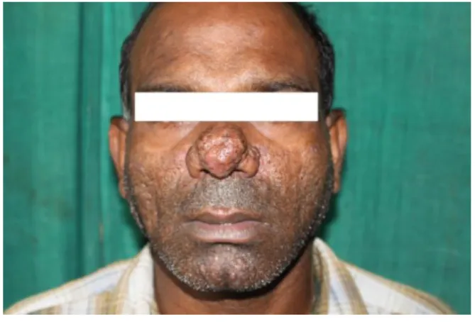
Facial telangiectasia and rosacea
Vascular laser can be used to deal with facial telangiecta-sia. CO2 and Erbium:YAG can be used to treat
rhi-nophyma (Figure 5). PDL remains the gold standard for the treatment of vascular lesions, though erythema and purpura may persist for a couple of weeks after treat-ment.
Figure 3. Hemangioma of the upper eyelid
Facial laser surgery
144
Laser for tattoo on the face
Traumatic tattoos can be due to carbon, graphite, etc. They can be removed with Q-switch Alexandrite laser[33]. Tattoos applied by a professional are deeper and are more difficult to remove. Exogenous ink is the chromo-phore. Quality switch lasers have been used traditionally to treat these tattoos. Multiple settings are required for complete removal of pigments. The appropriate lasers for different colours of tattoo are as follows:
1. Black and dark blue pigment: QS ruby, QS Nd:YAG (1,064 nm) and QS Alexandrite[34] 2. Orange and red brown: 1064-nm QS Nd:YAG
doubled in frequency 3. Red: Nd:YAG[35]
4. Green pigment: Alexandrite[35]
5. Purple and violet: Q-switched ruby laser[35] 6. Light coloured/pale: QS Alexandrite and QS
Nd:YAG laser[36] 7. Newer modalities:
a. Multi-pass treatment - multiple passes in one session[37]
b. Picosecond laser
Black, dark blue and red can all be removed with QS ruby, Nd:YAG and Alexandrite lasers.
Laser for unwanted hair on the face
Alexandrite (755 nm), diode (800 nm) and ruby (694 nm) lasers can be used to remove unwanted hairs on the face (Figure 6). Cutaneous hyper/hypopigmentation is more in these short wavelength lasers as compared to longer wavelength laser such as Nd:YAG (1,064 nm) laser. IPL and Q-switched Nd:YAG are other options for hair re-moval with good results.
Patients should be educated about the multiple treat-ment sessions and minor side effects such as itching, edema and redness after the procedure. People with higher Fitzpatrick skin types are more responsive to di-ode and Nd:YAG.
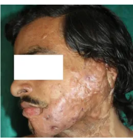
Laser for facial scar
Facial scars can be a result of trauma, post-surgery, post-burn or after any inflammatory process (Figures 7
and 8). Though many invasive and non-invasive treat-ment options are available, they are associated with re-currence, side effects, as well as failure rates.
Specific lasers for different scars
1. Post-burn scars: CO2 laser
2. Immature post-burn scar: Nd:YAG (1,064 nm)
3. Fibrotic scar: Fractional laser
4. Hyperpigmentation and discolouration in post-burn scars: IPL and Q-switch
5. Hypertrophic scars[38]: Nd:YAG (1,064 nm) and CO2
6. Post-traumatic scars[39]: Erbium glass (1,540 nm)
IPL, PDL and Erbium glass (1,540 nm) have shown de-cent results.
Figure 5. Rhinophyma of the nose
Figure 6. Black hairy nevus of the face
Facial laser surgery
Figure 8. Post-burn hypertrophic scar on the face
Lasers for cylindroma
The Nd:YAG laser[40] and CO2 laser[41] can be used to
treat cylindroma (Figure 9) in the head and neck region[40,41].
Figure 9. Cylindroma of the head and neck region
Conclusion
Lasers have become a powerful tool for many procedures on the face. Proper training of the care provider along with the staff and maintaining acceptable standards in the clinic makes the process safe. The risk-benefit ratio, the usefulness and the adverse effects of the procedure should not only be kept in mind at the time of proce-dure but are also explained to the patient and document-ed in detail prior to the procdocument-edure. The safety of mdocument-edical
personnel and the patient are of paramount importance and laser safety protocols should be followed every time the procedure is performed. Caution should be taken when evaluating the patient with unrealistic expectations.
Author contributions
Both S Harsh and SB Patil contributed to the drafting of manuscript.
Conflict of interest
The authors declare no potential conflict of interest with respect to the research, authorship, and/or publication of this article.
References
1. Shampo MA, Kyle RA, Steensma DP. Aleksandr Prokhorov—Laser and Masers. Mayo Clin Proc 2011; 86(5): e33. doi: 10.4065/mcp.2011.0194.
2. Omi T, Numano K. The role of CO2 laser and fractional CO2 laser in dermatology. Laser Ther 2014; 23(1): 49–60. doi: 10.5978/islsm.14-RE-01.
3. Sorokin PP and Lankard JR Stimulated emission ob-served from an organic dye, chloro-aluminum phthalo-cyanine. IBM J Res Develop 1966; 10(2): 162–163. doi: 10.1147/rd.102.0162.
4. Anderson RR, Parrish JA. Selective photothermolysis: Precise microsurgery by selective absorption of pulsed radiation. Science1983; 220(4596): 524–527. doi: 10.11 26/science.6836297.
5. Smalley PJ. Laser safety: Risks, hazards, and control measures. Laser Ther 2011; 20(2): 95–106. doi:10.5978/islsm.20.95.
6. Cevenini E, Invidia L, Lescai F, Salvioli S, Tieri P, et al. Human models of aging and longevity. Expert Opin Biol Ther 2008; 8(9): 1393–1405. doi: 10.1517/147125 98.8.9.1393.
7. Escoffier C, Rigal J, Rochefort A, Vasselet R, Leveque JL,
et al. Age related mechanical properties of human skin: An in vivo study. J Invest Dermatol 1989; 93(3): 353–357. doi: 10.1016/0022-202X(89)90058-4.
8. Watson REB, Craven NM, Griffiths CE, Kang S, Jones CJ, et al. A short term screening protocol, using fibrillin-1 as reporter molecule for photoaging. J Invest Dermatol 2001; 116(5): 672–678. doi: 10.1046/j.1523-1747.20 01.01322.x.
Harsh S and Patil SB
146
doi:10.18282/jsd.v2.it1.137 10. Preissig J, Hamilton K, Markus R. Current laser
resur-facing technologies: A review that delves beneath the surface. Semin Plast Surg 2012; 26(3): 109–116. doi: 10.1055/s-0032-1329413.
11. Kauvar ANB, Geronemus RG. Histology of laser resur-facing. Dermatol Clin 1997; 15(3): 459–467. doi: 10.1016/S0733-8635(05)70454-X.
12. Bernstein LJ, Kauvar ANB, Grossman MC, Geronemus RG. Scar resurfacing with high-energy, short-pulsed and flashscanning carbon dioxide lasers. Dermatol Surg 1998; 24(1): 101–108. doi: 10.1111/j.1524-4725.1998.tb040 60.x.
13. Nanni CA, Alster TS. Complications of carbon dioxide laser resurfacing. An evaluation of 500 patients. Dema-tologig Surgery.1998;24: 315–320
14. Avram DK, Goldman MP. The safety and effectiveness of single-pass Erbium:YAG laser in the treatment of mild to moderate photodamage. Dermatol Surg 2004; 30(8): 1073–1076. doi: 10.1111/j.1524-4725.2004.30330.x. 15. Newman JB, Lord JL, Ash K, McDaniel DH. Variable
pulse erbium:YAG laser skin resurfacing of perioral rhytides and side-by-side comparison with carbon dioxide laser. Lasers Surg Med 2000; 26(2): 208–214. doi: 10.1002/(SICI)1096-9101(2000)26:2<208::AID-LSM12> 3.0.CO;2-R.
16. Hamilton MM, Hobgood T. Emerging trends and tech-niques in male aesthetic surgery. Facial Plast Surg 2005; 21: 324–327. doi: 10.1055/s-2006-939512.
17. Kono T, Groff WF, Sakurai H, Takeuchi M, Yamaki T, et al. Comparison study of intense pulse light versus a long-pulse pulsed dye laser in the treatment of facial skin rejuvenation. Ann Plast Surg 2007; 59(5): 479–483. doi: 10.1097/SAP.0b013e3180327943.
18. Moody BR, McCarthy JE, Hruza GJ. Collagen remodel-ling after 585-nm pulsed dye laser irradiation: An ultra-sonographic analysis. Dermatol Surg 2003; 29: 997–1000.
19. Kawada A, Asai M, Kameyama H, Sangen Y, Aragane Y,
et al. Videomicroscopic and histopathological investiga-tion of intense pulsed light therapy for solar lentigines. J Dermatol Sci 2002; 29(2): 91–96. doi: 10.1016/S0923-18 11(02)00014-2.
20. Weiss RA, Weiss M, Beasley K. Rejuvenation of pho-toaged skin: 5 years results with intense pulsed light of the face, neck and chest. Dermatol Surg 2002; 28(12): 1115–1119. doi: 10.1097/00042728-200212000-00004. 21. Wall TL. Current concepts: Laser treatment of adult
vas-cular lesions. Semin Plast Surg 2007; 21(3): 147–158. doi: 10.1055/s-2007-991183.
22. Baez F, Reilly LR. The use of light-emitting diode
thera-py in the treatment of photoaged skin. J. Cosmet Derma-tol 2007; 6(3): 189–194. doi: 10.1111/j.1473-2165.20 07.00329.x.
23. Trelles MA, Allones I. Red light-emitting diode (LED) therapy accelerates wound healing post-blepharoplasty and periocular laser ablative resurfacing. J. Cosmet Laser Therapy 2006; 8(1): 39–42. doi: 10.1080/14764170 600607731.
24. Tanaka Y, Matsuo K, Yuzuriha S. Objective assessment of skin rejuvenation using near-infrared 1064-nm neodym-ium: YAG laser in Asians. Clin Cosmet Investig Dermatol 2011; 4: 123–130. doi: 10.2147/CCID.S22841.
25. Lee JH, Roh MR, Lee KH. Effects of infrared radiation on skin photo-aging and pigmentation. Yonsei Med J 2006; 47(4): 485–490. doi: 10.3349/ymj.2006.47.4.485. 26. Ghaninejhadi H, Ehsani A, Edrisi A, Gholamali F, Akbari
Z, et al. Solar lentigines: Evaluating pulsed dye laser (PDL) as an effective treatment option. J Lasers Med Sci 2013; 4(1): 33–38.
27. Vachiramon V, Panmanee W, Techapichetvanich T, Chanprapaph K. Comparison of Q-switched Nd:YAG la-ser and fractional carbon dioxide lala-ser for the treatment of solar lentigines in Asians. Lasers Surg Med 2016; 48(4): 354–359. doi: 10.1002/lsm.22472.
28. Muccini JA Jr, O’Donnell FE Jr, Fuller T, Reinisch L. Laser treatment of solar elastosis with epithelial preserva-tion. Lasers Surg Med 1998; 23(3):121–127. doi: 10.1002/(SICI)1096-9101(1998)23:3<121::AID-LSM1>3 .0.CO;2-R.
29. Jih MH, Kimyai-Asadi A. Laser treatment of acne vul-garis. Semin Plast Surg 2007; 21(3): 167–174. doi: 10.1055/s-2007-991185.
30. Alster TS, McMeekin TO. Improvement of facial acne scars by the 585 nm flashlamp-pumped pulsed dye laser. J Am Acad Dermatol 1996; 35(1): 79–81. doi: 10.1016/S0190-9622(96)90501-0.
31. Dixon JA, Huether S, Rotering R. Hypertrophic scarring in argon laser treatment of port-wine stains. Plast Reconst Surg 1984; 73(5): 771–777. 10.1097/00006534-198405 000-00009.
32. Hsiao YC, Chang CJ. Update on flashlamp pumped pulsed dye laser treatment for port wine stains (capillary malformation) patients. Laser Ther 2011; 20(4): 265–272. doi: 10.5978/islsm.11-RE-01.
Facial laser surgery
Fitzpatrick RE, et al. Comparison of the Q-switched Al-exandrite, Nd:YAG, and ruby lasers in treating blue-black tattoos. Dermatol Surg 1999; 25(1): 10–14. doi: 10.10 46/j.1524-4725.1999.08122.x.
35. Zelickson BD, Mehregan DA, Zarrin AA, Coles CC, Hartwig P, et al. Clinical, histologic, and ultrastructural evaluation of tattoos treated with three laser systems. La-sers Surg Med 1994; 15(4): 364–372. doi: 10.1002/lsm.19 00150406.
36. Fitzpatrick RE, Lupton JR. Successful treatment of treatment-resistant laser-induced pigment darkening of a cosmetic tattoo. Lasers Surg Med 2000; 27(4): 358–361. doi:10.1002/1096-9101(2000)27:4<358::AID-LSM9>3.0. CO;2-0.
37. Kossida T, Rigopoulos D, Katsambas A, Anderson RR.
Optimal tattoo removal in a single laser session based on the method of repeated exposures. J Am Acad Dermatol 2012; 66(2): 271–277. doi: 10.1016/j.jaad.2011.07.024. 38. El Taweel AI, Abd El-Rahman SH. Assessment of
frac-tional CO2 laser in stable scars. Egypt J Dermato Venerol 2014; 34(1): 74–80. doi: 10.4103/1110-6530.137317. 39. Venkatram, George EP. Treatment of posttraumatic scar
with ablative fractional erbium: YAG laser. Cosmetol & Oro Facial Surg 2016; 2(1): 104.
40. Tarsted M, Molin L. Nd:YAG laser for effective treatment of multiple cylindroma of scalp. J Cosmet Laser Ther 2004; 6(1): 41–43. doi: 10.1080/14764170410030804. 41. Brooke–Spiegler syndrome: Treatment of cylindromas
with CO2 laser. Dermatol Surg 2000; 26(9): 877–882 doi: 10.1046/j.1524-4725.2000.00 034.x.