Original Research Article
Evaluation of eustachian tube function in chronic suppurative otitis
media with reference to its treatment outcome
Sucheta Gupta
1, Mohit Goel
2*, Padam Singh Jamwal
3INTRODUCTION
Eustachian tube is the only tube connecting the middle ear and the nasopharynx and has long been recognized as main organ ventilating middle ear. Thus middle ear is then connected to atmospheric space, and middle ear pressure is regulated only at that movement.
Within this system, the Eustachian tube has at least three physiologic functions with respect to the middle ear. (1) Equilibration of air pressure (ventilation) in the middle ear with atmospheric pressure, (2) protectionfrom naso-pharyngeal sound pressure and secretions, (3) drainage (and clearance) into the nasopharynx of unwanted secretions and secretions produced within the middle ear.1
ABSTRACT
Background: The study was undertaken to find out Eustachian tube function in safe type of chronic suppurative otitis media and to study the comparison of graft uptake in normal, partially impaired and grossly impaired Eustachian tube function in safe type of chronic suppurative otitis media.
Methods: The present prospective study was conducted in the Department of Otorhinolaryngology and Head and Neck surgery, Sri Maharaja Gulab Singh Hospital, Jammu during the period from November 2016 to October 2017. Patients were diagnosed clinically and also audiometrically by pure tone audiometry and impedance audiometry. Eustachian tube function test- Toynbee test was done in all the patients.
Results: Out of 20 patients with normal Eustachian tube function, 19(95%) showed graft uptake. Out of 20 patients with partially impaired Eustachian tube function, 18 (90%) showed graft uptake. Out of 20 patients with grossly impaired Eustachian tube functions 13 (65%) showed graft uptake. Comparison of mean values of pre and post- operative air-bone (AB) gap with respect to normal, partially impaired and grossly impaired Eustachian tube functions is highly significant statistically with (p<0.001).
Conclusions: As seen in our study, functioning Eustachian tube is an important requirement for optimum outcome of myringoplasty. Testing the functions of Eustachian tube before surgery provides a possibility of predicting the possible outcome of myringoplasty or tymapanoplasty. This is also concluded that a partially functioning Eustachian tube should not be considered to be a contraindication to these surgeries as in many of these cases graft uptake and AB gap closure was good (90%).
Keywords: Eustachian tube, Tympanoplasty, Toynbee test
1Department of Health, Government of Jammu and Kashmir, Jammu and Kashmir, India
2Department of ENT, Dr. Rajendra Prasad Government Medical College, Tanda, Kangra, Himachal Pradesh, India 3Department of ENT, Government Medical College, Jammu, Jammu and Kashmir, India
Received: 31 August 2019
Revised: 02 December 2019
Accepted: 03 December 2019
*Correspondence:
Dr. Mohit Goel,
E-mail: [email protected]
Copyright: © the author(s), publisher and licensee Medip Academy. This is an open-access article distributed under the terms of the Creative Commons Attribution Non-Commercial License, which permits unrestricted non-commercial use, distribution, and reproduction in any medium, provided the original work is properly cited.
The drainage function is enhanced by the pump-like action of the tube during active opening and closing.2
There appear to be two types of Eustachian tube dysfunction which could result in otitis media: obstruction and abnormal patency.
Functional obstruction can be the result of denervation or direct interference with the action of tensor veli palatine muscle, such as can occur with neurologic disease, trauma, surgery or tumour.3,4 Mechanical obstruction of
the Eustachian tube may be intrinsic or extrinsic, or both.
Tubal malfunction is the most important factor in the
development and persistence of otitis media.
Measurement of the Eustachian tube function (ETF) is therefore an important part of the medical and surgical evaluation of the middle ear disease.5
Causes of Eustachian tube obstruction
Upper respiratory infection; allergy; sinusitis; nasal polyps; deviated nasal septum; hypertrophic adenoids; nasopharyngeal tumour or mass, cleft palate; submucous cleft palate; down syndrome and functional obstruction.
ETF tests were valsalva, politzer, catheterization, Toynbees test, tympanometry (also called inflation deflation test), radiological test with radio opaque dye, saccharine or methylene blue test, sonotubometry.
Properly functioning Eustachian tube is an integral part of a normally functioning middle ear and the existence of a good tubotympanic mucociliary drainage constitutes a favourable prognostic factor in the outcome of reconstructive surgery of middle ear.
Objectives of this study are to find out ETF in safe type of chronic suppurative otitis media, to compare graft uptake rate in normal, partially impaired and grossly impaired ETF in safe type of chronic suppurative otitis media and to compare the hearing improvement in ears with taken up drum in normal, partially impaired and grossly impaired ETF.
METHODS
The present prospective study was conducted in the Department of Otorhinolaryngology and Head and Neck surgery, Sri Maharaja Gulab Singh Hospital, Jammu during the period from November 2016 to October 2017. The study was conducted after taking approval of institutional Ethics committee. The patients attending ENT outpatient department were enrolled in the study.
Patients were diagnosed clinically and also
audiometrically by pure tone audiometry (PTA) and impedance audiometry. The ETF was determined by Toynbee test on tympanogram.
Inclusion criteria
All patients in the age group of 15-60 years, patients having a central type of perforation, dry ear preferably for a period of 3 months, patients without any evidence of upper respiratory tract infection, patients not having any evidence of intracranial complications, patients capable of performing various ETF tests, children, patients having good cochlear reserve were included in this study. General and systemic examinations shall be within normal limits.
Exclusion criteria
Age of greater than 15 and less than 60 years, cases with attic perforation or retraction, cases with infected middle or external ear; cases with bleeding diathesis, cases with diabetes mellitus, pregnancy, cases with poor air-bone gap on PTA were excluded.
Procedure
A detailed history of patients was taken with thorough clinical examination including otoscopy and tuning fork tests. PTA and impedance audiometry were done routinely in all patients. Other supportive investigations like complete hemogram, blood for sugar, urea and creatinine and radiology of paranasal sinuses was performed in all patients to exclude any abnormality and thereby preparing them for surgery. Detailed information of each patient was recorded in a performa. ETF test- Toynbee test was done in all the patients. An equal number of patients with normal, partially impaired and grossly impaired ETF were considered for surgery. Preoperative counselling was done in all patients to explain advantages and disadvantages of surgery. Post operatively the patients were followed weekly for 1 month and then monthly at 2 months and 3 months and hearing evaluation was done using pure tone audiometry and impedance audiometry.
The purpose of undertaking the ETF tests is to assess its physiological profile. It is the physiologically functioning and not anatomical patency of Eustachian tube that is required for maintaining the normal function of middle ear.
Toynbees test
partially neutralised with each swallow. The air pressure of middle ear was monitored and recorded graphically by impedance audiometer. Any change of pressure during swallowing was recorded as a step ladder type of graph. Normally the positive or negative middle ear pressure should be partially neutralised with each swallow and in 3 or 4 swallows the pressure should totally neutralise, i.e. it should reach 0 mm of water pressure. If some residual pressure persists even after 5 swallows, the tubal function was considered to be partially impaired. If positive or negative pressure built up by impedance audiometer cannot be neutralised at all by repeated swallowing, then the ETF was considered to be grossly impaired.
All the patients of safe type of Chronic suppurative otitis media with normal, partially impaired and grossly impaired ETF tests were considered for surgical procedure: myringoplasty or tympanoplasty was done under local or general anaesthesia by postaural or endaural or transcanal approach. Grafts used were temporalis fascia, tragal perichondrium, conchal perichondrium, temporalis fascia with partial thickness conchal cartilage.
Post operatively patients were started on suitable antibiotics for 1 week along with analgesics, antihistaminics, and multivitamins.
Mastoid dressing was changed on 2nd postoperative day
and dressing applied. The sutures were removed on 8th
post-operative day.
The patients were reviewed weekly after discharge for first three weeks and then reviewed monthly after 1 month, 2 months and 3 months. Patients were evaluated postoperatively using otoscopy.
Post operatively hearing evaluation was done in all operated patients using pure tone audiometry at 1 month, 2 months, and 3 months and compared with the preoperative evaluation. On the basis of ear findings following myringoplasty/tympanoplasty, patients were divided into two groups. (1) Successful outcome, defined as healed graft with good middle ear function, (2) graft failure or perforation secondary to otitis media during follow up period were considered as failures.
Statistical analysis
The information collected was compiled, tabulated and analysed. Descriptive statistics were used for the demographic profile of subjects and summarized as mean +standard deviation (SD). Paired sample T test was performed for continuous data to find out the significance of difference of means of various measures. A p value of less than 0.05 (p<0.05) was considered statistically significant. The results were tabulated and data was analysed using Statistical Package for Social Sciences (SPSS) computer software program version 20.
RESULTS
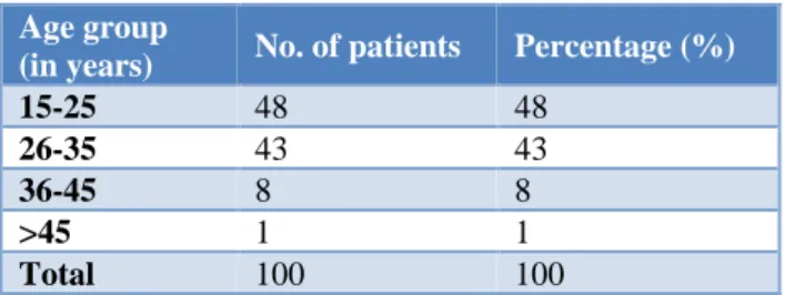
A total of 100 patients were enrolled in the present study. The patients were in the age group of 17-49 years with mean age of 26.66±6.39 years. Maximum patients were in the age group of 17-35 years (91%). Age wise distribution of cases is given in Table 1.
Table 1: Age distribution of patients.
Age group
(in years) No. of patients Percentage (%)
15-25 48 48
26-35 43 43
36-45 8 8
>45 1 1
Total 100 100
Table 2: Sex distribution of patients.
Sex No. of patients Percentage (%)
Female 45 45
Male 55 55
Total 100 100
Female patients were less in number as compared to male. 55% were males and 45% were females and M:F ratio was 1.2:1. Sex distribution of patients is given in Table 2.
There was history of hearing loss in 76 patients. History of discharge in 24 patients and no history of tinnitus in any patient. Presenting symptoms in patients shown in Table 3.
Table 3: Distributions of patients according to presenting symptoms.
Symptoms No. of patients Percentage (%)
Hearing loss 76 76
Discharge 24 24
Tinnitus 0 0
Total 100 100
Table 4: Distribution of patients according to age of onset of disease.
Age of onset
(in years) No. of patients Percentage (%)
15-20 34 34
21-25 30 30
26-30 19 19
31-35 11 11
36-40 5 5
41-45 0 0
>45 1 1
Maximum patients have age of onset of disease between 15-35 years with mean age of onset 24.01±6.29 years. Distribution of patients according to age of onset of disease is given in Table 4.
Table 5: Distribution of patients according to preoperative AB gap.
Preoperative of AB gap (in dB)
No. of
patients Percentage (%)
11-15 4 4
16-20 38 38
21-25 47 47
26-30 11 11
Total 100 100
Out of 100 patients 85 (85%) had pre-operative AB gap in the range of 16-25 db. With mean air-bone (AB) gap of 20.87±4.08. Preoperative AB gap of patients is shown in Table 5.
Table 6: Distribution of patients according to Eustachian tube functions.
ETF No. of
patients Percentage (%)
Normal 34 34
Partially impaired 34 34
Grossly impaired 32 32
Total 100 100
Out of 100 patients 34 patients have normal, 34 patients have partially impaired and 32 patients have grossly impaired ETFs. Distribution of patients according to ETFs is shown in Table 6.
Out of 100 patients, surgery was conducted only in 60 patients, as shown in Table 7. Equal number of patients with normal, partially impaired and grossly impaired
ETFs were taken. Number of 20 from each group (33.33%).
Table 7: Distribution of patients according to ETF in which we conducted surgery.
ETF No. of
patients Percentage (%)
Normal 20 33.33
Partially impaired 20 33.33
Grossly impaired 20 33.33
Total 60 100
All the patients were operated under local anaesthesia. The approach used in maximum patients i.e. 55 (91.66%) was post aural while transcanal in 4 (6.66%) patients and end aural in 1 patient (1.66%).
Graft used was temporalis fascia graft in 54 (90%) patients, tragal perichondrium in 5 (8.33%) patients, and conchal perichondrium in 1 (1.66%) patient.
Out of 20 patients with normal ETF, 19 (95%) showed graft uptake. Out of 20 patients with partially impaired ETF, 18 (90%) showed graft uptake. Out of 20 patients with grossly impaired ETFs 13 (65%) showed graft uptake. Table 8 shows the comparison of graft uptake in normal, partially impaired and grossly impaired ETFs. Comparison is significant with (p=0.024).
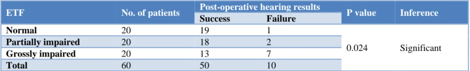
Out of 20 patients with normal ETF, 19 (95%) showed success in respect to hearing with failure of only 1 case. Out of 20 patients with partially impaired ETF 18 (90%) patients showed success in respect to hearing with failure of 2 cases and out of 20 patients with grossly impaired ETF, 13 (65%) patients showed success with failure of 7 cases. Table 9 is showing the hearing results in operated patients with respect to ETF which is significant (p=0.024).
Table 8: Comparison of graft uptake in normal, partially impaired and grossly impaired ETF in operated patients.
ETF No. of patients Graft uptake Percentage (%)
P value Inference
Normal 20 19 95
Partially impaired 20 18 90
0.024 Significant
Grossly impaired 20 13 65
Total 60 50 100
Chi Square=7.44, p=0.024.
Table 9: Hearing results in operated patients with respect to ETF.
ETF No. of patients Post-operative hearing results P value Inference
Success Failure
Normal 20 19 1
0.024 Significant
Partially impaired 20 18 2
Grossly impaired 20 13 7
Total 60 50 10
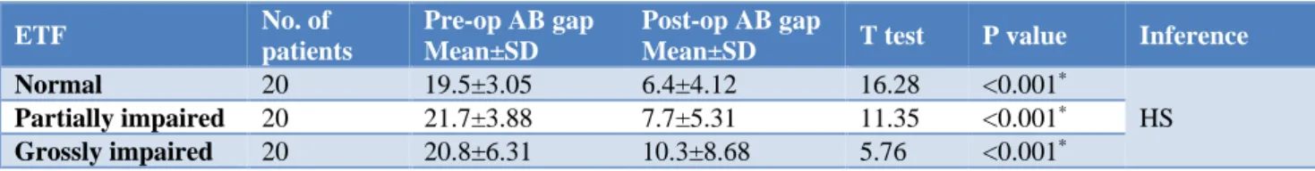
Table 10: Comparison of mean values of pre and post-operative AB Gap in different grades of ETF using paired t test.
ETF No. of
patients
Pre-op AB gap Mean±SD
Post-op AB gap
Mean±SD T test P value Inference
Normal 20 19.5±3.05 6.4±4.12 16.28 <0.001*
HS
Partially impaired 20 21.7±3.88 7.7±5.31 11.35 <0.001* Grossly impaired 20 20.8±6.31 10.3±8.68 5.76 <0.001* *P value is significant.
Comparison of mean values of pre and post-operative AB gap with respect to normal, partially impaired and grossly impaired ETFs. Comparison is highly significant statistically with (p<0.001) as shown in Table 10.
DISCUSSION
Eustachian tube dysfunction is widely recognised as the triggering factor of middle ear disease. ETF tests are not routinely done. ETF’s role in successful outcome of surgeries for chronic otitis media has become a topic of interest in recent years.
Recent studies show that the success rates of tympanoplasty were found to be lesser when surgery was done in ears having poor tubal function compared to ears with normal ETF. These studies have made surgeons realise the importance of Eustachian tube and testing the functioning of Eustachian tube.
Age and sex distribution
In our study the average age at the time of surgery was 26.66±6.39 years the range was 17-49 years which strongly correlates with research by Antony et al.6 The
majority of patients in their study were young adults between the age group 0f 20-29 years. The mean age group was 27.18±11.65 years. In our study of the 100 patients, 45% were females and 55% were males. However, in study conducted by Prasad et al of the 86 patients, 45 were males and 41 were females with no significant difference in sex ratio which is comparable with our study.7
ETFs
We categorised ETF as normal, partially impaired and grossly impaired. Priya et al and Saravanan et al have also categorised the ETFs of their patients in a similar manner.8,9
Relationship of graft uptake with respect to ETF tests
In the present study 20 patients each of normal, partially impaired and grossly impaired ETFs were subjected to myringoplasty. In patients with normal ETF, graft uptake was seen in 95% with failure of graft uptake in only 1 (5%) patient. In partially impaired ETF, graft uptake was seen in 90% patients with failure of graft uptake in only 2 (10%) patients while in grossly impaired ETF, graft
uptake was seen only in 65% patients with failure of 7 (35%) cases. This comparison was statistically significant with p value (0.024) depicting that ETF is important in predicting the outcome of myringoplasty or tympano-plasty, our results correlates with study conducted by Priya et al who found correlation between ETF and graft uptake was statistically significant with p<0.01 (i.e. 0.0005). 100% success rate was seen in normal ETF, 98% in partially impaired ETF, and 76% in grossly impaired ETF.8 Our results also correlates with Cohn et al. who
assessed ETF by using impedence audiometry (Toynbees test). In their study those with normal ETF, showed a graft uptake of 95%, 75% in partially impaired ETF and 69% graft uptake in totally impaired ETF.10 Although our
study don’t match with a study conducted by Tadke et al who showed graft uptake of (94.87%) in normal ETFs with failure of 2 (5.12%) cases and in impaired ETF uptake was (85.71%) with failure of 3 (14.28%) cases but they found no significant association between ETF and successful graft uptake (p>0.05).11
Hearing evaluation before and after surgery
In our study out of 20 patients with normal ETF, 19 (95%) showed success in respect to hearing with failure of only 1 (5%) case. Out of 20 patients with partially impaired ETF, 18 (90%) showed success in respect to hearing with failure of 2 (10%) cases and out of 20 cases with grossly impaired ETF, 65% (13) patients showed success with failure of 7 (35%) cases. Mean pre-operative AB gap in patients with normal ETF was 19.5±3.05. Mean preoperative AB gap in patients with partially impaired ETF was 21.7±3.88 and mean pre-operative AB gap in patients with grossly impaired ETF was 20.8±6.31. Mean post–operative gap in patients with normal ETF was 6.4±4.12. Mean post-operative AB gap in patients with partially impaired ETF was 7.7±5.31 and mean post –operative AB gap in patients with grossly impaired ETF was 10.3±8.68. P value came out to be (<0.001) showing the relationship highly significant which means that the patients with poorer ETF had a higher AB gap pre-operatively. Our hearing results are comparable with Singh MN et al which showed overall graft uptake rate in their study 90%and hearing improvement in terms of AB gap with in 0-15 db in 83.3%. 28 (93%) cases had a patent Eustachian tube, out of these 89.3% had a successful outcome, whereas 2 (7%) had a blocked Eustachian tube, out of which 100% had an unsuccessful outcome.12 However study by Antony et al which
PTA average of the patients was 30.69±13.997 db.6 Mean
PTA average at 3 months was 24.84±10.09 and at 6 months 25.37±10.52 db. Impedence audiometry for Eustachian tube showed that of the 50 cases, 68% of patients were having patent ETF and 32% patients were having obstructed ETF. 62.5% cases with obstructed ETF had successful graft uptake and 37.5% patients had reperforation after 6 months which showed that good ETF is required for obtaining a good outcome during tympanoplasty.
CONCLUSION
This study reveals that functioning Eustachian tube is an important requirement for optimum outcome of myringoplasty. Testing the functions of Eustachian tube before surgery provides a possibility of predicting the possible outcome of myringoplasty or tymapanoplasty. This is also concluded that a partially functioning Eustachian tube should not be considered to be a contraindication to these surgeries as in many of these cases graft uptake and AB –gap closure was good (90%).
Funding: No funding sources Conflict of interest: None declared
Ethical approval: The study was approved by the Institutional Ethics Committee
REFERENCES
1. Bluestone CD, Paradise JL, Beery QC. Physiology of the Eustachian tube in the pathogenesis and management of middle ear effusions. Laryngoscope. 1972;82:1654-70.
2. Honjo I, Kumazawa T, Honda K. Simple Impedance
Test for Eustachian Tube Function. Arch
Otolaryngol. 1981;107:221-3.
3. Myers EN, Beery QL, Bluestone CD, Rood SR, Sigler B. Effect of certain head and neck tumours and their management on the ventilator function of the Eustachian tube. Ann Otol Rhinol Laryngol. 1984;114:1-16.
4. Takahara T, Sando I, Bluestone CD, Myers EN. Lymphoma invading the anterior Eustachian tube:
Temporal bone histopathology of functional tubal
obstruction. Ann Otol Rhinol Laryngol.
1986;95:101-5.
5. Mittal VK. Assessment of Eustachian Tube
Functions in patients with ear drum perforation. Indian J Otolaryngol. 1985;37:23-5.
6. Antony A, Deviprasad, Kallikkadan HH.
Tympanometric Assessment of Eustachian tube function before and after tympanoplasty. IOSR– JDMS. 2015;14:42-7.
7. Prasad KC, Hegde MC, Prasad SC, Meyappan H. Assessment of Eustachian tube function in tympanoplasty. Otolaryngol Head Neck Surg. 2009;140(6):889-93.
8. Priya K, Karthikeyan P, Coumare VN, Sambandan AP. Evaluation of Eustachian tube function in chronic suppurative otitis media (tubotympanic type) with reference to its treatment outcome. Indian J Otol. 2012;18:179-83
9. Saravanan V, Kumar RV, Vasudevan M, Vineetha K. Study of Eustachian tube function in normal adults and those with middle ear disease. IOSR-JDMS. 2017;16:76-80.
10. Cohn AM, Schwaber MK, Anthony LS, Jerger JF. Eustachian tube function and tympanoplasty. Ann Otol. 1979;88:339347.
11. Tadke KR, Lahane VJ, Wakode PT. Role of
impedance audiometry in evaluation of Eustachian tube function and its correlation with tympanoplasty surgery outcome: Our experience. IOSR-JDMS. 2017;16:45-9.
12. Singh MN, Hamam PD, Lyngdoh NC, Priyokumar OS. Evaluation of hearing status in pre and post- operative endoscopic type 1 tympanoplasty and its influencing factors. J Med Soc. 2014;28:166-70.