Address for correspondence Dr. Talat Masood Akbar Department of Dermatology Lahore Medical and Dental College, Ghurki Trust Teaching Hospital, Lahore. Email: [email protected]
Original Article
Allergic contact dermatitis in hand eczema: a
clinico-etiological study
Introduction
Hand eczema is a common condition, often with a long-lasting and relapsing course.1 It causes
discomfort, pain, distress and adversely affects social interaction in patients. Studies reported a 1-year prevalence of 9.7% in the general population,2 and an incidence rate reported to be
5.5-8.8 per 1000 person-years.3,4
Etiology of hand eczema includes external, as well as, endogenous factors, such as exposure to skin irritants and contact allergens, and individual variations in the effectiveness of skin barrier functions.5 Environmental risk factors
contribute relatively more to the disease than genetic factors6 and it is a frequently reported
occupational dermatosis.7,8
Clinical patterns of hand eczema are variable and it may be classified into different
Talat Masood Akbar, Muhammad Nadeem*, T.S. Haroon
Department of Dermatology, Lahore Medical and Dental College, Lahore * Department of Dermatology, Fatima Jinnah Medical University, Lahore
Abstract
Objective To identify the various morphological types of hand eczema and to detect the common etiological sensitizers in our community by patch testing, and attempt to correlate them.Methods One hundred patients of hand eczema presenting to the department of dermatology, Mayo hospital, were enrolled in the research study after informed consent. After taking a careful history and complete examination, they were classified according to clinical presentation, and patch tested with European standard series. Patches were applied to the upper back in accordance with the principles of patch testing. Results were read at 48, 72 and 120 hours after removal of patches, and interpreted according to the International Contact Dermatitis Research Group criteria. Data was analyzed and interpreted.
Results Patch testing results indicated that 46% of patients had positive allergic reactions. The commonest allergens detected were nickel (21%), chromate (13%), neomycin (12%) and cobalt (11%). Nickel sensitivity was more common in females (19%), while chromate was more common in males (11%). Attempt at clinicoetiological correlation showed few patterns, as different allergens could cause various morphological presentations. However, nickel sensitivity was noted in ring eczema (75%) and pompholyx (44%), while no allergens were detected in apron and hyperkeratotic palmar eczema.
Conclusion A high allergic sensitivity (46%) was noted in our patients of hand eczema. Different allergens could cause varied morphology and no specific clinicoetiological patterns can be defined. Therefore, a patch test should essentially be done in all hand eczema patients, to detect an etiological allergen and for directing better management of our patients.
Key words
morphological types, but there are few correlations with their aetiology.9,10,11 An atopic
diathesis and exposure to irritants can be suspected on history, but there are no readily available tests, simple or reliable enough to confirm the condition. Allergic contact dermatitis, on the other hand, may be proved scientifically by a patch test if it is properly applied and correctly interpreted.12
Although clinical descriptions are still our focus, disease classification has moved from morphology to etiology, as for many other diseases. It is the rule rather than the exception that diseases, in spite of having similar etiologies, may show great variations clinically.13 This also applies to hand eczema.
In this study, hand eczema patients were studied to define their clinical patterns in our community, to determine common sensitizers by patch testing and attempt to correlate clinical patterns to etiology.
Methods
A hundred patients of hand eczema, presenting sequentially to the Department of Dermatology, Mayo Hospital, Lahore and fulfilling the inclusion criteria were enrolled in the study. Inclusion criteria were: patients of either sex, over 12 years of age, having eczema either only involving the hands or predominantly involving the hands, not having taken steroids, immunosuppressives or antihistamines for two weeks. Pregnant and lactating females were excluded from the trial.
After informed consent, a detailed history with special reference to atopy, type of work performed, and exposure to allergens was taken. A meticulous clinical examination was done. Patients of hand eczema were classified into morphological types: discoid eczema,
pompholyx, fingertip eczema, dry palmar eczema, ring eczema, gut eczema, apron eczema, recurrent focal palmar peeling, hyperkeratotic palmar and patchy vesiculosquamous eczema.14
Details were recorded on a specially designed proforma. Investigations performed included microscopic examination for fungus and histopathological examination of biopsy specimen if needed.
A patch test was then applied to each patient. The battery employed was the European Standard Series (Chemotechnique Diagnostics AB®, Malmo, Sweden) in standard chambers (Van der Bend square chambers). Patches were applied to the upper back in accordance with the principles of patch testing, as described by Fisher.12 Patients were advised to avoid exposure
to water or exercise, which could cause sweating and displace the patches. After 48 hours the patches were removed, chambers location was marked by a hypoallergenic indelible skin marker, tape erythema was allowed to settle down, and then a reading taken. Results were also read at 72 and 120 hours after removal of patches, and interpreted according to the International Contact Dermatitis Research Group criteria (ICDRG).15
Results were reviewed, related to the history and clinical presentation. The patients were then advised about appropriate management and issued exposure lists of allergens where required.
Data were analyzed and statistical analysis was performed on SPSS 12. P value of <0.05 was taken as significant.
Results
number was the same as percentages: 71 female and 29 male. Female to male ratio was 2.4:1, which was highly significant (p<0.0001). The youngest patient was 12 years, and the oldest 70 years. A majority of patients were in their third decade. Mean age for females was 25.7, while it was 43.7 for males (p<0.001).
Occupations, with sex distribution are noted in
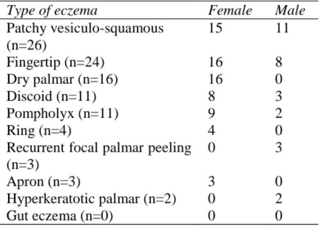
Table 1. A total of 36 patients had a history of atopy. The frequency of various clinical types is depicted in Figure 1, and their sex distribution in Table 2.
Allergic reactions on Patch testing were recorded in 46% patients, of which 27% were females and 19% males. The difference in positivity in both sexes, in relation to the numbers presenting, was statistically significant (p=<0.05, SD=+0.109), indicating that males had a higher rate of allergic reactions.
The commonest sensitizer detected (Figure 2)
was nickel (21%). The next in frequency were chromate (13%), neomycin (12%) and cobalt (11%). Sensitivity to fragrance mix (6%) and paraphenylenediamine (4%) was also noted. Balsam of Peru, sesquiterpene lactone mix, wool alcohol and parabens (1%) were least frequently detected.
Table 1 Occupations of hand eczema patients.
Occupation Female Male
Housewives 48 -
Students 16 -
Office workers - 9
Teachers 4 4
Skilled workers - 7
Masons - 4
Physicians 1 2
Cleaners 2 -
Dispensers - 1
Engineers - 1
Traders - 1
Figure 1 Frequency of clinical presentations (n=100).
Figure 2 Patch test results: sensitizers.
Figure 3 Sex distribution of common allergen.
Nickel sensitivity was seen more commonly in females (19%) than in males (2%), (p<0.001). In contrast, chromate allergy was more common in males (11%) than females (2%), (p<0.001).
Positivity in clinical types (Table 3) indicated that the highest rates of positive reactions were seen in the patchy vesiculo-squamous eczema, while no sensitivity was detected in apron and hyperkeratotic type.
Table 2 Distribution of clinical types of hand eczema (n=100=%).
Type of eczema Female Male
Patchy vesiculo-squamous (n=26)
15 11
Fingertip (n=24) 16 8
Dry palmar (n=16) 16 0
Discoid (n=11) 8 3
Pompholyx (n=11) 9 2
Ring (n=4) 4 0
Recurrent focal palmar peeling (n=3)
0 3
Apron (n=3) 3 0
Hyperkeratotic palmar (n=2) 0 2
Gut eczema (n=0) 0 0
Table 3 Positivity in clinical types (n=100).
Type of eczema N (%)
Patchy vesiculo-squamous (n=26) 18 (70)
Fingertip (n=24) 10 (42)
Dry palmar (n=16) 6 (37.5)
Discoid (n=11) 4 (36)
Pompholyx (n=11) 4 (36)
Ring (n=4) 3 (75)
Recurrent focal palmar peeling (n=3) 1 (33)
Apron (n=3) -
Hyperkeratotic palmar (n=2) -
Gut eczema -
morphological types of hand eczema. Three out of four positive patients of ring eczema were allergic to nickel. Patch test-positive patients of pompholyx were sensitive to nickel, alone or in combination with other allergens. There was no significant pattern to the other types. No sensitivity was detected in apron and hyperkeratotic palmar eczema.
Relevance to the eczema was noted in 38 patients.
Discussion
The incidence of hand eczema was significantly higher in females than males with a gender ratio of 2.4 (p<0.0001). It has been similarly reported in studies from different countries.1,10,11,15-17
Meding et al.1 reported a ratio of 2.05, while
Agner et al.15 had a ratio of 1.5. In contrast,
another study by Sarwar et al.18 from Pakistan
documented a lower ratio of females to males at 1:1.23.
Females presented at a younger age compared to males, with a mean age 25.7 years to 43.7 years. This is in accordance with other studies.10,12 This
coincides with the period of childcare and assuming household duties involving wet work.
Considering occupations, most of our females were housewives (48%) and students (16%). Only 8% females were gainfully employed. In contrast, the males had a wide variety of occupations (Table 1), which exposes them to various allergens.
The frequency of morphological types of hand eczema (Figure 1) provides a useful baseline for further studies in our population. The pattern in our study was different from that seen in Denmark.11
Patch test results demonstrated positive reactions in 46% patients. This is similar to results reported by Sarwar et al.18 at 48.6%, while
higher rates were noted by Diepgen et al.10 and
Agner et al.13 at 63%. Veien et al.16 in their study
recorded 33% positivity.
The commonest allergen was nickel at 21%. This is in accordance with another study on eczema in our community by Nadeem et al.19 at
21%, Rani et al.21 at 25%, and worldwide.5,16,17
Nickel allergy was significantly higher in females than males (p<0.001). Rates of sensitivity maybe higher due to early exposure to nickel in ear piercing and artificial jewellery.20,21 Hand eczema in nickel-sensitive
Chromate was detected at 13%, more commonly in males (11%) compared to females (2%), (p<0.001). Veien et al.16 noted a chromate
positivity of 3.4% in females (attributed to sensitization from footwear) and 1.6% in males. Studies from Denmark show a remarkable decreasing sensitivity to chromates over the years after addition of ferrous sulphate to cement during its manufacture, but no such measure is followed in our country.
Neomycin sensitivity was noted in 12%, which is much higher than in studies from European countries. Higher rates in our country indicate the common practice of prescribing the antibiotic topically, and easy over-the-counter availability. The trend should be curtailed.
Positive reaction to cobalt at 11% reflects its co-sensitivity with nickel and chromate. Fragrance mix positivity at 6% is similarly reported in other studies.16 Other allergens were recorded at
lower rates.
Personal and family history of atopy was noted in 36%. Atopic dermatitis is known to predispose to development of hand eczema.3
However, contact sensitization in atopics is still debated; there are studies from Norway and USA that found it to be a risk factor22,23 and
others negate it.24 Sixteen of our atopic patients
(44%) had positive patch tests to a variety of allergens, but no significant inferences could be drawn about a relationship to pattern of hand eczema or allergen.
Attempt at clinicoetiological correlation revealed that each morphological type of hand eczema could have different etiological factors and that there is no simple relationship between clinical type and etiology. This is similar to conclusions reached by other researchers.11,17,25,26,27
Conclusion
A high allergic sensitivity (46%) was noted in our patients of hand eczema. Different allergens could cause various patterns and no specific relationship could be established between etiological factors and morphology. Patch testing by a trained physician therefore is mandatory in all cases of hand eczema to detect an etiological allergen, for qualified counseling and better management of our patients.
It is recommended that our cement industry should, by law, be enforced to add ferrous sulphate in manufacturing cement, to reduce incidence of chromate induced allergic hand eczema.
References
1. Meding B, Wrangsjo K, Jarvholm B. Fifteen-year follow-up of hand eczema: persistence and consequences. Br J Dermatol. 2005;152:975-80.
2. Meding B, Jarvholm B. Hand eczema in Swedish adults: changes in prevalence between 1983 and 1996. J Invest Dermatol. 2002;118:719-23.
3. Lerbaek A, Kyvik KO, Ravn H, Menné T, Agner T. Incidence of hand eczema in a population-based twin cohort: genetic and environmental risk factors. Br J Dermatol. 2007;157:552-7.
4. Meding B, Jarvholm B. Incidence of hand eczema - a population-based, retrospective study. J Invest Dermatol. 2004;122:873-7. 5. Bryld LE, Hindsberger C, Kyvik KO, Agner
T, Menné T. Risk factors influencing the development of hand eczema in a population-based twin sample. Br J Dermatol. 2003;149:1214-20.
6. Lerbæk A, Kyvik K, Bryld LE, Menné T, Agner T. Heritability of hand eczema is not explained by comorbidity with atopic dermatitis. J Invest Dermatol. 2007;127:1632-40.
8. Funke U, Fartasch M, Diepgen TL. Incidence of work- related hand eczema during apprenticeship: first results of a prospective cohort study in the car industry. Contact Dermatitis. 2001;44:166-72. 9. Warshaw E, Lee G, Storrs FJ. Hand
dermatitis: a review of clinical features, therapeutic options, and long-term out- comes. Am J Contact Dermat. 2003;14:119-37.
10. Diepgen TL, Andersen KE, Brandao FM, Bruze M, Bruynzeel DP, Frosch P et al. A cross-sectional, multicentre study of the aetiology and morphology of hand eczema. Br J Dermatol. 2009;160:353-8.
11. Johansen JD, Hald M, Andersen BL, Laurberg G, Danielsen A, Avnstorp C et al. Classification of hand eczema: clinical and aetiological types. Based on the guidelines of the Danish Contact Dermatitis Group. Contact Dermatitis. 2011;65:13-21.
12. Fischer AA. The role of patch testing. In: Fischer AA, ed. Contact Dermatitis. Philadelphia: Lea and Febiger; 1986. P. 9-29.
13. Gøtzsche PC. Rational Diagnosis and Treatment. Evidence-Based Clinical Decision-Making, 4th edn. Chichester: John Wiley & Sons; 2008.
14. Ingram JR. Eczematous disorders. In: Griffiths CEM, Barker J, Bleiker T, Chalmers R, Creamer D, eds. Rook’s Textbook of Dermatology. 9th edn. Oxford, UK: Wiley Blackwell; 2016. P. 39.11-39.18. 15. Wilkinson DS, Fregert S, Magnussen B, Bandmann HJ, Calnan CD, Cronin E et al. Terminology of contact dermatitis. Acta Derm Venereol. 1970; 50:287-92.
16. Veien NK, Hattel T, Laurberg G. Hand Eczema: causes, course and prognosis I. Contact Dermatitis. 2008;58:330-4.
17. Agner T, Andersen KE, Brandao FM, Bruynzeel DP, Bruze M, Frosch P et al. Contact sensitization in hand eczema patients- relation to subdiagnosis, severity and quality of life: a multi-centre study. Contact Dermatitis. 2009;61:291-6.
18. Sarwar U, Asad F, Rani Z, Khurshid K, Pal SS. Frequency of allergic contact dermatitis in hand eczema patients with European standard and corticosteroid series. J Pak Assoc Dermatol. 2013;23:289-94.
19. Nadeem M, Akbar TM, Haroon TS. Patch testing with the European Standard series: our experience at Mayo Hospital, Lahore. J Pak Assoc Dermatol. 2001;11:7-12.
20. Thyssen JP, Menne T, Johansen JD. Nickel release from inexpensive jewellery and hair clasps purchased in an EU country- are consumers sufficiently protected from nickel exposure? Sci Total Environ. 2009;407:5315-8.
21. Rani Z, Tufail F, Asad F, Khurshid K,
Sarwar U, Pal SS. Frequency of allergic
contact dermatitis in patients with chronic eczema. Annals KEMU. 2010;16:SI:55-58. 22. Dotterud L K, Smith-Sivertsen T. Allergic
contact sensitisation in the general adult population: a population-based study from Northern Norway. Contact Dermatitis. 2007;56:10-5.
23. Ruff CA, Belsito DV. The impact of various patient factors on contact allergy to nickel, cobalt, and chromate. J Am Acad Dermatol. 2006;55:32-9.
24. Scha ̈fer T, Bo ̈hler E, Ruhdorfer S, Weigl
L, Wessner D, Filipiak B et al.
Epidemiology of contact allergy in adults. Allergy. 2001:56:1192-6.
25. Cronin E. Clinical pattern of hand eczema in women. Contact Dermatitis. 1985;13:153-61.
26. Magina S, Barros MA, Ferreira JA, Mesquita-Guimarães J. Atopy, nickel sensitivity, occupation and clinical patterns in different types of hand dermatitis. Am J Contact Dermat. 2003;14:63-8.