http://www.ijorl.com pISSN 2454-5929 | eISSN 2454-5937
Original Research Article
Sphenoid sinus mucoceles
Amarnath Devarasetty1
*, Kiran Natarajan
1, Thangammal Begum
2, Anand kumar R. S.
1,
Mohan Kameswaran
1INTRODUCTION
Approximately two-thirds of all mucoceles involve the frontal sinuses, the majority of the remainder involve the ethmoidal labyrinth. Sphenoidal mucoceles occur rarely and have an incidence of 1%.1 These lesions tend to extend and expand along the path of least resistance. The slow and silent expansion of a mucocele may be unsuspected until bone is eroded and it impinges on adjacent structures. Sphenoid sinus mucoceles have extremely variable clinical presentation because of the various anatomical relationships. The most common
presenting symptom is headache, which is most often described as frontal or retro orbital in nature and is found in roughly 70% of patients. The second most common symptom is visual disturbance, which is found in 65% of patients.1 Mucocele-like formation leading to neurological symptoms has been reported in prolactin-secreting pituitary adenomas.2 An endoscopic endonasal marsupialisation is the surgical approach of choice in most of the cases.3 This article aims to emphasize the clinical symptoms and signs, which together with imaging can enable prompt diagnosis and appropriate management of sphenoid mucoceles.
ABSTRACT
Background: Mucoceles are cyst-like lesions lined with respiratory epithelium that most commonly produce bone destruction within the paranasal sinuses. The spread of mucoceles is variable; hence they may cause different symptoms. In a majority of patients, mucoceles involve the frontal sinuses and the ethmoidal labyrinth. Sphenoidal mucoceles are rare and have an incidence of 1%. Prompt management of sphenoidal mucoceles is recommended in order to avoid potentially devastating complications. Our objective was to analyze the various presenting symptoms & signs of sphenoid mucoceles in twelve patients and the outcomes of management.
Methods: Twelve patients were diagnosed with sphenoidal mucocele over an 18 year period (January 1998 - December 2015). All patients underwent diagnostic nasal endoscopy, CT / MRI scans prior to management.
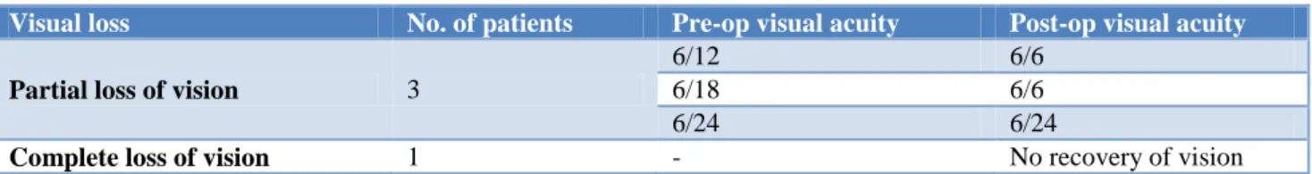
Results: Twelvepatients with sphenoid mucocele presented with variable signs & symptoms to a tertiary ENT care facility. All patients were successfully managed by endoscopic sinus surgery. Two out of three patients with diminished vision had complete recovery of vision after the procedure. One patient with complete loss of vision had no recovery after endoscopic sinus surgery.
Conclusions: Imagingplays a vital role in diagnosis of sphenoid mucoceles.The close proximity of the optic nerve to the sphenoid sinus makes it very vulnerable to get involved. Earlydiagnosis and management of sphenoid mucoceles is crucial.
Keywords: Sphenoid mucocele, Imaging, Endoscopic sinus surgery
Department of ENT, 1Madras ENT Research Foundation, Chennai, 2Revathi Medical Centre, Tiruppur, Tamil Nadu, India
Received: 20 February 2018
Revised: 24 April 2018
Accepted: 11 June 2018
*Correspondence:
Dr. Amarnath Devarasetty,
E-mail: [email protected]
Copyright: © the author(s), publisher and licensee Medip Academy. This is an open-access article distributed under the terms of the Creative Commons Attribution Non-Commercial License, which permits unrestricted non-commercial use, distribution, and reproduction in any medium, provided the original work is properly cited.
METHODS
Twelve patients were diagnosed with mucocele of the sphenoid sinus over an 18 year period (January 1998 to December 2015) at Madras ENT Research Foundation, a tertiary care otolaryngology facility in South India. All patients diagnosed to have sphenoid sinus mucoceles were included in the study. The exclusion criteria included patients with frontoethmoid mucoceles and sinus disease due to other causes. In all patients a detailed history taking and clinical examination was done. Symptoms such as nasal obstruction, nasal discharge, headache, double vision, decreased vision, symptoms of endocrine disturbance were noted. All patients underwent diagnostic nasal endoscopy. The diagnosis of sphenoid mucocele was by imaging- CT/MRI in all patients. On imaging, sinus expansion, bone erosion, clival involvement, destruction of sella turcica were specifically looked for. After obtaining consent, all patients were managed by endoscopic sinus surgery and none of the patients required an external procedure. Chi-square test was used to analyse the outcomes of treatment.
RESULTS
Twelve patients were diagnosed with mucocele of the sphenoid sinus over an 18 year period. Majority of patients were male (80%) and the average age was 46 years. Headache was reported by 11 patients. Otorhinolaryngologic history of nasal congestion and nasal discharge was obtained in 7 patients. Ophthalmic evaluation was recommended for all patients. Diplopia due to oculomotor palsy was present in 3 patients. Diminished vision was reported in 3 patients. Complete loss of vision was present in 1 patient (Table 1). None of our patients had any symptoms of an endocrine disturbance. In all patients, diagnostic nasal endoscopy was unremarkable. Examination findings included enlarged sphenoid sinus on CT scans in all patients. Bone
erosion was seen in 11 patients. One patient had coexisting mucocele with fungal sinusitis. None of the patients had clival involvement or sella turcica destruction. Diagnosis in all the patients was by a CT/MRI scan. All patients were managed by endoscopic sinus surgery (Figures 1–4). One patient had fungal debris coexisting with sphenoid mucocele. This was successfully managed by endoscopic sinus surgery (Figures 5–8). Diplopia improved in two patients after the surgery. Vision recovered completely in 2 of the 3 patients who complained of diminished vision. These patients underwent surgery within a week after onset of diminished vision. The third patient had diminished vision for a period of 3 weeks before the procedure. The patient with complete loss of vision did not regain his vision after the procedure (Table 2). Chi square test was done and proved statistically significant for most of the symptoms (Table 1). All patients were counselled about post-operative follow-ups and the follow-up period ranged from 12 months to 3 years. None of the patients had a recurrence of mucocele.
Figure 1: Mucocele in the right sphenoid sinus causing loss of vision in the right eye proptosis & a painful
eye.
Table 1: Pre-operative and post-operative symptoms of sphenoid mucocele and statistical test.
Symptoms Pre-operative Post-operative Chi-square test
Headache 11 1 0.000045
Nasal congestion 7 0 Not applicable
Nasal discharge 7 1 0.009
Double vision 3 1 0.27
Diminished vision 3 1 0.27
Complete loss of vision 1 1 Not applicable
Table 2: Pre-op and post-op visual acuity in patients with sphenoid mucocele.
Visual loss No. of patients Pre-op visual acuity Post-op visual acuity
Partial loss of vision 3
6/12 6/6
6/18 6/6
6/24 6/24
Figure 2: Coronal CT scans of showing right sphenoid mucocele.
Figure 3: MRI showing right sphenoid mucocele.
Figure 4: Right sphenoid mucocele being cleared by endoscopic sinus surgery.
Figure 5: CT scan of a patient with coexisting mucocele and fungal sinusitis.
Figure 6: MRI scan of the patient with coexisting mucocele and fungal sinusitis.
Figure 7: Mucocele being cleared in a patient with coexisting mucocele and fungal sinusitis.
Figure 8: Fungal debris being cleared in the patient with sphenoid mucocele.
DISCUSSION
approximately 1.0% of all paranasal sinus mucoceles. According to a study by Rombaux et al, 1% to 8% of the paranasal mucoceles involve the sphenoid sinus.3 Sphenoid sinus mucoceles were originally described by Berg in 1889.4 They are very rare in children, especially in children less than 12 years of age when pneumatization of the sphenoid sinus is completed.5
The anatomical location of the sinus makes early diagnosis of any kind of its pathology difficult. Pathologies involving isolated sphenoid sinus include inflammatory disease like sinusitis, tumors (benign or malignant), mucocele, fungal diseases and cerebrospinal fistula. The most frequent sphenoid pathology is inflammation, being responsible for 61% to 80% of all patients. The etiology of mucoceles is not clear, however, prior sinus diseases, allergic history have been implicated.6 Mechanical obstruction of sinus ostia from trauma, nasal polyposis, allergic rhinitis, and chronic sinusitis is the most common cause proposed for mucocele formation. Chronic non-invasive fungal sinusitis has also been associated with the formation of mucoceles. Mucoceles are characterized by the presence of pseudo stratified ciliated columnar epithelium on histopathology.7
The mucocele is characterized clinically by a silent initial period, of undetermined duration, followed by a period in which its expansion causes deformity and complications. Clinical presentation depends on location and direction of expansion and presence of infection. The sphenoid sinus maintains intimate anatomic relationships with the first six cranial nerves, with the dura mater, internal carotid arteries, cavernous sinus, pituitary, superior orbital fissure, optic chiasma, sphenopalatine nerve, and nerve of the pterygoid canal.Cystic dilatation with compression of these structures determines the signs and symptoms observed.4 Sphenoidal mucoceles generally tend to spread more frequently in an anterior–inferior fashion with invasion of the ethmoid, the nasal fossae and the nasopharynx. It may show upward invasion with destruction of the sellar floor. There may be an invasion in the orbital cavity when spreading occurs sideways. More rarely, the middle cranial fossa is invaded through the lateral wall and the posterior cranial fossa through the posterior wall.1
The common presenting symptoms mentioned are progressively worsening headache, visual loss, decreased visual acuity, visual field defect, diplopia, rhinorrhoea, and nasal congestion.8 If a mucocele becomes infected, clinical presentation is similar to acute sinusitis, with potential extension of infection into adjacent spaces. Visual symptoms & signs include diplopia, ocular muscle paresis, exophthalmus and complete visual loss. Most often, patients complain of diplopia, but progression to a complete orbital apex syndrome has been reported in only 8% of cases. Oculomotor nerve involvement was reported to account for 70% of ocular palsies.1 In our series 25%(3 patients) had oculomotor palsy. Some patients even present with acute onset of significant visual impairment
and headache.9 A rapid loss of vision is generally the result of spread of infection and inflammation from the mucocele to the optic nerve.10 The prognosis for the recovery of the visual loss is poor if the onset is sudden or if there is no light perception preoperatively. A gradual loss of vision occurs following circulatory disorders of the optic nerve caused by prolonged mucocele pressure that erode the bone and put the nerve in jeopardy.11 Sudden bilateral blindness has also been reported.12 Heybroek et al have also reported a case of sphenoidal mucocele presenting with bilateral visual disturbance along with pituitary gland dysfunction but without nasal or sinus complaints.13 Similarly a rare case of acute third cranial nerve palsy was also reported.14 A sphenoid mucocele may cause erosion in the sellar base, and may result in endocrine dysfunction through the disruption of the arterial feeding of the hypothalamo-pituitary axis due to mechanical compression.15 Sphenoid mucocele in a child causing meningitis and a brain abscess has been reported.16 Permanent central diabetes insipidus has been reported as a complication of sphenoid sinus mucocele.15 Differential diagnosis has to made with chronic sphenoid sinusitis, a fungus ball, benign neoplasms such as inverted papilloma, and rarely with malignant neoplasms.17
Diagnosis is usually made by history, physical findings and imaging modalities. High resolution Computed Tomography (CT) and Magnetic Resonance Imaging (MRI) are the diagnostic investigations of choice.18 The CT scan features of mucocele will show a homogenous, non-enhancing, expansile sinus mass completely filling the potential sinus cavity expanding or remodeling surrounding bone margins. Mucoceles generally do not enhance with contrast, but acutely inflamed mucopyoceles may show enhancement.19 The content of the sinus is variable, depending on the degree of hydration, ranging from near water attenuation to hyper-attenuating as secretions become increasingly thick and dehydrated. On MRI, the signal intensity of mucoceles varies in accordance with fluid content, presence of a proteinaceous component or hemorrhage. They usually have low signal intensity on T1 weighted images and a signal void on T2 weighted image sequence due to inspissated debris.20 On T1W MRI, low signal is noted (most common) if water rich content is present, high signal if content is protein rich. On T2W MRI,water rich content gives a high signal (most common) and protein rich content a low signal. In T1 C+ (Gd) images, enhancement if present, only occurs at the periphery. Colonisation with fungus can lead to very low signal on both T1 and T2 weighted sequences, mimicking a normal aerated sinus. In our series, 1 patient (8%) had fungal infection coexisting with the sphenoid mucocele. It is possible to make the differential diagnosis with neoplasms, since the mucocele does not undergo any enhancement with the administration of contrast.21
and may be approached with conventional methods or by nasal endoscopy. Marsupilaization by partial removal of the anterior and inferior walls of the mucocele via an endoscopic endonasal approach has been mentioned to be the treatment of choice for various reasons.22 This approach prevents recurrences and complications in most of the cases. The endoscopic approach represents the gold standard for sphenoid sinus mucocele treatment because it allows the best view of the sphenoid sinus with lesser trauma, avoids a scar, allows better preservation of mucociliary function, restoring of function and a higher compliance from the patient. Disadvantages include difficult management of eventual intraoperative complications (massive bleeding).23 Early intervention may result in improvement of visual symptoms as well.24 The prognosis is poor in patients who have already lost their eyesight before undergoing any kind of surgical intervention. Early detection of the condition can prevent development of serious and lifelong complications. Treatment of a sphenoid sinus mucocele extending into the middle cranial fossa and the pterygomaxillary fissure using an endoscope combined with a navigating system has been reported.25
CONCLUSION
Isolated involvement of sphenoid sinus is a rare entity. Mucoceles distort local anatomy and exert pressure on adjacent structures as they enlarge. The presence of a mucocele should be suspected in any patient presenting with headaches or visual disturbances. Early surgical intervention is vital to avoid complications.
Funding: No funding sources Conflict of interest: None declared
Ethical approval: The study was approved by the Institutional Ethics Committee
REFERENCES
1. Akan H, Cihan B, Celenk. Sphenoid sinus mucocele causing third nerve paralysis: CT and MR findings. Dentomaxillofacial Radiol. 2004;33:342–4.
2. Abe T, Ludecke D. Mucocele like formation leading to neurological symptoms in prolactin secreting pituitary adenomas under dopamine agonist therapy. Surg Neurol. 1999;52:274-9.
3. Rombaux P, Bertrand B, Eloy P, Collet S, Daele J, Bachert C, et al. Endoscopic endonasal surgery for paranasal sinus mucoceles. Acta Otorhinolaryngol Belg. 2000;54(2):115-22.
4. Costa LS, Resende LAL. Sphenoid Sinus Mucocele: An Infrequent Finding. Arch Neurol. 1984;41(8):897-8.
5. Haloi AK, Ditchfield M, Maixner W. Mucoceles of sphenoid sinus. Pediatr Radiol. 2006;36:987-90. 6. Di Girolamo S, Cannizzaro P, Picciotti P, Nardi C.
Ophthalmoplegia and ptosis as onset symptoms of an isolated primary mucocele of the sphenoid sinus. J Oral Maxillofac Surg. 2002;60:1500-2.
7. Lund VJ. Fronto-ethmoidal mucoceles: a histopathological sinuses. J Laryngol Otol. 1991;105:921-3.
8. Strek P, Zagólski O, Skadzieñ J, Oleoe K. Sphenoid sinus pyocele with intracranial extension managed under endoscopic guidance. A report of an extremely rare case: BENT. 2007;3:149-51.
9. Haloi AK, Ditchfield M, Maixner W. Mucoceles of sphenoid sinus. Pediatr Radiol. 2006;36:987-90. 10. Wurster CE, Levine TM, Sisson GA. Mucocele of
the sphenoid sinus causing sudden onset of blindness. Otolaryngol Head Neck Surg. 1986;94:257-9.
11. Levy J, Monos T, Puterman M. Bilateral consecutive blindness due to SSM with unilateral partial recovery. Can J Ophthalmol. 2005;40(4):506-8.
12. Casteels I, De Loof E, Brock P, Jorissen M. Sudden blindness in a child: presenting symptom of a sphenoid sinus mucocele. Brit J Ophthalmol. 1992;76:502-4.
13. Heylbroeck P, Watelet JB, Delbeke P, Van Cauwenberge P, Bachert C. Vision impairment as presenting symptom of a sphenoidal mucocele. Rhinol. 2003;41:187-91.
14. Vaphiades MS, Roberson GH. Sphenoid sinus mucocele presenting as a third cranial nerve palsy. J Neuroophthalmol. 2005;25:293-4.
15. Salehi PP, Chithiwala Z, Balough B. Atypical presentation of a sphenoid sinus mucocele: vision loss, partial hypopituitarism, adrenal insufficiency and SIADH. BENT. 2017;13:327–34.
16. Malard O, Gayet-Delacroix M, Jegoux F, Faure A, Bordure P, Beauvillain de Montreuil C.. Spontaneous Sphenoid Sinus Mucocele Revealed by Meningitis and Brain Abscess in a 12-Year-Old Child. Am J Neuroradiol. 2004;25(5):873-5. 17. Grillone GA, Kasznica P. Isolated sphenoid sinus
disease. Otol Clin N Am. 2004;37:435-51.
18. Gondim J, Pinheiro I, Tella OI Jr. Neurosurgical treatment of sphenoidal mucocele by endonasal transseptal endoscopic approach: report of two cases. Arq Neuropsiquiatr. 2002;60(2-A):299-302. 19. Som PM. CT of the paranasal sinuses. Neuroradiol.
1985;27:189-201.
20. Sinha BK, Adhikari P. Sphenoid sinus mucocele with blindness: A rare presentation. Nepal Med Coll J. 2008;10(3):204-6.
21. Tinoco P, Pereira JCO, Filho RCL, Silva FBC. Nasoendoscopic Treatment of the Sphenoid Sinus Mucoceles. Int Arch Otorhinolaryngol. 2009;13(3):336-9.
22. Moriyama H, Hesaka H, Tachibana T, Honda Y. Mucoceles of ethmoid and sphenoid sinus with visual disturbance. Arch Otolaryngol Head Neck Surg. 1992;118:142-6.
24. Nerurkar NK, Bradoo R, Muranjan S, Khare M. Sphenoid sinus mucocele with unilateral blindness. Ann Otol Rhinol Laryngol. 2004;113:294-6. 25. Nagatani T, Saito K, Yoshida J. Treatment of a
sphenoid mucocele using an endoscope combined with a navigating system: a case report. J Clin Neurosci. 2002;8(5):456-60.