Department of Pharmacology, University of North Carolina Chapel Hill, School of Medicine, Chapel Hill 27514 NC, USA. e-mail: [email protected]
N
eurotransmitter receptors are essential for mediating theeffects of neurotransmitters in the brain and peripheral nervous system. There are generally considered to be two types of neurotransmitter receptors: ionotropic and metabotropic. While ionotropic receptors are typically ligand-gated ion chan-nels, through which ions pass in response to a neurotransmitter, metabotropic receptors need G proteins and second messengers to indirectly modulate ionic activity in neurons. G protein–coupled receptors (GPCRs) represent the largest family of metabotropic receptors, although receptor tyrosine kinases1 and guanylate cyclase receptors2,3 can also be considered metabotropic receptors4. GPCRs also constitute the largest family of druggable targets5,6 encoded by the human genome, and 34% of medications approved by the US Food and Drug Administration have GPCRs as their main thera-peutic target7,8, especially those used for the treatment of neuropsy-chiatric disorders6,9,10 (Table 1).
There are many distinct chemical classes of neurotransmitters, including the following: (1) small molecules (e.g., glutamate, nor-epinephrine and serotonin); (2) neuropeptides (e.g., enkephalins, endorphins and neurokinins); and (3) others, including metabolites (e.g., endocannabinoids, ATP, ADP and adenosine) and gases (e.g., nitric oxide). Although the precise number of brain neurotransmit-ters is not known with certainty, it is likely that hundreds of distinct neurotransmitters exist, including many ‘orphan’ neuropeptides11. Each neurotransmitter system has distinct cellular and region-spe-cific distribution patterns in the brain; these can be visualized at the mRNA level with the Allen Brain Atlas12 or via genetically encoded markers with the GENSAT (Gene Expression Nervous System Atlas) resource13. Almost all neurotransmitters transduce their sig-nals at least in part by activating GPCRs, and the regional and cellu-lar distributions of brain GPCR mRNAs are well described12,14. As is the case with neurotransmitters, each of the metabotropic receptors for neurotransmitters has a distinct regional and cellular distribu-tion in the brain12,14 and elsewhere14.
GPCRs modulate synaptic transmission via so-called ‘slow synaptic transmission’15, which occurs in a time frame of seconds to minutes in the central nervous system. Metabotropic receptors such as GPCRs may be found pre- and post-synaptically. Pre-synaptic Gi-coupled GPCRs, such as the 5-HT1A and 5-HT1B serotonin
receptors16,17, inhibit neurotransmitter release via Gβ/γ-mediated activation of inhibitory channels, such as G protein–coupled inwardly rectifying potassium channels18, and via the inhibition of vesicle-docking SNARE-like proteins19,20 (Fig. 1). Post-synaptically, GPCRs can enhance neuronal excitability via Gs- and Gq-coupled
GPCRs, which have complex actions mediated by second messen-gers and protein kinases21,22 (Fig. 1).
This Review will discuss how structural insights into neurotrans-mitter GPCRs have transformed the understanding of these recep-tors, focusing on example GPCRs that are targets of drugs approved for use in the treatment of neuropsychiatric disorders. The past decade has witnessed remarkable progress in elucidating the struc-ture and function of GPCR family, with perhaps 60 or so GPCRs having their structures determined by X-ray crystallography7 or cryo-EM7. Additionally, various intermediate signaling states, ranging from inactive to active, have been elucidated for exem-plar GPCRs6. Finally, progress has been made on the use of such structural information for structure-guided and structure-inspired neuropsychiatric drug discovery8. This Review will provide an over-view of the understanding of how structure informs the function of representative neurotransmitter GPCRs. It will also show how an understanding of structure elucidates neuropharmacology and will highlight therapeutic challenges and opportunities for neuro-transmitter-targeted GPCRs.
Structural genomics of neurotransmitter-targeted GPCRs
Structural genomics has been defined as the approach that proposes ‘determining protein structures on a genome-wide scale’23, which boils down to ultimately determining the three-dimensional struc-ture of every protein encoded by the human genome. Obtaining GPCR structures has been historically challenging. Thus, although low-resolution structures of rhodopsin were available as early as 1993 (ref. 24), and useful models of helical arrangements were gen-erated through the use of that information25, the first bona fide high-resolution GPCR structure was not achieved until 2000, with rhodopsin26. The first non-opsin GPCR structures were obtained in 2007 (refs. 27–29), and since then, structures have been obtained, mainly via X-ray crystallography, for around 60 distinct human GPCRs7 (Fig. 2a). To generate useful structures, GPCR crystallog-raphy typically requires high-affinity ligands, which serve to stabi-lize the receptor in a distinct state suitable for crystallography. Such compounds not only need to be of high affinity (e.g., in the low nanomolar to picomolar range) but also need to have slow dissocia-tion rates30. Ideally, then, to obtain complete structural coverage of all GPCRs encoded by the human genome, one would need high-affinity, slowly dissociating ligands for each of them. Such ligands do not necessarily need to be specific, as, for example, the non-selective drug ergotamine has been used to obtain the structures of three different serotonin receptor subtypes: 5-HT1B (ref. 31, 5-HT2BMolecular pharmacology of metabotropic
receptors targeted by neuropsychiatric drugs
Bryan L. Roth
(ref. 32) and 5-HT
2C (ref. 33). Once structures are obtained, even with
non-selective ligands, the structural features for distinct pharmaco-logical properties can be elucidated (as in refs. 32,33). Furthermore, obtaining structures of related GPCRs frequently facilitates the structure-guided discovery and design of subtype-selective drugs34.
An analysis of available GPCR structures and cognate ligands, done with open-source databases of small molecules that bind known protein targets, along with databases of GPCR structural information, can be the first step toward determining the feasibil-ity of a comprehensive structural elucidation of the ‘GPCR-ome’. Chembl35, the KiDatabase36 and PubChem37 are databases that match compounds and their targets. Figure 2a shows the number of ligands with Chembl-annotated activity, plotted against a total of 393 non-olfactory GPCRs and, simultaneously, the number of structures available for each of these receptors (as of February 2019). Similar to previously published analyses6–8,38,39, in this analy-sis, around half of all human non-olfactory GPCRs are well anno-tated with respect to chemical matter, while the rest have few to no compounds associated with them in openly available databases35. With one exception—the receptor FZD4 (Frizzled 4), for which the structure of the seven-transmembrane domain (TM7) in the
ligand-free state has been solved40—those GPCRs for which there are reported structures typically have hundreds to thousands of annotated ligands (Fig. 2a). No clear relationship exists between the number of annotated ligands in Chembl and the number of distinct structures (Fig. 2b), although, as mentioned above, high-affinity, slowly dissociating ligands are typically key for obtaining structures. Thus, comprehensive elucidation of the structural genomics of GPCRs will require that suitable ligands be obtained for a large number of these receptors. Efforts are now ongoing to obtain these ligands, including the ‘Illuminating the Druggable Genome’ initiative (https://commonfund.nih.gov/idg), which has designated those GPCRs for which there is little chemical matter a top priority.
Taking into account the wealth of data noted above, and with new GPCR structures now appearing every week or so, what are the opportunities and challenges for the completion of a structural genomics survey for neurotransmitter GPCRs? Even for the 240 class A (Rhodopsin-like) GPCRs, which comprise most neurotrans-mitter receptors and for which there is the most structural cover-age, fewer than 50 distinct members have elucidated structures. Several classes — B2 (Adhesion), C (Glutamate), F (Frizzled) and T Table 1 | Representative metabotropic neurotransmitter receptors as targets for neuropsychiatric disorders and drugs of abuse
target class Neurotransmitter Receptor Representative
medication therapeutic indications or abuse Pharmacological action Structure determined (ref.)
GPCR Dopamine D2 dopamine Ropinirole Parkinson’s Disease Agonist 75
GPCR Dopamine D2 dopamine Nemonapride Schizophrenia Antagonist/
inverse agonist 75
GPCR Serotonin 5-HT2C serotonin Lorcaserin Obesity Agonist 33
GPCR Serotonin 5-HT1A serotonin Buspirone Anxiety and depression Partial agonist None reported
GPCR Serotonin 5-HT1B Ergotamine Migraine headaches Antagonist 31,32
GPCR Adenosine A2A adenosine Caffeine Alertness Antagonist 131
GPCR Norepinephrine β1 and β2
adrenergic Propranolol Post-traumatic stress disorder; anxiety disorders Inverse agonist 132,133 GPCR Serotonin 5-HT2A serotonin Pimavanserin Psychosis related to
Parkinson’s Disease Inverse agonist 134
GPCR Serotonin Many 5-HT
receptors LSD Hallucinogen use and abuse Agonist 70
GPCR Endocannabinoids CB1 cannabinoid Tetrahydrocannibinol Anorexia related to cancer chemotherapy; cannabis abuse
Agonist 135
GPCR GABA GABA-B Baclofen Spasticity-related
movement disorders Agonist None reported
GPCR Histamine H1-histamine Diphenhydramine Sedative Antagonist 41
GPCR Endorphins;
endomorphans μ-opioid Morphine Pain; abused opioid Agonist 59,136
GPCR Dynorphin κ-opioid Nalfurafine Itch Agonist 52,137
GPCR Dynorphin κ-opioid Salvinorin A Hallucinogen use and
abuse Agonist 52,137
GPCR Orexin OX1 orexin Suvorexant Insomnia Antagonist 138
GPCR Norepinephrine α2 adrenergic Clonidine Anxiety; opioid withdrawal Agonist None reported
GPCR S1P1 sphingosine
antagonist Fingolimod Multiple sclerosis Downregulates receptor 139 Receptor
tyrosine kinase
Nerve growth
factors TRK Entrectinib Cancers including astrocytoma Antagonist No full-length structure reported Guanylate
(Tastant 2), for example — have two reported structures (Glutamate and Frizzled) to no reported structures (Adhesion and Tastant).
Even among the thoroughly structurally annotated families of neurotransmitter receptors, such as the biogenic amine receptor family, structural annotation is still very sparse. Thus, only one (the H1 histamine receptor)41 of four histamine receptors has a reported structure, and there are no publicly available structures for α1 or
α2 adrenergic receptors. Finally, among 14 serotonin receptors17,
only 3 (5-HT1B, 5-HT2B and 5-HT2C)31–33 have published structures.
Thus, considerable opportunities exist for obtaining structures of representative members of each neurotransmitter GPCR subfam-ily. Additionally, even for neurotransmitter receptor families such as the muscarinic receptor family, which has acetylcholine as its neu-rotransmitter and for which four (M1, M2, M3 and M4)42–44 of five members have structures reported, the structural determinants for subtype selectivity are still to be elucidated and fully exploited for the creation of selective muscarinic agonists and antagonists45. For muscarinic receptors, in particular, it is clear that drugs targeting M1 and M4 muscarinic receptors may have special utility for the enhancement of cognition in Alzheimer’s disease and schizophre-nia, while interactions with M3 muscarinic receptors are associated with debilitating side effects46,47. In terms of creating subtype-selec-tive drugs, a variety of structure-guided approaches can be used, including targeting allosteric and orthosteric sites (as discussed in refs. 6,8,48). As the foregoing makes clear, although considerable progress has been made toward a comprehensive understanding of the structural genomics of human neurotransmitter GPCRs, a vast amount of work remains, which will require the integrated and coordinated efforts of structural biologists, pharmacologists, chem-ists and computational biologchem-ists.
Structural insights into activation and biased signaling
As with GPCR structural genomics, considerable insight into the structural features of neurotransmitter-targeted GPCR activation and biased signaling has been obtained. As for GPCR activation, the first structure of a GPCR in complex with hetereotrimeric G proteins was obtained in 2011 (ref. 49), and since then, several structures of nanobody-stabilized active states (e.g., β2 adrenergicreceptors50, M2 muscarinic receptors51 and κ- and μ-opioid recep-tors52,53) and heterotrimeric G protein–stabilized active states (e.g., calcitonin receptors54, CGRP (calcitonin gene–related peptide) receptors55,56, the 5-HT
1B serotonin receptor57,58, μ-opioid receptors59
and the GLP-1 (glucagon-like peptide-1) receptor60) have appeared, essentially all with neurotransmitter-targeted GPCRs. Furthermore, structures of arrestin-biased agonists at the receptor 5-HT2B
(refs. 32,61,62), arrestin-bound rhodopsin63 and G protein–biased agonists at the GLP-1 receptor64 have been reported. Finally, there are inactive-state structures stabilized by sodium (e.g., A2A
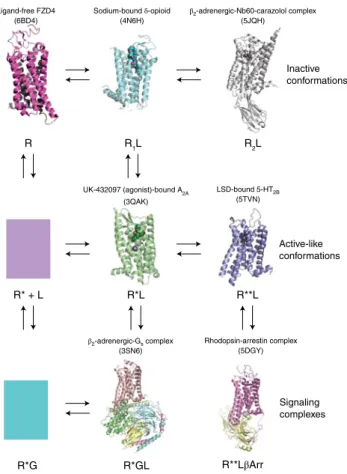
adenos-ine receptors65, δ-opioid receptors66 and D4 dopamine receptors34), negatively modulating allosteric nanobodies at the β2
adrener-gic receptor67 and the ligand-free basal structure of the receptor FZD440. Figure 3 depicts representative structures of these various states, along with a graphical representation of an extended ter-nary complex model of receptor activation that includes complexes stabilized by both G protein68 and β-arrestin69. The outward movement of TM6 is the hallmark for GPCR activation, as are several other canonical rearrangements49,53. These include rear-rangements of several microswitches32 such as the ‘P-I-F’ motif, formed by residues P5.50, I3.40 and F6.44 (Fig. 4). Here there is an
inward shift of P5.50, along with a rotamer switch of I3.40 and a large
inward movement of F6.44. Additional microswitches that undergo
rearrangement include the D(E)/RY motif in TM3 and the NPxxY motif in TM7. The salt bridge between D3.49 and R3.50 is typically
broken in active-state structures49,53. At the NPxxY motif, activation reveals a rotation of Y7.53. Additionally the sodium site collapses due
to rearrangements of key residues that coordinate sodium53 (Fig. 4). K+
K+
β γ
Gs P
SNAREs
cAMP PKA Na+
Na+ Gq
Ca++
Enhanced excitability Increased neurotransmitter release Diminished excitability
Inhibition of neurotransmitter release
Gi
β γ
Fig. 1 |Metabotropic receptors such as GPCRs modulate synaptic
transmission. This diagram shows several of the mechanisms by which metabotropic receptors such as GPCRs can modify synaptic transmission. Gi-coupled GPCRs can attenuate pre-synaptic release by activating various
channels, including inhibitory GIRKs (G protein–coupled inwardly rectifying potassium channels)18,128, and by inhibiting vesicle-release machinery, including SNARE proteins19. Post-synaptic G
q- and Gs-coupled GPCRs
can induce or potentiate neuronal firing via varous intracellular second messengers129 and secondary modulation of ion-channel activity21. PKA, protein kinase A.
A2A adenosine
(8,384 ligands; 44 structures)
β1 adrenergic
(2,919 ligands, 20 structures)
FZD4
(0 ligands, 1 structure)
A2A adenosine
(8,384 ligands; 44 structures)
D2 dopamine (9,304 ligands; 1 structure) NTSR1 neurotensin
(522 ligands; 8 structures)
GLP1RCHRM1ET -A
HRH4 FFAR1AVPR2 SSTR1 LPAR1 C3aRGPR34MCHR1CXCR6BAI2FZD10GPR115GPR157GPR32GPR82 LPAR6PAQR6
0 150 300 450 50,000 100,000 150,000
0 10 20 30 40 50
ChemBl compounds ( )
Structures
GPCR str
uctures ( )
ChemBl compounds
0 2,000 4,000 50,000 100,000 150,000
0 10 20 30 40 50
ChemBL
St
ru
ctures
393 Human GPCRs a
b
Finally, there is typically a large outward movement (of 10–15 Å) of the cytoplasmic end of TM6, which facilitates interactions with G proteins49 and other transducers53. In general, the ‘active-like’ receptor states in Fig. 3 show the various transitions mentioned above without the large outward movement of TM6.
Currently, there are two published structures of GPCRs in complex with β-arrestin-biased agonists, including LSD70 and ergotamine32 with the 5-HT
2B serotonin receptor, and one
struc-ture of a G protein–biased ligand in complex with hetereotrimeric G protein for the GLP-1 receptor64. The β-arrestin-biased agonist structures (Fig. 3) seem to represent an ‘intermediate’ state between the signaling complex states and the inactive states. Notably, the sodium site is collapsed, with rearrangements of key residues that stabilize the bound sodium ion, including (with the Ballesteros– Weinstein numbering convention71) Ser3.39 and Asp2.50 (Fig. 4a).
Notably, Ser7.45 rotates in to occlude the sodium pocket (Fig. 4a).
Additionally, a key residue (Leu209) in extracellular loop 2 (EL2) seems to form a ‘lid’ over LSD, retarding its dissociation and being essential for the recruitment of arrestin. A similar role for the same residue in EL2 was verified in studies of the D2 dopamine receptor and several other biogenic amine receptors61,62,72. Notably, the P-I-F and NPXXY motifs, along with conformational rearrangements in TM6 and TM7, are consistent with an intermediate activated state61 (Fig. 4b). Collectively, these rearrangements suggest that concerted conformational changes involving residues throughout the recep-tor, although they do not fully mimic the changes in the rhodop-sin–arrestin complex63, are responsible for the arrestin-biased conformation that favors binding of arrestin (Fig. 4b). In contrast, the GLP-1 complex with the G protein–biased agonist exendin-P5 shows the most pronounced difference in TM1, along with extra-cellular regions of TM6, TM7 and extraextra-cellular loop 3 (EL3), for which there is support by extensive mutagenesis (details in ref. 64).
The inactive states typically show a conserved sodium site, with sodium visualized in the structures of highest resolution34,65,66. Additionally, a conserved ‘ionic lock’ between D/E3.49 (of the D(E)/
RY motif) and N6.30, which stabilizes the ground state of several
GPCRs73,74, is occasionally seen. Remarkably, in an inactive state stabilized by Nb60 (nanobody 60) and carazolol67, this ionic lock is recapitulated by the nanobody. Additionally, in many inactive-state structures, the P-I-F, NPxxY and D(E)/RY motifs are all in a gener-ally conserved ‘inactive-like’ state.
The studies discussed above demonstrate clear progress in the understanding of the structural features required for differ-ent modalities of receptor activation and signaling. Clearly, much remains to be done in this direction, and the discovery of many novel permutations of these features is eagerly anticipated.
R*GL R**LβArr
R*G R
Inactive conformations
Active-like conformations
Signaling complexes
Sodium-bound δ-opioid
(4N6H) β2
-adrenergic-Nb60-carazolol complex (5JQH)
UK-432097 (agonist)-bound A2A
(3QAK)
LSD-bound 5-HT2B
(5TVN)
β2-adrenergic-Gs complex
(3SN6)
Rhodopsin-arrestin complex (5DGY)
R* + L R*L R**L
R1L R2L
Ligand-free FZD4 (6BD4)
Fig. 3 |Structural validation of the extended ternary complex model of GPCR action. The extended ternary complex model6,68,69 predicts the existence of multiple interconvertible GPCR states stabilized by ligands (agonists, neutral antagonists and inverse agonists) and transducers (G proteins, arrestins and other transducers). The various high-resolution structures here have provided validation for this schema. These states include the following: inactive states stabilized by allosteric nanobodies (RL2)67; active sodium-bound (R*L)70 and inactive sodium-bound states66 stabilized by agonists and inverse agonists, respectively; and coupled states (R*GL and R**LβArr). The model also provides for the possibility of biased signaling due to agonist-induced stabilization of distinct active and coupled states31,32 (e.g., R*L and R**L). Finally, spontaneously active states (R*) and coupled states (R*G), which have been demonstrated in many systems68, are predicted to exist, although structures of these are not yet available.
βArr, β-arrestin. Each state for which a structure is known is presented with the Protein Data Bank accession code in parentheses directly above and a description of the structure at top. Here, * and ** represent distinct active and coupled states.
Inactive sodium-bound D4 dopamine (5WIV) LSD-bound arrestin-biased 5-HT2B serotonin (5TVN) Ser3.39
Asp2.50
Ser7.45
Ser7.45
Ile3.40
Phe6.44
Pro5.50
Na+
Inactive sodium-bound D4 dopamine (5WIV) LSD-bound arrestin-biased 5-HT2B serotonin (5TVN) G protein active 5-HT1B serotonin (5V54)
a
b
Fig. 4 |Structural rearrangements associated with distinct GPCR states. a, Rearrangements within the sodium pocket associated with stabilization of an arrestin-biased state of the 5-HT2B serotonin receptor: key residues
involved in stabilizing sodium (Ser3.39 and Asp2.59), along with Ser7.45,
collapse to occlude the sodium site. Green indicates the inactive sodium-stabilized state of the D4 dopamine receptor; cyan shows the LSD-bound 5-HT2B receptor (key). b, Rearrangements in the P-I-F motif associated
Challenges and opportunities for drug design and discovery
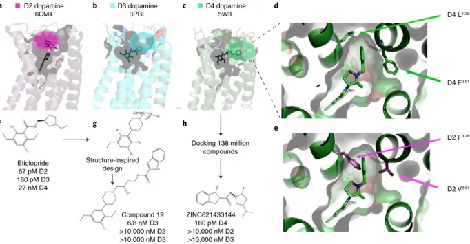
Selectivity. Given the large number of GPCR structures and reason-able coverage of some GPCR families, these structures should, at least in theory, be useful for the de novo design of selective drugs. This is particularly true when new ‘pockets’ are discovered, as was the case for the D275, D376 and D434 dopamine receptors. As shown in Fig. 5a–c, these structures revealed potentially unique binding surfaces that could be exploited for the design of selective ligands. Figure 5d shows a more detailed view of the D4 selectivity filter, while Fig. 5e shows how this is occluded in the D2 receptor34. Given that all three structures were obtained with chemically distinct and non-selective ligands, however, it is conceivable that the dif-ferent binding pockets might simply reflect the fact that difdif-ferent ligands with distinct chemical scaffolds (e.g., different chemotypes) engage different residues in the receptors. One way to test the hypothesis that these different binding pockets are pharmacologi-cally relevant is to create new ligands that are designed to engage these ‘selectivity filters’. For the D3 receptor, remarkable success was achieved77 with a compound with 1,700-fold selectivity for the D3 receptor and minimal off-target activity (Fig. 5). Starting with the seed compound eticlopride (Fig. 5f), which is a potent and non-selective D2 and D3 antagonist with weaker activity at D4, that group of researchers77 used a combination of docking and medici-nal chemistry to identify a potential modified scaffold predicted to target a putative selectivity region within the D3 receptor previously identified by mutagenesis and molecular modeling76 (Fig. 5g). Via this structure-inspired design approach, compound 19 (Fig. 5g) was eventually synthesized and was found to be a potent and selective D3 antagonist.
An alternative approach is to target such ‘selectivity filters’ via automated docking (Fig. 5c–e,h). In an initial proof-of-concept study, the D4 dopamine receptor selectivity filter was targeted by a docking screen of 600,000 commercially available compounds34 from the ZINC database78. A set of ‘seed’ compounds discovered
from the initial docking screen was further optimized by dock-ing and subsequent identification of analogs; that ultimately led to the highly selective D4 partial agonist compound 9-6-2434, which was >1,000-fold selective for the D4 receptor and lacked activity at 320 other GPCRs.
Subsequent to that, the computational approach has been enhanced by automated docking-based screening of ultra-large libraries in a screen with 138 million drug-like compounds. This in silico screening, wherein each compound was docked in
>100,000 conformations, required the analysis and scoring of 70 trillion docking events79 (Fig. 5h). In this study79, the previously identified D4 selectivity filter34 composed of the pocket formed by residues Leu3.28 and Phe2.61 (Fig. 5c–e) was again computationally
targeted. That docking screen led to the discovery of a large num-ber of chemically novel and highly selective D4 ligands. Of these, ZINC621433144 (Fig. 5h) was the most potent of the agonists tested, with a median effective concentration (EC50) of 180 pM and
selectivity for D4 over D2 and D3 (Fig. 5h). On the basis of the rate of true positive results among the predicted active compounds, the authors estimate that as many as 400,000 distinct D4-active com-pounds with more than 70,000 diverse chemotypes79 could exist in the library of 138 million compounds. Similar, albeit less com-putationally intensive, docking-based approaches have yielded selective ligands for the 5-HT1B serotonin receptor80, κ-opioid
receptor81, D2 dopamine receptor81 and other receptors (reviews available in refs. 8,82).
Structure-guided design of biased ligands
Functional selectivity, also known as ‘biased signaling’83, has been defined as the process by which “...ligands induce (or stabilize) unique, ligand-specific receptor conformations…result(ing) in dif-ferential activation of signal transduction pathways associated with that particular receptor”83. The phenomenon of functional selec-tivity occurs when different ligands at the same receptor lead to
D2 dopamine 6CM4
D3 dopamine 3PBL
D4 dopamine
5WIL D4 L3.28
D4 F2.61
D2 F3.28
D2 V2.61
Docking 138 million compounds Structure-inspired
design Eticlopride
67 pM D2 160 pM D3
27 nM D4
Compound 19 6/8 nM D3 >10,000 nM D2 >10,000 nM D3
ZINC621433144 160 pM D4 >10,000 nM D2 >10,000 nM D3
a b c
f g h
d
e
preferential activation of different G proteins (e.g., G protein–sub-type bias), a preference for G-protein signaling over arrestin (e.g., G-protein bias) or a preference for arrestin signaling over G pro-teins (e.g., arrestin bias). Reports of functional selectivity at GPCRs have appeared for many decades (an example of this is in ref. 84), and such bias is now recognized as a nearly universal phenomenon for GPCRs6. Indeed, there are now multiple reports of the discov-ery and optimization of arrestin-biased ligands85,86 and G protein– biased ligands87–89 for many GPCRs (reviews available in refs. 6,90,91). As mentioned above, so far there is only one structure of a GPCR (a GLP-1 receptor) in complex with a G protein–biased ligand (peptide exendin-P5) and hetereotrimeric G proteins64, a handful of structures of arrestin-biased ligands in complex with the 5-HT2B
serotonin receptor32,62,70, and no structures of arrestin-biased ligands in a GPCR–arrestin complex. Thus, although there is a paucity of structural information on GPCR functional selectivity, there have been several reports in which informed and structure-inspired design of neurotransmitter-targeted G protein and arres-tin-biased ligands have been achieved.
For example, the discovery of G protein–biased agonists for the
μ-opioid receptor has been reported92, although this discovery was accomplished without knowledge of any of the structural features responsible for biased signaling. In that study, molecular docking of 3 million compounds was performed against the inactive conforma-tion of the μ-opioid receptor targeting a highly conserved aspartic acid (Asp3.32) and engaging a conserved water network and a tyrosine
residue (Tyr3.33) putatively involved in selectivity and agonist activity.
Compounds with predicted activity were tested for functional activ-ity at G protein and arrestin signaling and, after several rounds of medicinal chemistry optimization, PZM21 was identified as a potent and efficacious G protein–biased agonist92. Similar G pro-tein–biased ligands have been discovered for μ-opioid receptors93,94 and κ-opioid receptors89,95, although these were not informed by structural determinants. On the other hand, via a combination of molecular dynamics and synthetic studies, a derivative of salvinorin A96 was discovered for μ-opioid receptors97 that is G protein biased and thus probably has less potential for abuse. Similarly, in a study of κ-opioid receptors, residues identified from structural studies as being essential for biased signaling ultimately led to the creation of novel structure-guided G protein–biased opioid agonists52.
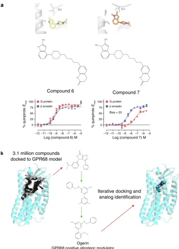
A structure-inspired approach72 for the creation of biased ligands at aminergic GPCRs, all of which are essential targets for the neu-rotransmitters serotonin, norepinephrine, dopamine and histamine, proved successful as well. For this study72, the authors took advan-tage of prior studies indicating that interactions with TM5 serine and other residues are essential for G-protein signaling62 at amin-ergic GPCRs, while interactions with EL2 residues can be essential for arrestin signaling61. Via a combination of molecular modeling, molecular dynamics simulations, automated docking and synthesis, the arrestin-biased compound 7 was discovered for the D2 dopa-mine receptor72 (Fig. 6a). As previous studies have shown that such arrestin-biased compounds may have efficacy in the treatment of schizophrenia and related neuropsychiatric disorders85,98, this could represent a useful approach for optimizing arrestin-biased medications for the treatment of neuropsychiatric disorders. Finally, it is also worth mentioning that biased signaling might be influenced at the level of recruitment of and receptor phosphoryla-tion by GPCR kinases99, and that this potentially can be influenced by GPCR ligands.
Orphan and understudied GPCRs. As previously mentioned, nearly 50% of GPCRs, most of which are highly expressed in the brain14, have a paucity of known ligands. Even for those neurotrans-mitter receptors that have been the subject of intensive investiga-tion, such as muscarinic and dopamine receptors, the identification of suitably selective ligands for the various subtypes continues to be
challenging. Thus, for example, there are no truly selective agonists for D5 dopamine or M5 muscarinic receptors, although a modestly selective D5 antagonist100 and a fairly selective M5 antagonist101 have been reported. Unfortunately, off-target pharmacology has not been reported for these compounds, so their selectivity over other GPCRs is unknown.
Although there are no published structures of orphan or under-studied brain GPCRs (oGPCRs38), the use of homology models and automated docking has provided a structure-inspired approach for ligand discovery. Thus, for example, an integrated approach using parallel screening, homology modeling, docking and analog synthesis led to ogerin — a selective positive allosteric modu-lator for GPR68 — and selective negative allosteric modumodu-lators for GPR65102 (Fig. 6b). The allosteric modulator ogerin was demonstrated to affect learning and behavior in mice, which indicates that GPR68 is a potentially important GPCR in the brain102. A similar approach led to the discovery of selective ago-nists for the oGPCR MRGPRX2, which is involved in pain and itch103 and binds many important neurotransmitters, including
HN
N N
O
HN
O
CH3
N
N N
O
HN
O
CH3
H3C
Compound 7
% quinpirole
Emax
Log (compound 6) M
HN N NH
O N+ O
-O O
N N
N NH2
H N
F F
OH H N H2N
N N
N 3.1 million compounds docked to GPR68 model
Compound 6
Ogerin
GPR68 positive allosteric modulator –12
0 25 50
G protein
β-arrestin
G protein
Bias = 20
β-arrestin
75 100
% quinpirole
Emax
0 25 50 75 100
–11 –10 –9 –8 –7 –6 –5
Log (compound 7) M –12 –11 –10 –9 –8 –7 –6 –5 a
b
Iterative docking and analog identification
Fig. 6 |Structure-inspired design of ligands for GPCRs to modulate synaptic transmission.a, Strategy for the design of ligands with arrestin bias for biogenic amine neurotransmitter receptors72 (details in text). Here, through the use of a combination of molecular dynamics simulations, docking and medicinal chemistry, compounds were created that were predicted to interact with a conserved EL2 residue that can impart arrestin bias. b, Computational strategy for the discovery of small-molecule probes with which to modulate oGPCRs such as GPR68102 Here, 3.1 million compounds were docked to a model of GPR68 and then, via iterative docking and testing of analogs, ogerin was identified as a selective GPR68 positive allosteric modulator.
substance P and other peptides103. These approaches are facilitated by parallel screening approaches in which hundreds of oGPCRs can be screened simultaneously102,104; active compounds are used to inform homology models for docking and subsequent discovery of new ligands102,103,105–107.
Genetic and model organism studies are also facilitating understanding of the potential therapeutic importance of oGP-CRs for neuropsychiatric disease. Thus, a published study has indicates that the oGPCR MRGPRX4 is involved in the pain and itch sensations mediated by bile acids108 Another study has shown that MRGPRX4 is also essential for the preference for menthol cigarettes in certain ethnic populations109. Given the distribution of MRGPRX4 in peripheral nerves, it is likely that MRGPRX4 and related receptors are neurotransmitter recep-tors involved in peripheral sensations such as pain and itch110. Databases devoted to the brain distribution of oGPCRs39,111,112 should facilitate discovery of their endogenous neurotransmit-ters, their function and neuropsychiatric implications for patho-genesis and treatment.
Polypharmacology. Historically, the most effective drugs for many complex neuropsychiatric diseases have targeted multiple GPCRs and other molecular targets9,113. It is now understood that this is probably because of the exceedingly complex genetic landscape of common diseases in which hundreds to thousands of genes might exert small effects114. This has led to the understanding that com-plex diseases are omnigenic rather than polygenic114, meaning that nearly every gene may ultimately exert a small effect on core disease pathways. Perhaps not surprisingly, the desire to discover increasingly selective drugs has resulted in lower overall success rates of drug-discovery screens115. Such a lack of success has led to the hypothesis that generating polypharmacological drugs with designated multiple targets represents a useful approach for the treatment of complex diseases113,116. For aminergic GPCRs, there has been some success in discovering the structural features responsible for polypharmacological activi-ties33, which comprise a series of nine semi-conserved residues (Asp3.32, Ile/Val3.33, Cys3.36, Thr3.37, Ala/Ser/Thr5.46, Trp6.48, Phe6.51,
Phe6.52 and EL2 Val/Ile/Leu). Although in theory,
structure-guided approaches should be helpful for the discovery and design of polypharmacological drugs, so far, this has not been entirely successful117,118
therapeutic and commercial opportunities for
structure-guided GPCR drug discovery
Heptares (acquired by Sosei in 2016)119, and Receptos (acquired by Celgene in 2015)120, along with Conformetrix121, have invested con-siderable resources in bringing GPCR structure-guided drug dis-covery to fruition. So far, publicly available information indicates that the exemplar compounds are targeted mainly to well-studied GPCRs for which selective ligands are difficult to obtain. Thus, for example, Heptares has advanced an M4-selective agonist to phase I trials, presumably for cognition enhancement (https://clinicaltrials. gov/ct2/show/NCT03244228?term=heptares&rank=4). Heptares has also brought a structure-guided mGlu5 negative allosteric modulator122 for metabotropic glutamate receptors to initial phase I trials, presumably for application to the central nervous system (https://clinicaltrials.gov/ct2/show/NCT03785054?term=heptare s&rank=2). Receptos, in partnership with Celgene, has advanced RPC1063, an S1P receptor agonist for the treatment of multiple sclerosis (https://www.accessdata.fda.gov/scripts/opdlisting/oopd/ detailedIndex.cfm?cfgridkey=611517), to phase III clinical trials.
Future directions
Given the wealth of structural information now available for many GPCRs, it is anticipated that structure-guided approaches will
eventually lead to neuropsychiatric medications with greater effi-cacy and fewer side effects. As on-target and off-target side effects, along with clinical effectiveness and safety, continue to be major drivers of drug failures in clinical trials123, insights into the struc-tural basis of ligand engagement could provide new opportunities for GPCR-based neuropsychiatric drug discovery. Thus, where a G protein–biased ligand might show greater efficacy and fewer side effects, insights into the structural basis of such bias could acceler-ate the discovery of novel chemotypes with biased signaling pat-terns. Indeed, this approach has been successful in the generation of biased tool compounds for many neurotransmitter receptors, including D4 dopamine receptors34,124, μ-opioid receptors92 and other receptors62,72. Although there are currently no structure-guided biased drugs in clinical trials, encouraging results have appeared for TRV130 as a G protein–biased opioid receptor ago-nist for pain125, albeit with some abuse liabilities, as manifested in preclinical studies126,127.
As is clear from the foregoing, the past decade has witnessed astounding progress in understanding of the structure and func-tion of metabotropic receptors for neurotransmitters. This increasing wealth of structural information, as well as advances in structure-guided and structure-inspired drug discovery, prom-ise to accelerate the discovery of drugs that target metabotropic receptors for neurotransmitters with improved efficacies and fewer side effects.
Received: 13 March 2019; Accepted: 15 May 2019; Published online: 3 July 2019
References
1. Ullrich, A. & Schlessinger, J. Signal transduction by receptors with tyrosine
kinase activity. Cell 61, 203–212 (1990).
2. Schulz, S., Chinkers, M. & Garbers, D. L. The guanylate cyclase/receptor
family of proteins. FASEB J. 3, 2026–2035 (1989).
3. Chinkers, M. et al. A membrane form of guanylate cyclase is an atrial
natriuretic peptide receptor. Nature 338, 78–83 (1989).
4. Purves, D. et al. Two families of postsynaptic receptors. in Neuroscience
2nd edn. (Sinauer Associates, 2001).
5. Overington, J. P., Al-Lazikani, B. & Hopkins, A. L. How many drug targets
are there? Nat. Rev. Drug Discov. 5, 993–996 (2006).
Ref. 5 is classic paper that provides estimates for the size of the ‘druggable’ genome.
6. Wacker, D., Stevens, R. C. & Roth, B. L. How ligands illuminate GPCR
molecular pharmacology. Cell 170, 414–427 (2017).
7. Hauser, A. S., Attwood, M. M., Rask-Andersen, M., Schiöth, H. B. &
Gloriam, D. E. Trends in GPCR drug discovery: new agents, targets and
indications. Nat. Rev. Drug Discov. 16, 829–842 (2017).
8. Roth, B. L., Irwin, J. J. & Shoichet, B. K. Discovery of new GPCR ligands to
illuminate new biology. Nat. Chem. Biol. 13, 1143–1151 (2017).
9. Roth, B. L., Sheffler, D. J. & Kroeze, W. K. Magic shotguns versus magic
bullets: selectively non-selective drugs for mood disorders and
schizophrenia. Nat. Rev. Drug Discov. 3, 353–359 (2004).
10. Marder, S. R. et al. Advancing drug discovery for schizophrenia.
Ann. NY Acad. Sci. 1236, 30–43 (2011).
11. Fricker, L. D. & Devi, L. A. Orphan neuropeptides and receptors: Novel
therapeutic targets. Pharmacol. Ther. 185, 26–33 (2018).
12. Lein, E. S. et al. Genome-wide atlas of gene expression in the adult mouse
brain. Nature 445, 168–176 (2007).
13. Gerfen, C. R., Paletzki, R. & Heintz, N. GENSAT BAC cre-recombinase driver lines to study the functional organization of cerebral cortical and
basal ganglia circuits. Neuron 80, 1368–1383 (2013).
14. Regard, J. B., Sato, I. T. & Coughlin, S. R. Anatomical profiling of G protein-
coupled receptor expression. Cell 135, 561–571 (2008).
15. Greengard, P. The neurobiology of slow synaptic transmission. Science 294,
1024–1030 (2001).
16. Berger, M., Gray, J. A. & Roth, B. L. The expanded biology of serotonin.
Annu. Rev. Med. 60, 355–366 (2009).
17. McCorvy, J. D. & Roth, B. L. Structure and function of serotonin G protein-
coupled receptors. Pharmacol. Ther. 150, 129–142 (2015).
18. Andrade, R., Malenka, R. C. & Nicoll, R. A. A. G protein couples serotonin
and GABAB receptors to the same channels in hippocampus. Science 234,
19. Blackmer, T. et al. G protein βγ directly regulates SNARE protein fusion
machinery for secretory granule exocytosis.Nat. Neurosci. 8, 421–425 (2005).
Ref. 19 is one of the first demonstrations that G-protein β- and
γ-subunits regulate vesicle-fusion machinery to inhibit synaptic
transmission.
20. Gerachshenko, T. et al. Gβγ acts at the C terminus of SNAP-25 to mediate
presynaptic inhibition. Nat. Neurosci. 8, 597–605 (2005).
21. Skeberdis, V. A. et al. Protein kinase A regulates calcium permeability of
NMDA receptors. Nat. Neurosci. 9, 501–510 (2006).
22. Carver, C. M. & Shapiro, M. S. Gq-coupled muscarinic receptor enhancement of KCNQ2/3 channels and activation of TRPC channels in
multimodal control of excitability in dentate gyrus granule cells. J. Neurosci.
39, 1566–1587 (2019).
23. Stevens, R. C. & Wilson, I. A. Tech.Sight. Industrializing structural biology.
Science 293, 519–520 (2001).
24. Schertler, G. F., Villa, C. & Henderson, R. Projection structure of
rhodopsin. Nature 362, 770–772 (1993).
Ref. 24 provided the first cryo-EM structure of a membrane protein.
25. Baldwin, J. M. The probable arrangement of the helices in G
protein-coupled receptors. EMBO J. 12, 1693–1703 (1993).
26. Palczewski, K. et al. Crystal structure of rhodopsin: A G protein-coupled
receptor. Science 289, 739–745 (2000).
Ref. 26 provided the first crystal structure of a GPCR.
27. Rosenbaum, D. M. et al. GPCR engineering yields high-resolution
structural insights into β2-adrenergic receptor function. Science 318,
1266–1273 (2007).
28. Rasmussen, S. G. F. et al. Crystal structure of the human β2 adrenergic
G-protein-coupled receptor. Nature 450, 383–387 (2007).
29. Cherezov, V. et al. High-resolution crystal structure of an engineered
human β2-adrenergic G protein-coupled receptor. Science 318,
1258–1265 (2007).
Refs. 27–29 provided the first crystal structures of non-opsin GPCRs.
30. Katritch, V., Cherezov, V. & Stevens, R. C. Structure-function of the G
protein-coupled receptor superfamily. Annu. Rev. Pharmacol. Toxicol. 53,
531–556 (2013).
31. Wang, C. et al. Structural basis for molecular recognition at serotonin
receptors. Science 340, 610–614 (2013).
32. Wacker, D. et al. Structural features for functional selectivity at serotonin
receptors. Science 340, 615–619 (2013).
33. Peng, Y. et al. 5–HT2C receptor structures reveal the structural basis of
GPCR polypharmacology. Cell 172, 719–730.e714 (2018).
34. Wang, S. et al. D4 dopamine receptor high-resolution structures enable the
discovery of selective agonists. Science 358, 381–386 (2017).
35. Gaulton, A. et al. ChEMBL: a large-scale bioactivity database for drug
discovery. Nucleic Acids Res. 40, D1100–D1107 (2012).
36. Roth, B. et al. Multiplicity of serotonin receptors: useless diverse molecules
or an embarrassment of riches? Neuroscientist 6, 252–262 (2000).
37. Wang, Y. et al. PubChem: a public information system for analyzing
bioactivities of small molecules. Nucleic Acids Res. 37, W623–33 (2009).
38. Roth, B. L. & Kroeze, W. K. Integrated approaches for genome-wide interrogation of the druggable non-olfactory G protein-coupled receptor
superfamily. J. Biol. Chem. 290, 19471–19477 (2015).
39. Oprea, T. I. et al. Unexplored therapeutic opportunities in the human
genome. Nat. Rev. Drug Discov. 17, 377 (2018).
40. Yang, S. et al. Crystal structure of the Frizzled 4 receptor in a ligand-free
state. Nature 560, 666–670 (2018).
41. Shimamura, T. et al. Structure of the human histamine H1 receptor
complex with doxepin. Nature 475, 65–70 (2011).
42. Haga, K. et al. Structure of the human M2 muscarinic acetylcholine
receptor bound to an antagonist. Nature 482, 547–551 (2012).
43. Kruse, A. C. et al. Structure and dynamics of the M3 muscarinic
acetylcholine receptor. Nature 482, 552–556 (2012).
44. Thal, D. M. et al. Crystal structures of the M1 and M4 muscarinic
acetylcholine receptors. Nature 531, 335–340 (2016).
45. Liu, H. et al. Structure-guided development of selective M3 muscarinic
acetylcholine receptor antagonists. Proc. Natl Acad. Sci. USA 115,
12046–12050 (2018).
46. Vardigan, J. D. et al. Improved cognition without adverse effects: novel M1 muscarinic potentiator compares favorably to donepezil and xanomeline in
rhesus monkey. Psychopharmacology (Berl) 232, 1859–1866 (2015).
47. Foster, D. J., Choi, D. L., Conn, P. J. & Rook, J. M. Activation of M1 and M4 muscarinic receptors as potential treatments for Alzheimer’s disease and
schizophrenia. Neuropsychiatr. Dis. Treat. 10, 183–191 (2014).
48. Thal, D. M., Glukhova, A., Sexton, P. M. & Christopoulos, A. Structural
insights into G-protein-coupled receptor allostery. Nature 559, 45–53 (2018).
49. Rasmussen, S. G. et al. Crystal structure of the β2 adrenergic receptor-Gs
protein complex. Nature 477, 549–555 (2011).
Ref. 49 provided the first structure of a GPCR in a complex in its active state with a G protein heterotrimer.
50. Rasmussen, S. G. et al. Structure of a nanobody-stabilized active state of the
β2 adrenoceptor. Nature 469, 175–180 (2011).
51. Kruse, A. C. et al. Activation and allosteric modulation of a muscarinic
acetylcholine receptor. Nature 504, 101–106 (2013).
52. Che, T. et al. Structure of the nanobody-stabilized active state of the κ
opioid receptor. Cell 172, 55–67.e15 (2018).
53. Huang, W. et al. Structural insights into µ-opioid receptor activation. Nature
524, 315–321 (2015).
54. Liang, Y. L. et al. Phase-plate cryo-EM structure of a class B GPCR-G-protein
complex. Nature 546, 118–123 (2017).
Ref. 54 provided the first cryo-EM structure of an active GPCR in complex with G protein.
55. Liang, Y. L. et al. Cryo-EM structure of the active, Gs-protein complexed,
human CGRP receptor. Nature 561, 492–497 (2018).
56. Draper-Joyce, C. J. et al. Structure of the adenosine-bound human
adenosine A1 receptor-Gi complex. Nature 558, 559–563 (2018).
57. García-Nafría, J., Nehmé, R., Edwards, P. C. & Tate, C. G. Cryo-EM
structure of the serotonin 5-HT1B receptor coupled to heterotrimeric Go.
Nature 558, 620–623 (2018).
58. García-Nafría, J., Lee, Y., Bai, X., Carpenter, B. & Tate, C. G. Cryo-EM
structure of the adenosine A2A receptor coupled to an engineered
heterotrimeric G protein. eLife 7, e35946 (2018).
59. Koehl, A. et al. Structure of the µ-opioid receptor-Gi protein complex.
Nature 558, 547–552 (2018).
60. Zhang, Y. et al. Cryo-EM structure of the activated GLP-1 receptor in
complex with a G protein. Nature 546, 248–253 (2017).
61. Wacker, D. et al. Crystal structure of an LSD-bound human serotonin
receptor. Cell 168, 377–389.e12 (2017).
62. McCorvy, J. D. et al. Structural determinants of 5-HT2B receptor activation
and biased agonism. Nat. Struct. Mol. Biol. 25, 787–796 (2018).
63. Kang, Y. et al. Crystal structure of rhodopsin bound to arrestin by
femtosecond X-ray laser. Nature 523, 561–567 (2015).
Ref. 63 provided the first crystal structure of a GPCR with arrestin.
64. Liang, Y. L. et al. Phase-plate cryo-EM structure of a biased agonist-bound
human GLP-1 receptor-Gs complex. Nature 555, 121–125 (2018).
65. Liu, W. et al. Structural basis for allosteric regulation of GPCRs by sodium
ions. Science 337, 232–236 (2012).
66. Fenalti, G. et al. Molecular control of δ-opioid receptor signalling. Nature
506, 191–196 (2014).
67. Staus, D. P. et al. Allosteric nanobodies reveal the dynamic range and
diverse mechanisms of G-protein-coupled receptor activation. Nature 535,
448–452 (2016).
68. Samama, P., Cotecchia, S., Costa, T. & Lefkowitz, R. J. A mutation-induced
activated state of the β2-adrenergic receptor. Extending the ternary complex
model. J. Biol. Chem. 268, 4625–4636 (1993).
69. Roth, B. L. DREADDs for neuroscientists. Neuron 89, 683–694 (2016).
70. Wacker, D. et al. Crystal structure of an LSD-bound human serotonin
receptor. Cell 168, 377–389.e312 (2017).
71. Ballesteros, J. A. & Weinstein, H. Integrated methods for the construction of three-dimensional models and computational probing of
structure-function relations in G protein-coupled receptors. Meth. Neurosci. 25,
366 (1995).
72. McCorvy, J. D. et al. Structure-inspired design of β-arrestin-biased ligands
for aminergic GPCRs. Nat. Chem. Biol. 14, 126–134 (2018).
73. Ballesteros, J. A. et al. Activation of the β2-adrenergic receptor involves
disruption of an ionic lock between the cytoplasmic ends of transmembrane
segments 3 and 6. J. Biol. Chem. 276, 29171–29177 (2001).
74. Shapiro, D. A. et al. Evidence for a model of agonist-induced activation of 5–HT2A serotonin receptors which involves the disruption of a strong ionic
interaction between helices 3 and 6. J. Biol. Chem. 18, 18 (2002).
75. Wang, S. et al. Structure of the D2 dopamine receptor bound to the atypical
antipsychotic drug risperidone. Nature 555, 269–273 (2018).
76. Chien, E. Y. et al. Structure of the human dopamine D3 receptor in
complex with a D2/D3 selective antagonist. Science 330, 1091–1095 (2010).
77. Kumar, V. et al. Highly selective dopamine D3 receptor (D3R) antagonists and partial agonists based on eticlopride and the D3R crystal structure:
new leads for opioid dependence treatment. J. Med. Chem. 59,
7634–7650 (2016).
78. Irwin, J. J. & Shoichet, B. K. ZINC—a free database of commercially
available compounds for virtual screening. J. Chem. Inf. Model. 45,
177–182 (2005).
79. Lyu, J. et al. Ultra-large library docking for discovering new chemotypes.
Nature 566, 224–229 (2019).
80. Rodríguez, D., Brea, J., Loza, M. I. & Carlsson, J. Structure-based
discovery of selective serotonin 5-HT(1B) receptor ligands. Structure 22,
1140–1151 (2014).
81. Negri, A. et al. Discovery of a novel selective κ-opioid receptor agonist
using crystal structure-based virtual screening. J. Chem. Inf. Model. 53,
82. Irwin, J. J. & Shoichet, B. K. Docking screens for novel ligands conferring
new biology. J. Med. Chem. 59, 4103–4120 (2016).
83. Urban, J. D. et al. Functional selectivity and classical concepts of
quantitative pharmacology. J. Pharmacol. Exp. Ther. 320, 1–13 (2007).
84. Roth, B. L. & Chuang, D.-M. Multiple mechanisms of serotonergic signal
transduction. Life Sci. 41, 1051–1064 (1987).
Ref. 84 provided the first explication of the principles of functional selectivity and biased agonism and antagonism.
85. Allen, J. A. et al. Discovery of β-arrestin-biased dopamine D2 ligands for
probing signal transduction pathways essential for antipsychotic efficacy.
Proc. Natl Acad. Sci. USA 108, 18488–18493 (2011).
86. Boerrigter, G. et al. Cardiorenal actions of TRV120027, a novel β
-arrestin-biased ligand at the angiotensin II type I receptor, in healthy and heart failure canines: a novel therapeutic strategy for acute heart failure.
Circ. Heart Fail. 4, 770–778 (2011).
87. Chen, X. et al. Discovery of G protein-biased D2 dopamine receptor partial
agonists. J. Med. Chem. 59, 10601–10618 (2016).
88. Chen, X. T. et al. Structure-activity relationships and discovery of a G
protein biased μ opioid receptor ligand, [(3-methoxythiophen-2-yl)methyl]
(2-[(9R)-9-(pyridin-2-yl)-6-oxaspiro-[4.5]decan-9-yl]ethyl)amine (TRV130),
for the treatment of acute severe pain. J. Med. Chem. 56, 8019–8031 (2013).
89. Lovell, K. M. et al. Structure-activity relationship studies of functionally
selective κ opioid receptor agonists that modulate ERK 1/2 phosphorylation
while preserving G protein over βarrestin2 signaling bias. ACS Chem.
Neurosci. 6, 1411–1419 (2015).
90. Wootten, D., Christopoulos, A., Marti-Solano, M., Babu, M. M. & Sexton, P. M. Mechanisms of signalling and biased agonism in G protein-coupled
receptors. Nat. Rev. Mol. Cell Biol. 19, 638–653 (2018).
91. Smith, J. S., Lefkowitz, R. J. & Rajagopal, S. Biased signalling: from
simple switches to allosteric microprocessors. Nat. Rev. Drug Discov. 17,
243–260 (2018).
92. Manglik, A. et al. Structure-based discovery of opioid analgesics with
reduced side effects. Nature 537, 185–190 (2016).
Ref. 92 reports structure-guided discovery of biased ligands.
93. Váradi, A. et al. Mitragynine/corynantheidine pseudoindoxyls as opioid
analgesics with μ agonism and δ antagonism, which do not recruit
β-arrestin-2. J. Med. Chem. 59, 8381–8397 (2016).
94. Schmid, C. L. et al. Bias factor and therapeutic window correlate to predict
safer opioid analgesics. Cell 171, 1165–1175.e1113 (2017).
95. White, K. L. et al. Identification of novel functionally selective κ-opioid
receptor scaffolds. Mol. Pharmacol. 85, 83–90 (2014).
96. Roth, B. L. et al. Salvinorin A: a potent naturally occurring
nonnitrogenous κ opioid selective agonist. Proc. Natl Acad. Sci. USA 99,
11934–11939 (2002).
97. Crowley, R. S. et al. Synthetic studies of neoclerodane diterpenes from salvia
divinorum: identification of a potent and centrally acting μ opioid analgesic
with reduced abuse liability. J. Med. Chem. 59, 11027–11038 (2016).
98. Park, S. M. et al. Effects of β-arrestin-biased dopamine D2 receptor ligands
on schizophrenia-like behavior in hypoglutamatergic mice.
Neuropsychopharmacology 41, 704–715 (2016).
99. Choi, M. et al. G protein–coupled receptor kinases (GRKs) orchestrate
biased agonism at the β2-adrenergic receptor. Sci Signal 11,
eaar7084 (2018).
100. Mohr, P., Decker, M., Enzensperger, C. & Lehmann, J. Dopamine/serotonin receptor ligands. 12(1): SAR studies on hexahydro-dibenz[d,g]azecines lead to 4-chloro-7-methyl-5,6,7,8,9,14-hexahydrodibenz[d,g]azecin-3-ol, the first
picomolar D5-selective dopamine-receptor antagonist. J. Med. Chem. 49,
2110–2116 (2006).
101. Gentry, P. R. et al. Discovery, synthesis and characterization of a highly muscarinic acetylcholine receptor (mAChR)-selective M5-orthosteric
antagonist, VU0488130 (ML381): a novel molecular probe. ChemMedChem
9, 1677–1682 (2014).
102. Huang, X. P. et al. Allosteric ligands for the pharmacologically dark
receptors GPR68 and GPR65. Nature 527, 477–483 (2015).
Ref. 102 reports structure-inspired discovery of allosteric ligands for an orphan GPCR.
103. Lansu, K. et al. In silico design of novel probes for the atypical opioid
receptor MRGPRX2. Nat. Chem. Biol. 13, 529–536 (2017).
104. Kroeze, W. K. et al. PRESTO-Tango as an open-source resource for
interrogation of the druggable human GPCRome. Nat. Struct. Mol. Biol. 22,
362–369 (2015).
105. Ngo, T. et al. Orphan receptor ligand discovery by pickpocketing
pharmacological neighbors. Nat. Chem. Biol. 13, 235–242 (2017).
106. Ngo, T. et al. Identifying ligands at orphan GPCRs: current status using
structure-based approaches. Br. J. Pharmacol. 173, 2934–2951 (2016).
107. Trauelsen, M. et al. Receptor structure-based discovery of non-metabolite
agonists for the succinate receptor GPR91. Mol. Metab. 6, 1585–1596 (2017).
108. Meixiong, J. et al. Identification of a bilirubin receptor that may mediate a
component of cholestatic itch. eLife 8, e44116 (2019).
109. Kozlitina, J. et al. An African-specific haplotype in MRGPRX4 is associated
with menthol cigarette smoking. PLoS Genet. 15, e1007916 (2019).
110. Dong, X., Han, S., Zylka, M. J., Simon, M. I. & Anderson, D. J. A diverse family of GPCRs expressed in specific subsets of nociceptive sensory
neurons. Cell 106, 619–632 (2001).
111. Ehrlich, A. T. et al. Expression map of 78 brain-expressed mouse orphan GPCRs provides a translational resource for neuropsychiatric research.
Commun Biol 1, 102 (2018).
112. Oprea, T. I. et al. Far away from the lamppost. PLoS Biol. 16,
e3000067 (2018).
113. Dar, A. C., Das, T. K., Shokat, K. M. & Cagan, R. L. Chemical genetic
discovery of targets and anti-targets for cancer polypharmacology. Nature
486, 80–84 (2012).
114. Boyle, E. A., Li, Y. I. & Pritchard, J. K. An expanded view of complex traits:
from polygenic to omnigenic. Cell 169, 1177–1186 (2017).
115. Shih, H. P., Zhang, X. & Aronov, A. M. Drug discovery effectiveness from
the standpoint of therapeutic mechanisms and indications. Nat. Rev. Drug
Discov. 17, 78 (2018).
116. Besnard, J. et al. Automated design of ligands to polypharmacological
profiles. Nature 492, 215–220 (2012).
117. Anighoro, A., Bajorath, J. & Rastelli, G. Polypharmacology: challenges and
opportunities in drug discovery. J. Med. Chem. 57, 7874–7887 (2014).
118. Weiss, D. R. et al. Selectivity challenges in docking screens for GPCR
targets and antitargets. J. Med. Chem. 61, 6830–6845 (2018).
119. Micklus, A. & Muntner, S. Biopharma deal-making in 2016. Nat. Rev. Drug
Discov. 16, 161–162 (2017).
120. Morrison, C. & Lähteenmäki, R. Public biotech in 2015 - the numbers.
Nat. Biotechnol. 34, 709–715 (2016).
121. Blundell, C. D. & Almond, A. Method for determining three-dimensional structures of dynamic molecules. UK patent 0718027.6 (2017).
122. Christopher, J. A. et al. Fragment and structure-based drug discovery for a class C GPCR: discovery of the mGlu5 negative allosteric modulator HTL14242 (3-chloro-5-[6-(5-fluoropyridin-2-yl)pyrimidin-4-yl]benzonitrile.
J. Med. Chem. 58, 6653–6664 (2015).
123. Harrison, R. K. Phase II and phase III failures: 2013-2015. Nat. Rev. Drug
Discov. 15, 817–818 (2016).
124. Lyu, J. et al. Ultra-large library docking for discovering new chemotypes.
Nature 566, 224–229 (2019).
125. Singla, N. K. et al. APOLLO-1: a randomized, controlled, phase 3 study of oliceridine (TRV130) for the treatment of moderate to severe pain following
bunionectomy. Spine J. 17, S2017 (2017).
126. Araldi, D., Ferrari, L. F. & Levine, J. D. Mu-opioid receptor (MOR) biased agonists induce biphasic dose-dependent hyperalgesia and analgesia, and
hyperalgesic priming in the rat. Neuroscience 394, 60–71 (2018).
127. Austin Zamarripa, C. et al. The G-protein biased mu-opioid agonist, TRV130, produces reinforcing and antinociceptive effects that are comparable
to oxycodone in rats. Drug Alcohol Depend. 192, 158–162 (2018).
128. Kunkel, M. T. & Peralta, E. G. Identification of domains conferring G
protein regulation on inward rectifier potassium channels. Cell 83,
443–449 (1995).
129. Alexander, G. M. et al. Remote control of neuronal activity in transgenic
mice expressing evolved G protein-coupled receptors. Neuron 63,
27–39 (2009).
Ref. 129 was the first paper to demonstrate that designer receptors exclusively activated by designer drugs can activate neuronal activity.
130. Tang, L., Todd, R. D., Heller, A. & O’Malley, K. L. Pharmacological and functional characterization of D2, D3 and D4 dopamine receptors in
fibroblast and dopaminergic cell lines. J. Pharmacol. Exp. Ther. 268,
495–502 (1994).
131. Doré, A. S. et al. Structure of the adenosine A2A receptor in complex
with ZM241385 and the xanthines XAC and caffeine. Structure 19,
1283–1293 (2011).
132. Cherezov, V. et al. High-resolution crystal structure of an engineered human
β2-adrenergic G protein-coupled receptor. Science 318, 1258–1265 (2007).
133. Warne, T. et al. Structure of a β1-adrenergic G-protein-coupled receptor.
Nature 454, 486–491 (2008).
134. Kimura, K. T. et al. Structures of the 5-HT2A receptor in complex with
the antipsychotics risperidone and zotepine. Nat. Struct. Mol. Biol. 26,
121–128 (2019).
135. Hua, T. et al. Crystal structure of the human cannabinoid receptor CB1.
Cell 167, 750–762.e714 (2016).
136. Manglik, A. et al. Crystal structure of the µ-opioid receptor bound to a
morphinan antagonist. Nature 485, 321–326 (2012).
137. Wu, H. et al. Structure of the human κ-opioid receptor in complex with
JDTic. Nature 485, 327–332 (2012).
138. Yin, J. et al. Structure and ligand-binding mechanism of the human OX1
and OX2 orexin receptors. Nat. Struct. Mol. Biol. 23, 293–299 (2016).
139. Hanson, M. A. et al. Crystal structure of a lipid G protein-coupled receptor.
140. Misono, K. S. et al. Structure, signaling mechanism and regulation of the
natriuretic peptide receptor guanylate cyclase. FEBS J. 278, 1818–1829 (2011).
acknowledgements
The author thanks W. Kroeze for editing and comments. Work described in this Review was funded by grants and contracts from the US National Institute of Health as well as the Michael Hooker Distinguished Professorship.
Competing interests
The author declares no competing interests.
additional information
Reprints and permissions information is available at www.nature.com/reprints. Correspondence should be addressed to B.L.R.
Peer review information: Katarzyna Marcinkiewicz was the primary editor on this article and managed its editorial process and peer review in collaboration with the rest of the editorial team.
Publisher’s note: Springer Nature remains neutral with regard to jurisdictional claims in published maps and institutional affiliations.