Big
Idea
3
Genetics and Information Transfer
INVESTIGATION
8
BIOTECHNOLOGY:
BACTERIAL
TRANSFORMATION*
How can we use genetic engineering techniques to
manipulate heritable information?
■
BACKGROUND
Are genetically modified foods safe? There is ongoing debate about whether it is safe to eat fruit and vegetables that are genetically modified to contain toxins that ward off pests. For instance, biotechnologists have succeeded in inserting a gene (Bt) from the bacterium Bacillus thuringiensis into the corn genome. When expressed, the Bt toxin kills caterpillars and controls earworms that damage corn, but is the corn safe for human consumption?
Genetic information passed from parent to offspring via DNA provides for continuity of life. In order for information in DNA to direct cellular activities, it must be transcribed into RNA. Some of the RNAs are used immediately for ribosomes or to control other cellular processes. Other RNAs are translated into proteins that have important roles in determining metabolism and development, i.e., cellular activities and phenotypes (traits). When the DNA of a cell changes, the RNAs and proteins they produce often change, which in turn changes how that cell functions.
DNA inside a cell can change several ways. It can be mutated, either spontaneously or after the DNA replication machinery makes an error. Biotechnologists may cause an intentional mutation in a cell’s own DNA as a way to change how that cell behaves. The most powerful tool biotechnologists have, though, is the ability to transfer DNA from one organism to another and make it function there. With this tool, they can make cells produce novel protein products the cells did not make previously.
Examples of this powerful tool are all around us. Insulin that people take to control their blood sugar levels is often made from engineered bacteria. Some vaccines, as well as enzymes used for manufacturing denim jeans, are also made using engineered cells. In the near future, engineered bacteria and other cells being developed could help clean up spilled oil or chemicals, produce fuel for cars and trucks, and even store excess carbon dioxide to help slow global climate change. Can you think of other possible applications of genetic engineering? However, biotechnology and human manipulation of DNA raise several ethical, social, and medical issues, such as the safety of genetically modified foods. Can you think of other issues to consider?
This biotechnology depends on plasmids, small circles of DNA that were found first in bacteria. Plasmids allow molecular biologists to manipulate genetic information in a laboratory setting to understand more fully how DNA operates. Plasmids also let us move DNA from one bacterium to another easily.
In this investigation, you will learn how to transform Escherichia coli (E. coli) bacteria with DNA it has not possessed before so that it expresses new genetic information. Bacterial cells that are able to take up exogenous (external) genetic material are said to be “competent” and are capable of being transformed. You also will calculate transformation efficiency to find out how well the E. coli took up the “foreign” DNA. Using these techniques, you will have the opportunity to explore the field of biotechnology further. You might want to explore the following questions:
• What causes mutations in bacteria? Can mutations affect plasmids?
• What is the function of plasmids in bacteria?
• Do cells take up more plasmids in some conditions and less in others?
By learning and applying these fundamental skills, you will acquire the tools to conduct more sophisticated biotechnology investigations, including designing your own experiments to manipulate DNA.
This investigation also provides you with the opportunity to review, connect, and apply concepts that you have studied previously, including cell structure of bacteria; structure and function of cell membranes, enzymes, and DNA and RNA; transcription and translation; the operon model of the regulation of gene expression; evolution and natural selection; and interactions between organisms and their environment.
Interspersed within each investigation are supplemental activities designed to keep you on track and to provide opportunities for you to take a deeper dive into the concepts. Your teacher may assign these activities for homework or ask that you do them as you work through each investigation.
■
Learning Objectives
• To demonstrate the universality of DNA and its expression
• To explore the concept of phenotype expression in organisms
• To explore how genetic information can be transferred from one organism to another
• To investigate how horizontal gene transfer is a mechanism by which genetic variation is increased in organisms
• To explore the relationship between environmental factors and gene expression
• To investigate the connection between the regulation of gene expression and observed differences between individuals in a population of organisms
■
General Safety Precautions
Basic Sterile Technique
When working with and culturing bacteria, it is important not to introduce
contaminating bacteria or fungi into the experiment. Because these microorganisms are ubiquitous, i.e., you can find them everywhere — on fingertips, bench tops, lab tables, etc. — you must avoid these contaminating surfaces. When working with inoculation loops, bulb pipettes, micropipettes, and agar plates, do not touch the tips of them (or, in the case of agar, the surface itself) or place them directly onto contaminating surfaces. Be sure to wash your hands before beginning the procedure and after — and cover your sneezes. Do not eat, drink, apply cosmetics, or use personal electronic devices in the work area.
Working with E. coli
The host E. coli used in this investigation, plasmids, and the subsequent transformants created by their combination are not pathogenic (disease-causing) bacteria like the
E. coli O157:H7 strain that has been in the news. However, handling E. coli requires appropriate microbiological and safety procedures. Your teacher will provide instructions, but these practices include, but are not limited to, the following:
• Decontaminating work surfaces once a day and after any spill of viable material with a 10% household bleach solution
• Decontaminating all contaminated liquid or solid wastes before disposal [This can be done in an autoclave (20 minutes at 121°C) or in a 10% bleach solution (soaked for 20 minutes).]
• Washing your hands after handling organisms containing recombinant DNA and before leaving the lab
• Wearing protective eyewear and disposable gloves
• Not eating, drinking, applying cosmetics, or using personal electronic devices, such as iPods and cell phones, in the work area
■
THE INVESTIGATIONS
■
Getting Started
DNA provides the instructions necessary for the survival, growth, and reproduction of an organism. When genetic information changes, either through natural processes or genetic engineering, the results may be observable in the organism. These changes may be advantageous for the long-term survival and evolution of a species, but it also may be disadvantageous to the individuals who possess the different genetic information.
In bacteria, genetic variation does not happen by mutation alone. It also can be introduced through the lateral (horizontal) transfer of genetic material between cells. Some bacteria undergo conjugation, which is direct cell-to-cell transfer. Other bacteria acquire DNA by transduction (viral transmission of genetic information). The third route is transformation, which is uptake of “naked” DNA from the environment outside the cell.
(You may have previously studied transformation in a different context. In an experiment conducted in 1928, Frederick Griffith, seeking a vaccine against a virulent strain of pneumonia, suggested that bacteria are capable of transferring genetic information through transformation. Little did Griffith know that his work would provide a foundation for genetic engineering and recombinant DNA technology in the 21st century.)
The concept of cell transformation raises the following questions, among others:
• To transform an organism to express new genetic information, do you need to insert the new gene into every cell in a multicellular organism or just one? Which organism is best suited for total genetic transformation — one composed of many cells or one composed of a single cell?
• Can a genetically transformed organism pass its new traits on to its offspring? To get this information, which would be a better candidate for your investigation — an organism in which each new generation develops and reproduces quickly or one that does this more slowly?
• Based on how you answered the first two sets of questions, what organism would be a good choice for investigating genetic transformation — a bacterium, earthworm, fish, or mouse?
If your answer to the last question is “bacterium,” you are on the right track.Genetic transformation of bacteria most often occurs when bacteria take up plasmids from their environment. Plasmids are not part of the main DNA of a bacterium. They are small, circular pieces of DNA that usually contain genes for one or more traits that may be beneficial to survival. Many plasmids contain genes that code for resistance to antibiotics like ampicillin and tetracycline. [Antibiotic-resistant bacteria are responsible for a number of human health concerns, such as methicillin-resistant Staphylococcus aureas (MRSA) infections.] Other plasmids code for an enzyme, toxin, or other protein that gives bacteria with that plasmid some survival advantage.In nature, bacteria may swap these beneficial plasmids from time to time. This process increases the variation
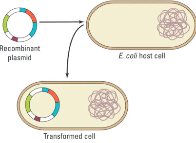
between bacteria — variation that natural selection can act on. In the laboratory, scientists use plasmids to insert “genes of interest” into an organism to change the organism’s phenotype, thus “transforming” the recipient cell. Using restriction enzymes, genes can be cut out of human, animal, or plant DNA and, using plasmids as vectors (carriers of genetic information), inserted into bacteria. If transformation is successful, the recipient bacteria will express the newly acquired genetic information in its phenotype (Figure 1).
Bio_S_Lab08_01
NOTE: Illustration requires less width than specified. Transformed cell
Recombinant
plasmid E. coli host cell
Figure 1. Transformation of Bacteria
In nature, the efficiency of transformation is low and limited to relatively few bacterial strains. Also, bacteria can take up DNA only at the end of logarithmic growth; at this time, the cells are said to be “competent.” In the lab, you have discovered several ways to increase the rate of transformation. Now, rather than just a few bacteria taking up a plasmid you want them to use, millions of bacteria can be transformed. The number of bacteria that take up a plasmid successfully is called the “transformation efficiency.” This is one of the values you will calculate in this lab unit.
In this investigation, you will use a predefined procedure to transform E. coli bacteria with a plasmid carrying a foreign gene. There are several different plasmids your instructor can choose from; you will be instructed about which one to work with for this unit.
E. coli is an ideal organism for the molecular geneticist to manipulate because it naturally inhabits the human colon and easily can be grown in a nutrient medium such as LB broth.
But what is E. coli’s natural or pre-transformation phenotype?
• Observe the colonies of E. coli grown on the starter LB/agar plate provided by your teacher to glean some information before you determine if any genetic transformation has occurred. What traits do you observe in pre-transformed bacteria? Record your observations in your laboratory notebook.
• Some bacteria are naturally resistant to antibiotics, but others are not. How could you use two LB/agar plates, some E. coli, and some ampicillin (an antibiotic) to determine how E. coli cells are affected by ampicillin?
• What would you expect your experimental results to indicate about the effect of ampicillin on the E. coli cells? Do you think that exposure to ampicillin will cause the
E. coli cells to evolve resistance to ampicillin? Why or why not?
• How will you be able to tell if host E. coli cells have been genetically transformed? (Hint: You will need some information from your teacher about the plasmid you will be using.)
■
Procedure
Your teacher will provide you with a plasmid containing one or more genes. The plasmid likely will contain the gene for resistance to ampicillin (pAMP), an antibiotic that is lethal to many bacteria, including E. coli cells. This transformation procedure involves the following three main steps to introduce the plasmid DNA into the E. coli cells and to provide an environment for the cells to express their newly acquired genes:
1. Adding CaCl2
2. “Heat shocking” the cells
3. Incubating the cells in nutrient broth for a short time before plating them on agar
Materials
Your Workstation
• E. coli starter plate prepared by your teacher
• Poured agar plates prepared by your teacher, most likely the following:
• 2 LB agar plates
• 2 LB/amp agar (LB agar contain-ing ampicillin) plates
• Transformation solution (CaCl2, pH 6.1) kept ice cold
• LB nutrient broth
• Sterile inoculation loops
• 100–1000 μL sterile bulb pipettes
• 1–10 μL micropipettes with sterile tips
• Microcentrifuge tubes
• Microcentrifuge tube holder/float
• Container full of crushed ice
• Marking pen
Common Workstation
• DNA plasmid (most likely pAMP at 0.005 μg/µL)
• 42°C water bath and thermometer
• 37°C incubator
• 20 μL adjustable-volume micropipettes and tips (optional)
• 10% household bleach
• Biohazardous waste disposal bags
In your lab notebook, record data, answers to questions, and any questions that arise during this part of the activity.
Step 1
Form lab teams, as instructed by your teacher. Familiarize yourself with sterile technique, materials and lab equipment, and safety procedures for handling bacteria and decontaminating the work area.Step 2
Label one closed microcentrifuge tube (micro test tube) “+ plasmid” and one tube“-plasmid.” (What do the “+” and “-” symbols mean?) Label both tubes with your group’s number (e.g., G2), and place them in the microcentrifuge tube holder/float.
Step 3
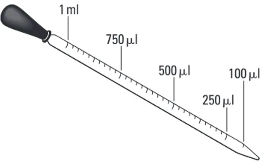
Carefully open the tubes and, using a 100–1000 μL bulb pipette with a sterile tip, transfer 250 μL of the ice cold transformation solution (CaCl2) into each tube. (Note that “μl” and “μL” are alternative symbols for the same volumetric measurement.)Bio_T_Lab08_02
750!l
500!l 250!l
100!l
1ml
Figure 2. Measuring Volume with a Pipette
Step 4
Place both tubes on (into) the ice.Step 5
Use a sterile inoculation loop to pick up a single colony of bacteria from your starter plate. Be careful not to scrape off any agar from the plate. Pick up the “+ plasmid” tube and immerse the loop into the CaCl2 solution (transforming solution) at the bottom of the tube. Spin the loop between your index finger and thumb until the entire colony is dispersed in the solution. Appropriately discard the loop.Step 6
Use a new sterile 100–1,000 μL micropipette to repeatedly pulse the cells in solution to thoroughly resuspend the cells. (Note that the clear transformation solution will become cloudy as the E. coli cells are suspended.) Place the tube back on the ice.Step 7
Using a new sterile inoculation loop, repeat Steps 5 and 6 for the “- plasmid” tube.CAUTION: Keep your nose and mouth away from the tip end when pipetting suspension culture to avoid inhaling any aerosol!
Step 8
Using a 1–10 μL micropipette with a sterile tip, transfer 10 μL of the plasmid solution directly into the E. coli suspension in the “+ plasmid” tube. Tap tube with a finger to mix, but avoid making bubbles in the suspension or splashing the suspension up the sides of the tube. Do not add the plasmid solution into the “- plasmid” tube! (Why not?)Step 9
Incubate both tubes (“+ plasmid” and “- plasmid”) on ice for 10 minutes. Make sure the bottom of the tubes make contact with the ice.Step 10
While the tubes are sitting on ice, label each of your agar plates on the bottom (not the lid) as directed by your teacher.Step 11
Following the 10-minute incubation at 0°C, remove the tubes from the ice and“heat shock” the cells in the tubes. It is critical that the cells receive a sharp and distinct shock! Make sure the tubes are closed tightly! Place the tubes into a test tubeholder/ float, and dunk the tubes into the water bath, set at 42°C, for exactly 50 seconds. Make sure to push the tubes all the way down in the holder so that the bottom of the tubes with the suspension makes contact with the warm water.
Step 12
When the 50 seconds have passed, place both tubes back on ice. For besttransformation results, the change from 0°C to 42°C and then back to 0°C must be rapid. Incubate the tubes on ice for an additional two minutes.
Step 13
Remove the holder containing the tubes from the ice and place on the lab counter.Using a 100–1,000 μL micropipette with sterile tip, transfer 250 μL of LB nutrient broth tothe “+ plasmid” tube. Close the tube and gently tap with your finger to mix. Repeat with a new sterile micropipette for the “- plasmid” tube.
Step 14
Incubate each tube for 10 minutes at room temperature.Step 15
Use a 10–1,000 μL micropipette with sterile tip to transfer 100 μL of thetransformation (“+ plasmid”) and control (“- plasmid”) suspensions onto the appropriate LB and LB/Amp plates. Be sure to use a separate pipette for each of the four transfers.
Step 16
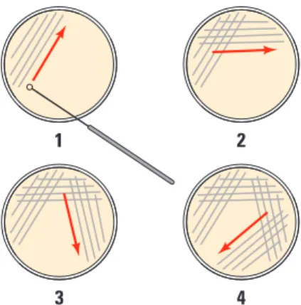
Using a new sterile inoculation loop for each plate, spread the suspensions evenly around the surface of the agar by quickly “skating” the flat surface of the sterile loop back and forth across the plate surface (Figure 3). Do not poke or make gashes in the agar! Your teacher might suggest that you use small sterile glass beads to spread the suspensions by gently rocking the beads across the surface of the agar. Allow the plates to set for 10 minutes.Bio_S_Lab08_03
3 4 1 2
Figure 3. Technique for Plating Bacteria on Agar
Step 17
Stack your plates and tape them together. Place the stack upside down in the 37°C incubator for 24 hours or as per instructed by your teacher.■
Analyzing Results
Think about these questions before collecting data and analyzing your results. Be sure to record your answers in your laboratory notebook.
1. On which of the plates would you expect to find bacteria most like the original non-transformed E. coli colonies you initially observed? Why?
2. If there are any genetically transformed bacterial cells, on which plate(s) would they most likely be located? Again, why?
3. Which plates should be compared to determine if any genetic transformation has occurred? Why?
4. What barriers might hinder the acquisition of plasmids?
5. How can the procedures described above (addition of CI2 and “heat shocking”) help facilitate the introduction of plasmids into the E. coli cells?
Consider the amount of bacterial growth you see on each plate. What color are the colonies?How many bacterial colonies are on each plate? Additional questions you might want to consider include the following:
1. Do your results support your original predictions about the “+ plasmid” transformed
E. coli cells versus “- plasmid” nontransformed cells?
2. Which of the traits that you originally observed for E. coli did not seem to become altered? Which traits seem now to be significantly different after performing the transformation procedure?
3. What evidence suggests that the changes were due to the transformation procedures you performed?
4. What advantage would there be for an organism to be able to turn on or off particular genes in response to certain conditions?
5. Was your attempt at performing a genetic transformation successful? If so, how
successful?
By calculating transformation efficiency, you can measure the success of your transformation quantitatively.
■
Calculating Transformation Efficiency
Your next task is to learn how to determine the extent to which you genetically transformed E. coli cells. This quantitative measurement is referred to as the
transformation efficiency. What is the importance of quantifying how many cells have been transformed? In many applications, it is important to transform as many cells as possible. For example, in some forms of gene therapy, cells are collected from the patient, transformed in the laboratory, and then put back into the patient. The more cells that are transformed to produce the needed protein, the more likely the therapy will work.
Calculating transformation efficiency gives you an indication of how effective you were in getting plasmids carrying new information into host bacterial cells. In this example, transformation efficiency is a number that represents the total number of bacterial cells that express the gene for ampicillin resistance divided by the amount of DNA plasmid used in the experiment. The transformation efficiency is calculated using the following formula.
Transformation efficiency = Total number of colonies growing on the agar plate Amount of DNA spread on the agar plate (in μg)
What two pieces of information will you need to calculate the efficiency of your transformation? Be sure to record all calculations.
1. Calculate the total number of transformed cells.
Observe the number of colonies visible on your LB/amp plate. Do not open the plate! Each colony on the plate can be assumed to be derived from a single cell. As individual cells reproduce, more and more cells are formed and develop into what is termed a colony. Thus, the most direct way to determine the total number of bacteria that were transformed with the plasmid is to count the colonies on the plate.
2. Calculate the amount of plasmid DNA in the bacterial cells spread on the LB/amp plate.
You need two pieces of information to find out the amount of plasmid DNA in the bacterial cells spread on the LB/amp plate: a) the total amount of DNA with which you began the experiment and b) the fraction of the DNA in the bacteria that actually got spread onto the LB/amp plate.
Once you determine this information, you will multiply the total amount of plasmid DNA used in the transformation times the fraction of DNA you spread on the LB/ amp plate.
a. Calculate the total amount (mass) of plasmid DNA.
The total amount (mass) of DNA with which you began the experiment is equal to the product of the concentration and the total volume used, or
DNA in μg=(concentration of DNA of μg/μL)x(volume of DNA in μL)
In this example, assume you used 10 μL of plasmid at a concentration of 0.005 pAMP μg/μL.
• Calculate the amount (mass) of plasmid DNA (pAMP) inμg per 1 μL of solution.
• Calculate the total amount of DNA used in this experiment. How will you use this information?
b. Calculate the fraction of plasmid DNA that actually got spread onto the LB/amp plate.
Since not all the DNA you added to the bacterial cells will be transferred to the agar plate, you need to calculate what fraction of the DNA was actually spread onto the LB/amp plate.
Fraction of DNA used= Volume spread on the LB/amp plate (μL) Total sample volume in test tube (μL)
Calculate the fraction of plasmid DNA you spread on the LB/amp plate. (Hint: Refer to the procedure and your notes. How many microliters of cells containing DNA did you spread onto the plate? What was the total volume of solution in the test tube? Did you add all the volumes?)
c. Calculate the micrograms of plasmid DNA that you spread on the LB/amp plate.
To answer this question, you multiply the total mass of plasmid DNA used times the fraction of plasmid DNA you spread on the LB/amp plate.
DNA spread in μg = Total amount of DNA used in μg x fraction of DNA used
What does this number tell you?
3. Calculate transformation efficiency.
Look at your calculations. Fill in the blanks with the correct numbers. Number of colonies on the LB/amp plate:
Micrograms of plasmid DNA spread on the plate: Now calculate the efficiency of the transformation.
Transformation efficiency = Total number of colonies growing on the agar plate Amount of DNA spread on the LB/amp plate (in μg) 4. What does this mean?
Transformation efficiency calculations result in very large, and very small, numbers. For both very large and very small numbers, scientists often use a mathematical shorthand referred to as scientific notation. For example, if the calculated
transformation efficiency is 1,000 bacteria/μg of DNA, they often report this as 103 transformants/μg.
How would scientists report 10,000 transformants/μg in scientific notation?
Suppose scientists calculated an efficiency of 5,000 bacteria/μg of DNA. How would they report this in scientific notation?
a. Report your calculated transformation efficiency in scientific notation.
b. What does your calculation of transformation efficiency mean?
c. Biotechnologists generally agree that the transformation protocol that you have just completed has a transformation efficiency of between 8.0 x 10² and 7.0 x 10³ transformants per microgram of DNA. How does your transformation efficiency compare? What factors could explain a transformation efficiency that was greater or less than predicted?
■
Evaluating Results
1. What are some challenges you had in performing your investigation? Did you make any incorrect assumptions?
2. What are some possible sources of error in the transformation procedure? If you had to repeat the procedure, what are ways to minimize potential sources of error?
3. Were you able to perform without difficulty the mathematical routines required to calculate transformation efficiency? Which calculations, if any, were challenging or required help from your classmates or teacher?
4. Can you suggest other preliminary activities that would have better prepared you to tackle the investigation?
5. Does a bacterial cell take in a plasmid with genes the cell already possesses? If so, would this affect your calculations?
■
Designing and Conducting Your Investigation
Think about these questions again for a minute.• What causes mutations in bacteria? Can mutations affect plasmids? How would you be able to tell if any observed changes in phenotypes are due to the expression of genes carried on plasmids and are not attributed to a possible mutagen?
• Do bacteria take up more in plasmid in some conditions and less in others? What conditions favor uptake, and which ones inhibit it?
• What other questions do you have about plasmids and transformation?
You can either design an investigation focusing on the information below OR design one based on a question(s) or observation you had as you worked through the genetic transformation you just conducted. Be sure that your experiment applies the science skills you acquired as you worked through this investigation. Make sure that your teacher approves your plan.
You should have noted satellites around the transformed colonies. (Satellites are smaller colonies that grow around the larger transformed colony.) What observations can you make about the satellites? Do they look like transformed bacteria? How can you tell if the satellites contain the plasmid? Design and conduct an experiment to determine if the E. coli satellite colonies from your genetic transformation experiment are transformed, too. Available to you are the same chemicals, supplies, and equipment you used in the previous investigation.
■
Where Can You Go from Here?
The background to this investigation asks you to think about several applications of genetic transformation, including genetically modified food and possible ethical, social, or medical issues raised by the manipulation of DNA by biotechnology. Why are these “issues”? What questions are posed by genetic engineering? In terms of what you have learned about biotechnology, how would you respond to the quote from Michael Crichton’s novel and film Jurassic Park: “Just because science can do something doesn’t mean that it should”?