THE EFFECTS OF MEDIAL UNLOADER BRACES AND LATERAL HEEL WEDGES IN THE GAIT BIOMECHANICS OF HEALTHY SUBJECTS
Sanjana Bhat
A thesis submitted to the faculty of the University of North Carolina at Chapel Hill in partial fulfillment of the requirements for graduation with honors in the Department of Exercise and
Sport Science.
Chapel Hill 2015
Approved by:
J. Troy Blackburn
Brian G. Pietrosimone
© 2015 Sanjana Bhat
ABSTRACT
Sanjana Bhat: The effects of medial unloader braces and lateral heel wedges inthe gait biomechanics of healthy subjects
(Under the direction of J. Troy Blackburn)
Orthotic devices such as medial unloader braces and lateral heel wedges have typically been used in patients with medial compartment knee osteoarthritis (OA) to force the knee into a neutral or valgus alignment and reduce loading on the medial compartment, but they may also be used in individuals following traumatic injuries or surgical repair procedures to protect cartilage. It was hypothesized that the medial unloader brace and lateral heel wedge would decrease knee adduction angle (KAA) and moment (KAM) in subjects without knee OA. Gait analysis performed on ten subjects with normal knee alignment and no history of knee injuries showed that the orthotic devices did not significantly affect KAA or KAM. These results indicate that the orthotic devices do not affect the medial compartment in the same way it does in knee OA patients, likely die to neutral rather than varus alignment in our sample of healthy subjects.
TABLE OF CONTENTS
LIST OF TABLES...………...vi
LIST OF FIGURES.…...………...vii
CHAPTER 1: INTRODUCTION………...1
CHAPTER 2: REVIEW OF LITERATURE………...7
Introduction.………...7
Knee Osteoarthritis………...7
The Epidemiology of Osteoarthritis………...7
Factors Leading to Knee OA………...8
Structures Affected by Knee OA………...13
Types of Knee OA...14
Treatment: What has been done to deal with knee OA...15
What has been done to slow OA progression?...15
What has been done to prevent it from occurring in healthy individuals...16
Methodological Considerations Behind the Study...21
Summary...22
CHAPTER 3: EXPERIMENTAL DESIGN AND METHODS...23
Subjects...23
Experimental Design...23
Assessments...23
Statistical Analysis...24
CHAPTER 4: RESULTS...25
Knee Adduction Angle (KAA)...25
Knee Adduction Moment (KAA)...25
CHAPTER 5: DISCUSSION...29
Limitations...31
Conclusions...32
LIST OF TABLES
Table 1. Peak Knee Adduction Angle (KAA)...26
LIST OF FIGURES
Figure 1. Peak Knee Adduction Angle...27
CHAPTER 1: INTRODUCTION
Knee osteoarthritis (OA) is a degenerative joint disease that refers to the breakdown of articular cartilage at the tibiofemoral joint and is the leading cause of pain and disability.1,2 Knee OA is more prevalent in the medial tibiofemoral joint compartment than the lateral because more load is placed onto this compartment during weight bearing activities.1 As a result, as knee OA progresses it creates great amounts of pain and disability during activities like walking that are essential to everyday life.1,3 Currently, one of the final clinical solutions is knee arthroplasty, or total joint replacement, but this approach is typically reserved for advanced or end-stage patients due to the high costs and associated risks. As it is, knee OA can lead to direct annual medical costs around $165 billion, or $5,700 per patient.4
There are two general types of knee OA: idiopathic and post-traumatic. Idiopathic, or primary, OA results from non-specific, gradual breakdown of articular cartilage and can be localized (affecting a particular joint) or generalized (affecting multiple joint regions).5 Conversely, post-traumatic, or secondary, OA results from a specific traumatic event.5 For example, knee OA can be caused by a complication of injuries to structures in the knee such as the anterior cruciate ligament (ACL) and menisci.6 The menisci improve joint stability and load distribution, and provide shock absorption and cartilage lubrication in the knee.7 The prevalence of knee OA due to an isolated ACL tear is only 13%, but is as high as 40% when combined with damage to the meniscus.8 A concomitant meniscus tear is present in up to 65% of ACL injuries, thus the risk of post-traumatic OA associated with ACL injuries is relatively high.6
effort to reduce the likelihood of developing knee OA. This includes performing meniscal repair, meniscectomy, mosaicplasty, or microfracture surgery. An injury to the meniscus can result in altered knee biomechanics which contribute to the development of knee OA. Meniscal injuries can be due to degeneration, trauma, and complications from having a discoid meniscus.9 One treatment for such injuries is by meniscal repair. Viability of meniscal repair depends on factors like vascularity of the location of the tear, type of tear, chronicity, and size.9 Meniscal repairs are typically performed over meniscectomies with the ideal situation being when there is an acute 1-2 cm longitudinal peripheral tear.9 The indications and results of meniscal repair, however, are controversial and provide mixed results. The clinical success rate for meniscal repair is 76% and based on improved pain, joint structure, and bilateral standing radiographs of the knee.10 Mensicectomy involves surgical removal of all or part of the meniscus, and, is commonly recommended when meniscal repair isn’t feasible.11 Meniscal repair is preferred to meniscectomy because of the uncertainty of results following a meniscectomy and overall better results. After undergoing a meniscectomy, patients are more likely to develop knee OA because of the decreased joint stability and load distribution that occurs due to the lack of part or all of the menisci.7 Additionally, there is a 75% decrease in intra-articular contact area that increases the risk of OA.10
involves stimulating mesenchymal cells from subchondral bone marrow to create a fibrin clot that forms a fibrocartilaginous regenerate.13 Mosaicplasty and MF surgery may reduce the risk of developing knee OA following traumatic injuries, but their long term impact are unknown.13
The breakdown of articular cartilage that leads to knee OA is thought to result, in part, from altered loading mechanics at the knee joint, particularly in the medial compartment. However, direct measurement of the load placed on the medial knee compartment in vivois not practical given the equipment needed and is thus not currently performed. Therefore, variables identified via biomechanical gait analysis are commonly used to estimate the load placed on the knee during walking gait. Walking is a critical part of human locomotion and occurs with great frequency, thus estimating the load placed on the knee during walking gait is a significant approach for understanding the repetitive, impulsive forces created that can lead to degenerative changes in articular cartilage.14
Peak knee adduction angle (KAA) and moment (KAM) are commonly used surrogate indicators of medial compartment loading.1 KAA refers to the frontal plane knee angle, and provides an indication of the proximity of medial tibiofemoral joint surfaces (i.e. greater knee varus motion approximates the medial joint surfaces).1 Greater approximation of these
articulating surfaces leads to greater compressive force being placed on the articular cartilage. KAA typically displays two peaks, the first of which occurs during early stance of gait and is associated with the presence, severity, and progression of knee OA.15
more rapid progression of knee OA.16,17 These findings suggest that factors which reduce KAA and KAM may reduce the risk of developing knee OA.
Limiting loading of the medial knee compartment (i.e. KAA and KAM) is critical for reducing the load placed on articular cartilage. Medial/valgus unloader braces are designed to force the knee into a more neutral or valgus frontal plane alignment to reduce the load on the medial compartment, and lateral heel wedges can be inserted in shoes to produce similar effects.1,19 These devices produce a valgus moment about the knee to decrease the varus alignment and loading that lead to knee OA.20 Studies that have evaluated the effects of these orthotic devices generally report that they are effective for reducing KAM and KAA when used in combination.1,16,21 The results of using these orthotic devices in isolation are similarly promising, as lateral wedges reduce the peak KAM, and similar reductions in KAM and KAA have been reported with medial unloader braces.1,22 As such, medial unloader braces and lateral heel wedges may be effective methods for slowing the progression of knee OA.
these orthotic devices due to the fact that they do not display chronic gait adaptations or pathological varus deformity, and do not possess chronic pain that requires compensatory gait strategies, similar to individuals who have experience traumatic cartilage injuries or surgical repair.
The limited research evaluating the effects of these orthotic devices in healthy subjects report favorable results. Medial unloader braces have demonstrated an decrease KAM21,23and KAA during gait.21 Lateral heel wedges produced similar results in addition to reducing the medial compartment contact force in computer simulations.24 These results suggest potential benefits of using such orthotic devices following cartilage trauma and surgical repair. However, it is unclear which of these devices is most effective for reducing KAA and KAM. Therefore, the purpose of this study was to evaluate and compare the effects of medial knee unloader braces and lateral heel wedges on gait biomechanics associated with knee OA development (KAA and KAM) in healthy individuals via the following specific aims:
1. To determine the effects of a medial unloader brace on peak KAM and peak KAA during
walking gait in healthy subjects.
Hypothesis 1: The medial unloader brace will reduce the peak KAM and peak KAA.
2. To determine the effects of a lateral heel wedge on peak KAM and peak KAA during
walking gait in healthy subjects.
Hypothesis 2: The lateral heel wedge will reduce the peak KAM and peak KAA.
3. To compare the effects of the medial unloader brace and lateral heel wedge on peak
Hypothesis 3: The medial unloader brace will be more effective in reducing peak KAM and peak KAA than the lateral heel wedge.
CHAPTER 2: REVIEW OF LITERATURE
Introduction
The purpose of this literature review is to review studies that help deepen knowledge and identify any deficits in our understanding on the topic of knee osteoarthritis. This review in particular focuses on the effects of using medial unloader braces and lateral heel wedges on the gait biomechanics of subjects with no history of knee injuries. It will evaluate how orthotic devices like medial unloader braces and lateral heel wedges can be used to potentially prevent joint degeneration and knee osteoarthritis. Secondly, this review gives evidence of the
effectiveness of braces in injured and healthy populations to show how they can be effectively used as cost effective and simpler alternatives to knee arthroplasty if knee osteoarthritis is developed. Lastly, it will evaluate the importance of determining whether the orthotic devices affect gait biomechanics in healthy subjects. This is important as it will help show whether orthotic devices can be used to correct joint dynamics in individuals at heightened risk for developing knee OA.
Knee Osteoarthritis
The Epidemiology of Osteoarthritis
to the aging population and rise in obesity.26,27 In addition, it is ranked eleventh in the world of causes of years living with a disability.28 It is a degenerative joint disease that damages articular cartilage at a joint and the tissues around it.25 Individuals suffering from it experience symptoms of pain, stiffness, and decreased range of motion in the joint.29
A loss of articular cartilage, problems in the remodeling of subchondral bone and
osteocyte formation, ligamentous laxity, weakening of the periarticular muscle and synovitis are all factors that contribute to OA.25 Pain is the most common symptom associated with OA and can affect one’s movement as a result as well.25It is seen mostly in hands, feet, facet joints and weight bearing joints like the knee and hip. OA leads to direct annual medical costs exceeding $321 billion of which knee OA, the most common type, accounts for $165 billion or $5,700 per patient.4 Knee OA has a doctor-diagnosed prevalence of 13.8% in populations over the age of 45.28 Of these people with knee OA, 10.9% have OA in another joint.28 Women also tend to be affected by OA more than men.28 The prevalence and debilitating nature of knee OA has made it important to find ways to prevent it and slow its progression.16
Factors leading to Knee OA
OA. People who suffer from one or more of these factors are at an increased risk of developing OA.
High BMI is a major risk factor for knee OA. With rates of obesity in the world
increasing, increasing rates of knee OA are to be expected as well.28This is demonstrated by the population risk for knee OA due to obesity set at 29%.8 For every 5 unit increase in BMI, the risk of knee OA increases by 35%.8 On the other hand, losing five pounds could decrease the chance for knee OA by 50% thus showing how reduced weight and load are important in helping curb knee OA.8
ACL rupture is another factor that has recently been shown to affect knee OA. When one tears the ACL, he/she also tends to damage articular cartilage, subchondral bone and collateral ligaments, and the menisci as well.8 The rupturing of the ACL is quite common in individuals who play high-risk sports and experience high valgus forces on their knees.8 While ACL ruptures on their own do accelerate the chances of experience knee OA, the damage to other structures is what may significantly increase the development of OA. This damage in turn can lead to the development of OA in a significant number of people as early as ten years after the initial ACL rupture occurred.8 Even after ACL reconstruction surgery, the knee could be impacted due to the lack of a functional ACL which affects the static and dynamic loading on the injured knee.8 This could lead to long term changes in the cartilage that is in direct contact during weight bearing activities,8and could increase the risk of developing knee OA. The prevalence of knee OA due to an ACL tear is only 13% but is between 21 and 40% when
tears leave people at a higher chance of developing knee OA.
Menisci help improve joint stability and load distribution, and provide shock absorption and cartilage lubrication in the knee.7 Therefore, any injury to a meniscus can alter knee
biomechanics and increase the risk of knee OA.30 Meniscal injuries can be due to degeneration, trauma, and from having a discoid meniscus.7,9 Meniscal degenerative pathology is correlated with degenerative cartilage changes which in itself can lead to knee OA.7 These injuries thus commonly require surgical repair which also increases the risk of developing knee OA.31
One treatment for this is meniscal repair. Performing meniscal repair may depend on factors like vascularity of the location of the tear, type of tear, chronicity, and size.9 In other words, an acute longitudinal tear in the red zone is more likely to be repaired than a chronic flap tear in the white zone of the meniscus.9 In this case, meniscal repair is typically used when there is an acute 1-2 cm longitudinal peripheral tear that is fixed during ACL reconstruction in
particular.9Meniscal repair can be done in open or arthroscopically, but arthroscopic surgery is preferred because it involves a small incision, has early recovery and rehabilitation.9The advantage of performing meniscal repair is also that it retains native anatomy by suturing the meniscus back into place to keep its stability and function intact.32Meniscal repair overall has a clinical success rate of 76% for meniscal repair based on improved pain, joint structure, and bilateral standing radiographs of the knee despite it being a relatively unproven method to repairing the meniscus.10 Meniscal repair procedures do have a failure rate that ranges between 5% and 43% that subsequently leads to undergoing a meniscectomy to address continued complications.32
of developing knee OA increases 6-fold.33 This is more likely to happen because of the decreased joint stability and load distribution that occurs in the knee due to the damage to the menisci.7,34 The pressure placed on the joint may be increased by 85% during flexion and contact pressure by 100-200%.10 However, unlike a total meniscectomy, a partial meniscectomy is has more favorable results. Like meniscal repair, it attempts to preserve the meniscus and has an 88% success rate among patients.34Despite that, meniscal repair is preferred to any kind of a meniscectomy since meniscus-deficient knees can increase the risk of developing OA.35
In addition to meniscal tears, meniscal extrusion is seen as one of the strongest predictors of developing OA.30 Meniscal extrusion refers to the extending of the meniscus beyond the tibial margin.36 When this occurs, there are typically larger tears in the meniscus that affect the
meniscus’s ability to take on load.30,36 In this case, the meniscus may also be displaced from the tibial articular cartilage.37 Varus alignment of the lower limb and joint space narrowing, two factors that contribute to knee OA, are also seen to cause meniscal extrusion.37 It is unknown whether meniscal extrusion occurs prior to or after knee OA development.37 Regardless, meniscal extrusion may still cause cartilage degeneration, which leads to knee OA, to occur.30
progression in those with damage to the meniscus.
Gait biomechanics are another factor that can lead to knee OA. Injuries like those to the meniscus can affect gait biomechanics and place more stress on the medial compartment of the knee. One way that these changes are studied is to examine the loading mechanics in individuals with knee OA. While it is not currently practical to directly measure load place on the knee in vivo, variables like peak knee adduction moment (KAM) and knee adduction angle (KAA) can
be used as surrogate indicators of load placed on the medial compartment.1,38
KAM is typically used to estimate the magnitude of dynamic medial joint loading.16,38 It is determined by the ground reaction force (GRF) and the lever arm to the center of the knee joint in the frontal plane.17 The presence of a KAM thus means that the medial aspect of the tibia that articulates with the inferior aspect of the medial femoral condyle is being loaded in
compression.18 KAM can be used to explain the limb’s alignment, bone mineral density of the proximal tibia and the progression and development of knee OA.17 In people suffering from medial compartment knee OA, the KAM tends to be large during the stance phase.38 This larger KAM means that the knee is in more of a varus alignment, and may have potentially lost bone mineral density and medial joint space.16,17 This, as a result, may all cause rapid progression of knee OA
In regards to these two variables, greater KAA and KAM are both associated with knee OA. Alterations in these variables in response to pain and other stimuli influence the progression of knee OA and can negatively affect the knee joint in the process. Finding out how these
variables are affected in healthy subjects and while treating individuals with knee OA is essential to testing the effectiveness of a particular treatment.
Structures Affected by Knee OA
Knee OA is an all-encompassing degenerative joint disease: it affects all components within the knee joint including hyaline articular cartilage, subchondral bone, ligaments, and the menisci.39 Breakdown of the cartilage, bone lesions, and/or ligament and meniscal tears can all contribute to knee OA. Damage to or malalignment of any of these structures can leave one vulnerable to knee OA. This is also the same reason it can be difficult to prevent knee OA from developing.
Another factor seen in radiographic knee OA patients is the decrease in joint space that may occur in response to the damage of any of the structures described above. This decrease in joint space is associated with osteophytes, which are also known to be involved in subchondral schlerosis and pain in the knee joint.25,40 Osteophytes are bony outgrowths that are found near the joint typically at the anterior part of the tibial plateau.41,42 The size of osteophytes is often used to grade the severity of knee OA with small osteophytes pointing to the possibility of narrowing of joint space and large osteophytes with severe narrowing of joint space, bony end deformation, and schlerosis.25,42,43 The larger and more abundant number of osteophytes in the knee is indicative of the increased severity of knee OA.
things that physicians look for especially when using radiography to diagnose knee OA.39 MRI on the other hand is able to identify damage to subchondral bone, the synovium, ligaments, and the other soft tissues involved in knee OA to potentially give a more definitive picture of the structures involved in knee OA.39
Types of Knee OA
There are two ways that knee OA can be developed: idiopathically or post-traumatically. Idiopathic, or primary OA, results from the non-specific, gradual breakdown of articular
cartilage and can be localized (affecting one joint) or generalized (affecting multiple joint regions).5 This type of OA does not originate from one particular event, thus it tends to take longer to develop than post-traumatic OA.5 Idiopathic knee OA results from repeated loads placed on the joint that accumulate over time as a result. Other factors discussed already like obesity may also make one more prone to develop idiopathic knee OA.
Treatments: What has been done to deal with knee OA?
What has been done to slow OA progression?
There is no effective treatment for reversing knee OA other than joint arthroplasty. Treatments thus revolve around trying to keep the condition from getting worse and relieving pain. As a result, conservative treatments of knee OA involve pharmacological and
non-pharmacological solutions to reduce pain, improve mobility, and limit functional impairment in the joint.45,46 After trying to correct those issues, treatments often focus on trying to stop the progression of the disease and improving muscular strength to help individuals try to lead normal lives despite having knee OA.46 As knee OA can be caused by changes in gait biomechanics, it is important to approach it holistically and not only find ways to reduce pain, but also try to isolate the mechanisms that lead to and progress the development of knee OA. A primary objective for slowing knee OA progression is to reduce the load on the medial compartment during weight-bearing tasks.
Pharmacological treatments for knee OA involve the use of oral analgesics like acetaminophen, non-steroidal anti-inflammatory drugs (NSAIDs) like ibuprofen, topical analgesics, neutraceuticals like glucosamine, and corticosteroid injections.46 Which pain
relievers are used is based on the severity of the pain felt and other symptoms like inflammation that may occur as well. NSAIDs are seen as the most effective in helping these symptoms and are often the first pain medicine given to patients,47but corticosteroid injections may also be used for short-term relief if necessary.46
Much of the changes that can be made to slow the progression of knee OA are
OA, lifestyle changes that can be made include losing weight, altering daily activity, and engaging in non-pharmacological treatments including exercise, using thermal modalities, acupuncture, transcutaneous electrical nerve stimulation, shoe insoles/wedges, walking aids like canes, and knee braces. The effectiveness of each of these methods in preventing the progression of knee OA varies by individual and in general, but they are moderately effective for reducing the symptoms of knee OA. .
Knee OA can also be treated by choosing surgical options like arthroscopy, osteotomy, and arthroplasty.48 The reasons behind choosing the surgical route to treat knee OA involves location and severity of the damage, individual situation, and other risk factors.48 Despite the option of surgery being available to patients, the non-surgical treatments described above are preferred, and surgery is only recommended if those options fail to give relief to the patient and/or are not effective in slowing the progression of knee OA.48 There are different techniques involving arthroscopy but it is widely seen as rather ineffective and with short-term benefits that help only select populations like those dealing with meniscal tears.48 Osteotomy is typically used more in younger patients with knee OA who do not need total knee replacement, and involves removing the bone to change alignment and relieve weight placed on the joint.48 The final and more drastic way to treat knee OA is through knee arthroplasty, or knee replacement surgery. This surgery is safe but is only recommended when all other options have failed because of the irreversibility of the procedure and the risks such as infection.
What has been done to prevent it from occurring in healthy individuals?
include engaging in quadriceps strengthening programs, performing exercises to increase flexibility, balance, muscle strength and endurance in general to help prevent injury that may lead to knee OA, and maintaining a healthy weight.49
Healthy individuals in relation to those with knee OA refer to those who do not have any bony malalignments or pain associated with knee OA. As a result, this includes those who have immediately had a traumatic knee injury such as an ACL tear or meniscal tear, as well those who have already undergone cartilage repair surgeries like microfracture (MF) and mosaicplasty, that may increase the risk of developing knee OA. The quick fix after traumatic injuries to prevent the development of knee OA is often times just to undergo ACL reconstruction or a
meniscectomy to help them regain mechanical stability in the joint. However, simple ACL reconstruction and mensicectomy is often not a long term solution to the problem and those individuals often are still prone to developing knee OA.
One way to correct the cartilage defects and breakdown that can progress to knee OA is by mosaicplasty. It is a procedure that involves getting osteochrondral grafts from a less
important weight bearing area of the articular surface to the cartilage defect and drilling holes at the defect site.12,13 This procedure is effective when resurfacing osteochondral defects in the knee and rebuilding an articular surface.12,13 Problems can occur since a graft used from another area may not match the knee cartilage it is replacing and may also negatively affect the area the graft was taken from.12 It is, however, a technique that is used to try to prevent the progression of knee OA following cartilage injuries and has a success rate in doing so of 83-92% in the short and mid-term.13
Another way to correct cartilage defects and breakdown is through the use of
subchondral bone marrow to create a fibrin clot that forms a fibrocartilaginous regenerate.13 This regenerate is not as structurally sound as the native articular cartilage but the addition of fibrocartilage to the joint improves knee function and relieves pain.13 It is seen as a procedure that can be especially highly successful if rehabilitation protocols are strictly followed to heal the cartilage.50 There is evidence that shows that an MF procedure can improve pain and function in 80% of patients under the age of 45 after a 7-17 year follow-up.50,51 Like with mosaicplasty, undergoing a MF procedure is thus a rather good way to help prevent the development of knee OA in individuals following traumatic injuries, but the long term impact of such procedures is still relatively unknown.13
Despite the relative success of mosaicplasty and MF procedures, the uncertainty of its impact in the long term does mean that protective measures may be needed in these individuals to not only enhance the effects of the surgery but also ensure long term success. One such protective measure that can be used in individuals following a traumatic injury and corrective cartilage surgeries is the use of orthotic devices. In this case, medial unloader braces and lateral heel wedges may be used in slowing the development of medial compartment knee OA.
The Role of Bracing
more neutral or valgus frontal plane alignment to reduce the load on the medial compartment.24,44 Using these medial unloader braces can thus help reduce KAM and shift the loads from the medial compartment to the lateral compartment.19 They can decrease the load on the medial compartment by about 11% but more data is still needed to further validate these statistics.24
Lateral heel wedges can be inserted in shoes to produce similar effects.1 These devices produce a valgus moment about the knee to decrease this varus alignment.20 They are designed to reduce KAM by relatively shifting the center of pressure of the GRF laterally in comparison to the center of the knee joint.24 Doing this in turn changes the varus moment in order to produce a more valgus moment and alleviate loading in the medial compartment.52
Studies looking at the effects of these orthotic devices on individuals with knee OA show that they are most effective in reducing KAM and KAA when used in combination.1,16,21 The results of using these orthotic devices separately are just as encouraging as they respectively reduce the peak KAM and KAA in those with knee OA.1,22 As such, medial unloader braces and lateral heel wedges are seen as effective methods used to slow the progression of knee OA.
These orthotic devices may also be effective for protecting healing cartilage, potentially reducing the risk of knee OA. They can improve tibiofemoral alignment by shortening the moment arm at the knee and foot, and thus reducing KAM as well.46 As a result, medial unloader braces in particular are able to positively impact the distance one can walk, gait velocity, pain, muscle strength, and frontal stability.46
There are no significant differences noticed in KAM between the group that wore the lateral heel wedges and the group that wore the medial unloader braces after a period of 6 weeks either.54 While more research is needed to validate this information, it does show the limitations that these orthotic devices can present. It shows a need to perform more research on these devices with knee OA patients and in those without knee OA to see if the devices can be effective in slowing the progression of knee OA in that population. Yet, the majority of the literature in this area has been conducted in individuals already diagnosed with knee OA.
In a study involving healthy male runners in particular, there was a reduction in KAM noticed after the use of a valgus unloader brace which shows how the use of braces may also help those who do not have knee OA.23 Overall, it has been seen that medial unloader braces have been demonstrated to decrease KAM21,23and KAA during gait.21 Lateral heel wedges are also effective in decreasing KAM and KAA while also reducing the medial compartment contact force in computer simulations.24On the other hand, there is also contradicting data regarding medial unloader braces in healthy individuals that shows that there was no significant difference in KAM between the braced and unbraced conditions.55
thing to consider with medial unloader braces and lateral heel wedges is the weighing the benefits of using the devices with comfort. Despite the success of these devices in those with knee OA, medial unloader braces that have large angulations and lateral heel wedges with greater wedge height are associated with less comfort, which can thus decrease its effectiveness in those with or without knee OA respectively.16
Methodological Considerations Behind the study
This study focuses on the role of medial unloader braces and lateral heel wedges in reducing medial knee compartment loading in healthy subjects. Due to the lack of research regarding how these braces influence gait biomechanics in healthy individuals, it is unclear if they could provide benefits to those following surgical cartilage repair procedures. Individuals more prone to developing knee OA include people with traumatic injuries like meniscal tears and ACL tears and more chronic issues like obesity that progress cartilage breakdown. It is thus important to see whether these orthotic devices can effectively limit loading on the medial compartment of the knee in such vulnerable populations that do not yet have varus deformity in the joint due to knee OA.
. The use of healthy subjects in particular in this study is helpful though as their knees exhibit similar qualities to those with cartilage injuries who have not yet developed knee OA. It may also show whether the same effects of reducing medial compartment loading from using the orthotic devices in those with knee OA can be extended to those without knee OA as well. The main problem for these individuals lie in the instability that comes from the injuries they suffer that can alter biomechanics and loading patterns in the knee without initially affecting bony alignment.7Seeing how the devices can be used to limit varus loading on the knee joint and therefore protect injured cartilage in the medial compartment as it heals can be essential when trying to prevent the development of knee OA in such individuals.
Summary
Knee OA is a debilitating disease that occurs as a result of cartilage breakdown in the knee joint. While there are methods like meniscal tear surgeries, meniscectomies, mosaicplasty, and microfracture surgery that may be used to help to fix menisci and cartilage, and keep the damage from progressing into knee OA, there are no fool-proof solutions to the problem as of now. The main purpose of this study therein lies with finding a way to prevent the development of knee OA in such individuals who do not yet have the bony deformity and varus alignment seen in those with knee OA. Testing orthotic devices like medial unloader braces and lateral heel wedges to see if they can be effective in trying to protect the joint in such cases is important. The use of healthy subjects to do this is essential as these individuals would not have the bony deformities associated with knee OA, therefore being similar to those that have damaged
CHAPTER 3: EXPERIMENTAL DESIGN AND METHODS
Subjects
Ten healthy individuals (4 males and 6 females) between the ages of 18 and 35 years were recruited for this study. Subjects were required to not have a history of lower extremity injury in the 6 months prior to participation, osteoarthritis or symptoms related to osteoarthritis, or any traumatic knee joint injuries. Each subject was required to read and sign an informed consent form prior to participation.
Experimental Design
This investigation utilized a randomized controlled crossover design. Three-dimensional walking gait biomechanics were captured as subjects complete four testing conditions
(control/no device, unloader brace at 50% max load, lateral heel wedge, and unloader brace at 100% max load) in a single testing session. The order of the conditions was determined via an unbalanced Latin square. Subjects were fitted for an adjustable medial unloader brace (Bledsoe Z-12, Bledsoe Brace Systems, Grand Prairie, TX), and the lateral heel wedge consisted of a 3” wool felt inserted into the shoe (Hapad Inc., Bethel Park, PA).
Assessments
along a 6 m walkway at a comfortable, self-selected pace.1,16,20,45,55 All data were sampled from the right leg, and motion capture sensors were placed on the sacrum, lateral side of the thigh on the IT band, medial side of the tibia, and the dorsum on the foot via double sided tape. Gait speed was monitored via an infrared timing system, and subjects were required to remain ±10% of the mean speed obtained from at least 5 practice trials. The five practice trials were carried out to determine the exact distance of the start to ensure the subjects’ right heel-strikes hit the force plate each time without noticeably altering gait patterns and speed. Subjects then performed 5 valid trials for each testing condition. Three-dimensional marker positions were sampled at 100 Hz using electromagnetic motion capture system and low-pass filtered at 10 Hz. Ground reaction forces were sampled at 1,000 Hz and low-pass filtered at 75 Hz.
Peak KAA and KAM were measured during the first 50% of the stance phase of gait (heel strike to toe off). Heel strike was identified as the instant when the vertical ground reaction force (vGRF) exceeds 20 N, and toe off was identified as the instant when the vGRF returns below 20 N. Customized computer software (LabVIEW, National Instruments, Inc., San Antonio, TX) was utilized to calculate these measures. The joint moments (KAM) were
normalized to the product of body weight (BW) and height (Ht) of the subject (BW*Ht) and then averaged for each subject for statistical analysis.1,16,20,45
Statistical Analysis
CHAPTER 4: RESULTS
Ten subjects who met the inclusion criteria for the study completed all testing conditions, and were included in the final analyses. The subjects (4 males and 6 females) had a mean mass of 68.7 (±16.5) kg, height of 168.3 (±12.9) cm, and age of 21.6 (±2.95) years.
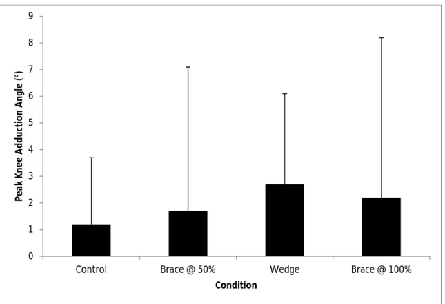
Knee Adduction Angle (KAA)
The orthotic devices did not significantly affect peak KAA (F1.367, 12.304 = 0.487, p =
0.556). Peak KAA ranged 1.2-2.7° across conditions, but did not differ significantly. Descriptive statistics for peak KAA are listed in Table 1 and Figure 1.
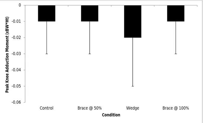
Knee Adduction Moment (KAM)
Similar to KAA, the orthotic devices did not affect peak KAM (F1.155, 10.393 = 0.729, p =
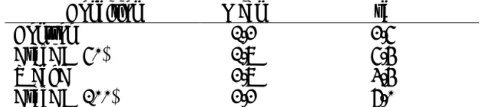
Table 1. Peak Knee Adduction Angle (KAA)
Condition Mean sd
Control Brace @ 50% Wedge
Brace @ 100%
1.2 1.7 2.7 2.2
2.5 5.4 3.4 6.0
Table 2. Peak Knee Adduction Moment (KAM)
Condition Mean sd
Control Brace @ 50% Wedge
Brace @ 100%
-0.01 -0.01 -0.02 -0.01
Figure 1. Peak Knee Adduction Angle
0 1 2 3 4 5 6 7 8 9
Control Brace @ 50% Wedge Brace @ 100%
Pe
ak
K
ne
e
Ad
du
ct
io
n
An
gl
e
(°
)
Figure 2. Peak Knee Adduction Moment
-0.06 -0.05 -0.04 -0.03 -0.02 -0.01 0
Control Brace @ 50% Wedge Brace @ 100%
Pe
ak
K
ne
e
Ad
du
ct
io
n
M
om
en
t (
xB
W
*H
t)
CHAPTER 5: DISCUSSION
The primary findings of this study demonstrated that medial unloader braces and lateral heel wedges did not alter KAA or KAM during walking gait in healthy subjects. These findings are contrary to our hypotheses and contrast with the decreases in KAA and KAM noted in individuals with knee OA.
The literature regarding the effects of medial unloader braces and lateral heel wedges on gait biomechanics in healthy individuals is equivocal. Our findings agree with those of Ebert et al.55who reported no influence of unloader braces on KAM. Conversely, Orishimo et al.,21 Pagani et al.,23and Kakihana et al.52,56who reported reductions in KAM and KAA with these orthotic devices. The discrepancies between the studies are likely due to differences in the orthotic devices, the testing procedures, and the individuals who were evaluated.
Orishimo et al.21used a different unloader brace (DonJoy OA Adjustor Medial Unloader Brace, DonJoy Orthopedics, Vista, CA) in a slightly older healthy population (32 ± 10 years) compared to our investigation, and reported decreases in KAM and KAA. This discrepancy in results likely can be attributed to the fact that the DonJoy brace induced as much as 20° of valgus correction when loaded maximally.57 Comparatively, the Bledsoe Z-12 OA brace we used provides a maximum of 3° of valgus correction.58 This dramatic difference in valgus loading capacity of the two unloader braces likely explains the difference in results.
the subjects used in our study. In combination, these data suggest that knee alignment influences the efficacy of medial unloader braces, as they reduce medial knee compartment loading in individuals with varus alignment but do not alter frontal plane knee biomechanics in individuals with neutral alignment. This result may be explained by the fact that varus alignment causes joint opening on the lateral side (e.g. greater space between the femoral and tibial contact surfaces). As such, the valgus moment provided by the unloader brace has the potential to alter knee alignment from varus to valgus by approximating the lateral tibial and femoral joint
surfaces. In comparison, the spacing between the joint contact surfaces on the lateral side of the joint in neutrally aligned knees is much smaller, thus limiting the additional approximation that can be caused by the orthotic device.
Kakihana et al.52,56reported significant decreases in KAM with lateral wedges. These investigators used 3° and 6° wedges, but only the 6° wedge decreased KAM. Additionally, these wedges spanned the entire length of the foot. We used a 7° wedge that was placed directly under the calcaneous, and only came into contact with the rearfoot. The foot possesses substantial capacity to adapt to different surfaces via motion between the rearfoot and forefoot segments. As such, the wedge used in our investigation may have produced calcaneal eversion that was countered by forefoot inversion to allow the plantar aspect of the foot to maintain contact with the ground. This compensation for the effects of the lateral heel wedge would have minimized the proximal effects on frontal plane knee biomechanics. Conversely, the wedges used by Kakihana et al. spanned the entire length of the foot, thus limiting compensatory motion between the forefoot and rearfoot, and potentially having a greater proximal effect on frontal plane knee biomechanics.
that matched that of the knee OA patients in their study. This pace may have altered the
subjects’ “normal” gait patterns. In contrast, our subjects walked at a comfortable, self-selected pace. As such, this unfamiliar task may partially explain the discrepancies across studies.
Similar to the effects of medial unloader braces, it is likely that lateral wedges may have minimal effects on neutrally aligned knees while still altering knee frontal plane knee
biomechanics in those with varus alignment. The risk of developing OA increases with age and affects the majority of people above the age of 65.59 An elderly individual is more likely to have cartilage degeneration in the knee than younger individual such as those in our investigation regardless of activity level because of the forces placed on the joint that accumulate over time.59 Even though the subjects assessed by Kakihana et al.52were healthy, they were elderly (average age 65 ± 2 years) and likely had mild degenerative changes in the knee cartilage that were asymptomatic. These mild degenerative changes in the knee cartilage could also allow for a greater joint opening on the lateral side that could make it easier for the wedge to produce valgus orientation at the knee. As such, the difference in populations in the two studies may have contributed to the differential results.
Limitations
OA development.60 Examining these relationships could be helpful when drawing conclusions about the devices’ effectiveness in healthy individuals.
Conclusions
REFERENCES
1. Hinman RS, Bowles KA, Metcalf BB, Wrigley TV, Bennell KL. Lateral wedge insoles for medial knee osteoarthritis: effects on lower limb frontal plane biomechanics. Clinical biomechanics. Jan 2012;27(1):27-33.
2. Clynes MA, Parsons C, Edwards MH, et al. Further evidence of the developmental origins of osteoarthritis: results from the Hertfordshire Cohort Study. Journal of developmental origins of health and disease. Aug 26 2014:1-6.
3. Peat G, McCarney R, Croft P. Knee pain and osteoarthritis in older adults: a review of community burden and current use of primary health care. Annals of the rheumatic diseases. Feb 2001;60(2):91-97.
4. Maetzel A, Li LC, Pencharz J, et al. The economic burden associated with osteoarthritis, rheumatoid arthritis, and hypertension: a comparative study. Annals of the rheumatic diseases. Apr 2004;63(4):395-401.
5. Altman R, Asch E, Bloch D, et al. Development of criteria for the classification and reporting of osteoarthritis. Classification of osteoarthritis of the knee. Diagnostic and Therapeutic Criteria Committee of the American Rheumatism Association. Arthritis and rheumatism. Aug 1986;29(8):1039-1049.
6. Nordenvall R, Bahmanyar S, Adami J, Mattila VM, Fellander-Tsai L. Cruciate Ligament Reconstruction and Risk of Knee Osteoarthritis: The Association between Cruciate Ligament Injury and Post-Traumatic Osteoarthritis. A Population Based Nationwide Study in Sweden, 1987-2009. PloS one. 2014;9(8):e104681.
7. Englund M, Roos EM, Lohmander LS. Impact of type of meniscal tear on radiographic and symptomatic knee osteoarthritis: a sixteen-year followup of meniscectomy with matched controls. Arthritis and rheumatism. Aug 2003;48(8):2178-2187.
8. Johnson VL, Hunter DJ. The epidemiology of osteoarthritis. Best practice & research. Clinical rheumatology. Feb 2014;28(1):5-15.
9. Yoon KH, Park KH. Meniscal repair. Knee surgery & related research. Jun 2014;26(2):68-76.
10. Mordecai SC, Al-Hadithy N, Ware HE, Gupte CM. Treatment of meniscal tears: An evidence based approach. World journal of orthopedics. Jul 18 2014;5(3):233-241.
12. Reverte-Vinaixa MM, Joshi N, Diaz-Ferreiro EW, Teixidor-Serra J, Dominguez-Oronoz R. Medium-term outcome of mosaicplasty for grade III-IV cartilage defects of the knee. Journal of orthopaedic surgery. Apr 2013;21(1):4-9.
13. Ulstein S, Aroen A, Rotterud JH, Loken S, Engebretsen L, Heir S. Microfracture
technique versus osteochondral autologous transplantation mosaicplasty in patients with articular chondral lesions of the knee: a prospective randomized trial with long-term follow-up. Knee surgery, sports traumatology, arthroscopy : official journal of the ESSKA. Jun 2014;22(6):1207-1215.
14. Liikavainio T, Isolehto J, Helminen HJ, et al. Loading and gait symmetry during level and stair walking in asymptomatic subjects with knee osteoarthritis: importance of quadriceps femoris in reducing impact force during heel strike? The Knee. Jun 2007;14(3):231-238.
15. Roda RD, Wilson JL, Wilson DA, Richardson G, Dunbar MJ. The knee adduction moment during gait is associated with the adduction angle measured during computer-assisted total knee arthroplasty. The Journal of arthroplasty. Jun 2012;27(6):1244-1250.
16. Moyer RF, Birmingham TB, Dombroski CE, et al. Combined effects of a valgus knee brace and lateral wedge foot orthotic on the external knee adduction moment in patients with varus gonarthrosis. Archives of physical medicine and rehabilitation. Jan
2013;94(1):103-112.
17. Kutzner I, Trepczynski A, Heller MO, Bergmann G. Knee adduction moment and medial contact force--facts about their correlation during gait. PloS one. 2013;8(12):e81036.
18. Hunt MA, Birmingham TB, Giffin JR, Jenkyn TR. Associations among knee adduction moment, frontal plane ground reaction force, and lever arm during walking in patients with knee osteoarthritis. Journal of biomechanics. 2006;39(12):2213-2220.
19. Briem K, Ramsey DK. The role of bracing. Sports medicine and arthroscopy review. Mar 2013;21(1):11-17.
20. Gaasbeek RD, Groen BE, Hampsink B, van Heerwaarden RJ, Duysens J. Valgus bracing in patients with medial compartment osteoarthritis of the knee. A gait analysis study of a new brace. Gait & posture. Jun 2007;26(1):3-10.
21. Orishimo KF, Kremenic IJ, Lee SJ, McHugh MP, Nicholas SJ. Is valgus unloader bracing effective in normally aligned individuals: implications for post-surgical protocols
22. Haladik JA, Vasileff WK, Peltz CD, Lock TR, Bey MJ. Bracing improves clinical outcomes but does not affect the medial knee joint space in osteoarthritic patients during gait. Knee surgery, sports traumatology, arthroscopy : official journal of the ESSKA. Jul 12 2013.
23. Fantini Pagani CH, Potthast W, Bruggemann GP. The effect of valgus bracing on the knee adduction moment during gait and running in male subjects with varus alignment. Clinical biomechanics. Jan 2010;25(1):70-76.
24. Shelburne KB, Torry MR, Steadman JR, Pandy MG. Effects of foot orthoses and valgus bracing on the knee adduction moment and medial joint load during gait. Clinical biomechanics. Jul 2008;23(6):814-821.
25. Clynes MA, Parsons C, Edwards MH, et al. Further evidence of the developmental origins of osteoarthritis: results from the Hertfordshire Cohort Study. Journal of developmental origins of health and disease. Dec 2014;5(6):453-458.
26. Le TK, Montejano LB, Cao Z, Zhao Y, Ang D. Healthcare costs associated with osteoarthritis in US patients. Pain practice : the official journal of World Institute of Pain. Nov 2012;12(8):633-640.
27. Zhang Y, Jordan JM. Epidemiology of osteoarthritis. Clinics in geriatric medicine. Aug 2010;26(3):355-369.
28. Turkiewicz A, Petersson IF, Bjork J, et al. Current and future impact of osteoarthritis on health care: a population-based study with projections to year 2032. Osteoarthritis and cartilage / OARS, Osteoarthritis Research Society. Jul 30 2014.
29. Kaufman KR, Hughes C, Morrey BF, Morrey M, An KN. Gait characteristics of patients with knee osteoarthritis. Journal of biomechanics. Jul 2001;34(7):907-915.
30. Badlani JT, Borrero C, Golla S, Harner CD, Irrgang JJ. The effects of meniscus injury on the development of knee osteoarthritis: data from the osteoarthritis initiative. The
American journal of sports medicine. Jun 2013;41(6):1238-1244.
31. Englund M, Guermazi A, Lohmander LS. The meniscus in knee osteoarthritis. Rheumatic diseases clinics of North America. Aug 2009;35(3):579-590.
32. Lyman S, Hidaka C, Valdez AS, et al. Risk factors for meniscectomy after meniscal repair. The American journal of sports medicine. Dec 2013;41(12):2772-2778.
34. Montgomery SR, Zhang A, Ngo SS, Wang JC, Hame SL. Cross-sectional analysis of trends in meniscectomy and meniscus repair. Orthopedics. Aug 2013;36(8):e1007-1013.
35. Abrams GD, Frank RM, Gupta AK, Harris JD, McCormick FM, Cole BJ. Trends in meniscus repair and meniscectomy in the United States, 2005-2011. The American journal of sports medicine. Oct 2013;41(10):2333-2339.
36. Costa CR, Morrison WB, Carrino JA. Medial meniscus extrusion on knee MRI: is extent associated with severity of degeneration or type of tear? AJR. American journal of roentgenology. Jul 2004;183(1):17-23.
37. Lee DW, Ha JK, Kim JG. Medial meniscus posterior root tear: a comprehensive review. Knee surgery & related research. Sep 2014;26(3):125-134.
38. Briem K, Snyder-Mackler L. Proximal gait adaptations in medial knee OA. Journal of orthopaedic research : official publication of the Orthopaedic Research Society. Jan 2009;27(1):78-83.
39. Crema MD, Roemer FW, Marra MD, Guermazi A. Magnetic resonance imaging assessment of subchondral bone and soft tissues in knee osteoarthritis. Rheumatic diseases clinics of North America. Aug 2009;35(3):557-577.
40. Conaghan PG, Felson D, Gold G, Lohmander S, Totterman S, Altman R. MRI and non-cartilaginous structures in knee osteoarthritis. Osteoarthritis and cartilage / OARS, Osteoarthritis Research Society. 2006;14 Suppl A:A87-94.
41. Osteophytes. In: Baert A, ed. Encyclopedia of Diagnostic Imaging: Springer Berlin Heidelberg; 2008:1441-1441.
42. Hayeri MR, Shiehmorteza M, Trudell DJ, Heflin T, Resnick D. Proximal tibial
osteophytes and their relationship with the height of the tibial spines of the intercondylar eminence: paleopathological study. Skeletal radiology. Sep 2010;39(9):877-881.
43. Culvenor A, Engen C, Øiestad B, Engebretsen L, Risberg M. Defining the presence of radiographic knee osteoarthritis: a comparison between the Kellgren and Lawrence system and OARSI atlas criteria. Knee Surgery, Sports Traumatology, Arthroscopy. 2014/07/31 2014:1-8.
44. Riordan EA, Little C, Hunter D. Pathogenesis of post-traumatic OA with a view to intervention. Best practice & research. Clinical rheumatology. Feb 2014;28(1):17-30.
45. Dessery Y, Belzile EL, Turmel S, Corbeil P. Comparison of three knee braces in the treatment of medial knee osteoarthritis. The Knee. Jul 27 2014.
47. Fraenkel L, Bogardus ST, Jr., Concato J, Wittink DR. Treatment options in knee osteoarthritis: the patient's perspective. Archives of internal medicine. Jun 28 2004;164(12):1299-1304.
48. Lutzner J, Kasten P, Gunther KP, Kirschner S. Surgical options for patients with osteoarthritis of the knee. Nature reviews. Rheumatology. Jun 2009;5(6):309-316.
49. Nicholson S, Dickman K, Maradiegue A. Reducing premature osteoarthritis in the adolescent through appropriate screening. Journal of pediatric nursing. Feb 2009;24(1):69-74.
50. Hurst JM, Steadman JR, O'Brien L, Rodkey WG, Briggs KK. Rehabilitation following microfracture for chondral injury in the knee. Clinics in sports medicine. Apr
2010;29(2):257-265, viii.
51. Steadman JR, Hanson CM, Briggs KK, Matheny LM, James EW, Guillet A. Outcomes after knee microfracture of chondral defects in alpine ski racers. The journal of knee surgery. Oct 2014;27(5):407-410.
52. Kakihana W, Akai M, Nakazawa K, Takashima T, Naito K, Torii S. Effects of laterally wedged insoles on knee and subtalar joint moments. Archives of physical medicine and rehabilitation. Jul 2005;86(7):1465-1471.
53. Kakihana W, Akai M, Nakazawa K, Naito K, Torii S. Inconsistent knee varus moment reduction caused by a lateral wedge in knee osteoarthritis. American journal of physical medicine & rehabilitation / Association of Academic Physiatrists. Jun 2007;86(6):446-454.
54. Duivenvoorden T, van Raaij TM, Horemans HL, et al. Do Laterally Wedged Insoles or Valgus Braces Unload the Medial Compartment of the Knee in Patients With
Osteoarthritis? Clinical orthopaedics and related research. Sep 30 2014.
55. Ebert JR, Hambly K, Joss B, Ackland TR, Donnelly CJ. Does an unloader brace reduce knee loading in normally aligned knees? Clinical orthopaedics and related research. Mar 2014;472(3):915-922.
56. Kakihana W, Akai M, Yamasaki N, Takashima T, Nakazawa K. Changes of joint moments in the gait of normal subjects wearing laterally wedged insoles. American journal of physical medicine & rehabilitation / Association of Academic Physiatrists. Apr 2004;83(4):273-278.
57. DonJoy OAdjuster Osteoarthritis Knee Brace http://www.betterbraces.com/donjoy-oadjuster-osteoarthritis-knee-brace. Accessed April 1, 2015.
59. Arden N, Nevitt MC. Osteoarthritis: epidemiology. Best practice & research. Clinical rheumatology. Feb 2006;20(1):3-25.