BINGE ALCOHOL CONSUMPTION DOES NOT INDUCE IL-10 IMMUNE RESPONSE IN THE VENTRAL TEMENTAL AREA OF THE MOUSE BRAIN
By Shay Daji
Senior Honors Thesis Biology Department
University of North Carolina at Chapel Hill
3/15/17
Approved: 4/08/17
Introduction
Binge drinking is a pattern of behavior in which excessive amounts of alcohol are consumed in a short period of time. The National Institute on Alcohol Abuse and Alcoholism defines a binge episode as a less than 2-hour period of time when a person’s blood alcohol concentration (BAC) reaches at least 0.08 grams percent weight per volume. For a male, this corresponds to 5 drinks in a two-hour time period and 4 drinks for a female in the same time period (NIAAA, 2004). There are many negative health effects that stem from this sort of binge drinking behavior such as liver disease, high blood pressure, neurological damage, stroke, and other cardiovascular diseases. There are also many negative psychological effects associated with binge drinking including changes in mood/personality, impaired attention span, depression, anxiety, sleep pattern disturbances, and addiction (Perkins et al. 2002). Despite these negative consequences, the prevalence of adults who binge drink on a regular basis is alarmingly high. One in six US adults or over 38 million people binge drink at least 4 times a month (CDC, 2013). Studies have also shown dramatic increases in the prevalence of binge drinking behavior on college campuses (NIAAA, 2014). With such pressing pervasiveness, it is important to investigate the
neurobiological mechanisms underlying the reinforcing behavior of binge alcohol consumption.
Binge drinking behavior has been shown to significantly increase the likelihood of developing alcohol addiction. Alcohol addiction is characterized by the compulsive need to obtain alcohol, the inability to control consumption, as well as the development of tolerance (Mello, 1973). Current alcohol research focuses on the factors that cause excessive drinking as well as the factors that lead to the development of dependence (Weiss and Porrino, 2002). Neuronal
try to understand why addiction develops and why addicts relapse. A number of studies have strongly implied a connection between dopamine systems and the incentivizing effects of drugs such as alcohol (Robinson and Berridge, 1993). Studies have demonstrated that addictive drugs increase dopamine neurotransmission by affecting the mesolimbic pathway (Di Chiara and Imperato, 1988). The mesolimbic pathway is a dopaminergic pathway that connects the ventral tegmental area (VTA) with the nucleus accumbens region and neurotransmission in this pathway facilitates reinforcement of rewarding stimuli and incentive salience. Furthermore, dysregulation of this reward pathway has been implicated with the development and maintenance of addiction (Robinson and Nestler, 2012). Studies have shown that low doses of ethanol activate and
increase the firing of the high concentrations of dopamine containing neurons in the VTA (Brodie et al. 1990, Young et al. 1992). The sensitivity to low concentrations of ethanol shown by VTA dopaminergic neurons suggests that they are involved in the reinforcing effects of alcohol addiction (Gessa et al. 1985). While dysfunction within the mesolimbic pathway as a factor contributing to addiction is widely accepted, the underlying cause of this dysfunction is unclear.
polarized cells (Arimoto et al. 2007). The M1 phenotype is characterized by the production of proinflammatory signals. Although crucial for fighting infections, many of the factors released by M1 microglia are damaging to neuronal cells (Fernandes et al. 2014). The M2 phenotype is characterized by the production of anti-inflammatory mediators and plays a part in tissue repair mechanisms and the cessation of inflammation. Imbalances in M1/M2 microglial activation, specifically M1 polarization, have been increasingly implicated in neurological and
neurodegenerative diseases (Tang et al. 2016).
Drug-induced activation of central-immune signaling has been demonstrated to contribute to abusive drug behavior by enhancing the engagement of the mesolimbic reward pathway (Coller et al. 2012). Neuroimmunomodulators such as lipopolysaccharides (LPS) have been shown to lead to long-lasting activation of proinflammatory brain immune signaling as well as a prolonged increase in high ethanol preference (Blednov et al. 2011). This further suggests that
2002; Balasingam et al. 1996). IL-10 has also been shown to limit immune responses in the brain by inhibiting the expression of major histocompatibility complex class II (MHC-II), halting antigen presentation to T-cells (Lobo-Silva et al. 2016). Additionally, IL-10 gene polymorphisms have been associated with alcoholism. This finding further support the hypothesis that
proinflammatory responses are linked to increased risk of alcohol dependence (Marcos et al. 2008). Interestingly, administration of IL-10 prior to a proinflammatory LPS injection has been shown to revert the binge behavioral effects of the LPS injection, demonstrating its potential as a therapeutic approach (Bluthe et al. 1999).
The “Drinking in the dark” (DID) method utilized in this study is a procedure that induces
alcohol-preferring strains to consume enough ethanol to reach BAC’s greater than 100 mg/dl (Thiele and Navarro, 2014). This method is different than common involuntary ethanol
consumption studies in that it produces pharmacologically relevant BAC’s in a time frame that can be set by the investigator. Furthermore, this pattern of heavy binge drinking more closely resembles the human behavior attempting to be modeled, giving the DID model more face validity than other alcohol disorder models. Another advantage of the DID model is that the mice are not forced to ingest the alcohol as they are in an involuntary ethanol consumption
The goal of this study was to determine the effects of binge ethanol consumption on the levels of expression of IL-10 in the VTA of the mesolimbic pathway utilizing the DID protocol. In this study an immunohistochemical assessment is used to measure IL-10 expression in the VTA. A lack of an IL-10 anti-inflammatory immune response or a decrease in basal IL-10 levels in the VTA could be a possible factor intensifying the inflammation of the mesolimbic reward pathway that underlies the behavioral maladaptations that lead to alcoholism.
Methods
Animal Handling
Six to eight-week-old C57BL/6J mice were obtained from the Jackson Laboratory (Bar Harbor, Maine). The average weight of each mouse upon arrival was between 20 – 25 grams. Prior to undergoing any procedures, the mice were given a week to become accustomed to their new environment. Mice were individually housed in an AALAC certified vivarium. The vivarium was kept at approximately 22°C on a 12-hour reversed light-dark cycle starting with lights off at 8:00am. During all experiments, mice had ad-libitum food and water access. All procedures used herein adhered to the National Institute of Health guidelines and were approved by the
University of North Carolina Institutional Animal Care and Use Committee.
Drinking in Dark
hours before the bottles were replaced with the homecage water bottles. On the fourth day of the procedure the mice have access to the ethanol for 4 hours instead of 2. Each 4 day DID
procedure is referred to as a “cycle.” The mice underwent either 1 or 3 DID cycles, with 3 days
of rest in between each. After the 4-hour testing period on the final test day, blood was collected from the mice using capillary tubes after a small incision was made on the tail. Blood samples were centrifuged and the serum was used to check the blood ethanol concentration (BAC) of each mouse using an alcohol analyzer (Analox Instruments, Lunenberg, MA) to make sure the mice were accurately modeling binge consumption behavior. For each mouse, BAC’s were run
in duplicates and averaged.
Immunohistochemistry
Upon completion of the DID procedure, the mice were anesthetically overdosed with a 0.15 mL intraperitoneal injection of ketamine (66.67 mg/mL) and xylazine (6.67 mg/mL) dissolved in 0.9% saline. They were then transcardially perfused with 1.0 M Phosphate Buffered Saline (PBS, pH = 7.4), which was immediately followed by a perfusion of 4% paraformaldehyde diluted in PBS. Extracted brains were postfixed in 4% paraformaldehyde for twenty-four hours. They were then sectioned coronally into 40 µm slices using a Leica VT 1000S vibratome (Leica
Microsystems, Nussloch, Germany). The brain sections were placed in a 1-in-4 series so that every fourth slice was used in the analysis. The slices were stored in ethylene glycol, a cryoprotectant, at -20˚C to preserve the tissue.
peroxide for 5 minutes to quench naturally occurring endogenous peroxidases. This was
immediately followed by another 3 rinses of PBS for 5 minutes each. Next, the slices were put in Standard Sodium Citrate (SSC) for 1 hour for antigen retrieval. This was followed by another round of three 5-minute PBS washes. The slices were then blocked from nonspecific binding using 4% goat block for 30 minutes (Sigma-Aldrich, Raleigh, NC). This was followed by another 3 five-minute PBS washes. Sections were incubated in goat IL-10 primary antibody (1:1000; R&D Systems; Minneapolis, MN) for 48 hours at 4˚C. Primary antibody was washed away using 3% rabbit serum (Sigma-Aldrich, Raleigh, NC). The sections were then incubated in biotinylated rabbit-anti goat secondary antibody, which was followed by
avidin-biotin-peroxidase complex (ABC elite kit, Vector Labs; Burlingame, CA). The complex was detected with chromagen 3,3’-diaminobenzidine tetrahydrochloride (Polysciences; Warrington, PA).
Processed sections were mounted onto glass slides and covered with SHUR/Mount™ coverslips (Triangle Biomedical Sciences; Durham, NC).
Image Acquisition and Analysis
An Axio Zoom.V16 microscope (Zeiss, Oberkochen, Germany) was used to capture high
determines the IL-10+ area and the total area of the highlighted brain region. The
immunoreactivity is presented as percent area, the IL-10+ area divided by the total regional area.
Statistics
All data were analyzed using Prism (GraphPad Software, Inc.; La Jolla, CA). One-way ANOVA tests were conducted comparing the water and ethanol groups and the water and sucrose groups for the VTA region. If a significant effect of treatment was determined, post-hoc Dunnett’s Multiple Comparison tests were used to compare sucrose or ethanol to the control water group. All data were reported as the mean ± standard error of the mean and analyses were considered significantly different if p < 0.05.
Results
Binge Alcohol Exposure Did Not Induce IL-10 Immunoreactivity in the VTA
Central immune signaling and the release of proinflammatory cytokines in the mesolimbic pathway has been shown to contribute to alcohol abuse and addiction. IL-10, a potent
anti-inflammatory immunoregulator, has been shown to limit inflammation in the brain. To determine the effects of binge ethanol consumption on the neuroinflammatory immune response in the mesolimbic reward pathway, immunohistochemical assessment was used to measure IL-10 expression in the VTA following binge drinking behavior.
do not generalize to rewarding solutions. However, ANOVA testing also indicated that EtOH levels had no effect on IL-10 expression for either the 1 or 3 DID cycle groups ([F = 1.352, p = 0.2762]; Figure 1B). These results demonstrate that IL-10 levels do not significantly change in response to binge alcohol consumption.
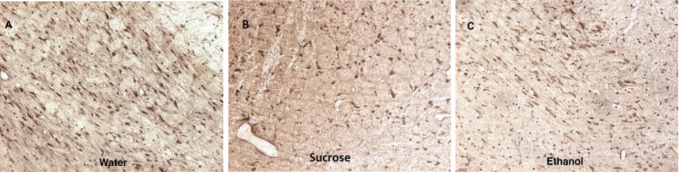
Figure 1. IL-10 Immunoreactivity visualized
IL-10 immunoreactivity visualized at 10x magnification in the VTA and can be seen as the dark spots on the tissue. IL-10 was visible in both the control and experimental group. These are representative images.
A B
Water v Sucrose
Water Suc-1 Suc-3
0.00 0.02 0.04 0.06 IL -1 0 I m m u n o re a c ti v it y (% A re a )
Water v EtOH
Figure 2. The effects of binge-like alcohol consumption on IL-10 immunoreactivity in the ventral tegmental area for water control groups, 1-DID cycle groups, and 3-DID cycle groups. Although the figures show trending, ANOVA testing showed no significant correlation between EtOH and IL-10 immunoreactivity.
Treatment Group
Consumption (g/kg/day)
BAC (mg/dL)
Ethanol 1 week 4.5 ± 0.4 54.1 ± 13.3
Ethanol 3 weeks 4.6 ± 0.3 61.3 ± 16.7
Sucrose 1 week 7.8 ± 0.4
Sucrose 3 weeks 7.7 ± 0.6
Table 1. Ethanol consumption and blood alcohol concentrations for both the 1-week and 3-week ethanol and sucrose groups. Data was collected following completion of the DID cycles. Values shown are averages and standard deviations of each test group.
Discussion
the mesolimbic dopamine reward pathway (Gessa et al. 1985). IL-10 is a major mediator of the neuroimmune response to binge levels of alcohol and has been shown to play a role in
controlling unresolved inflammation by regulating M1/M2 microglial polarization (Lobo-Silva et al. 2016). Administration of IL-10 prior to induced proinflammatory signaling has been shown to revert binge alcohol consumption behavior, however the effect of binge alcohol consumption on basal IL-10 immunoreactivity in the mesolimbic pathway has yet to be investigated. In order to determine the effects of binge ethanol consumption on the neuroinflammatory immune response in the mesolimbic reward pathway, immunohistochemical assessment was used to measure IL-10 expression in the VTA following a DID procedure.
After the DID cycles there was no significant increase in IL-10 immunoreactivity in the VTA. These results suggest that the lack of a neuroimmune anti-inflammatory response in the VTA could be a factor intensifying the inflammation of the mesolimbic reward pathway that underlies the behavioral maladaptations that lead to alcoholism.
However, it is too early to make any definitive conclusions because interactions between central immune signaling and alcohol response are extremely complicated with many interplaying factors. To achieve a global understanding of the homeostatic environment of the brain, it is necessary to understand how localized regions come together to work as system. The scope of this study was limited to the VTA of the mesolimbic reward pathway. The effects of binge ethanol consumption on central immune signaling and alcohol abuse can further be explored by investigating other regions of this pathway. Immunohistochemical assessment could be
alcohol addiction (Hyman et al. 2006). Additionally, there are many other proinflammatory and anti-inflammatory cytokines that are activated by alcohol exposure. Future studies could
investigate the role of other immunoregulators such as IL-1, IL-2, IL-6, IL-8, IL-12, and IL-13 which have all been shown to elevated in alcoholics (Gonzalez-Quintela, 2000). Understanding the roles each of these cytokines play and how they interact with each other can further elucidate the link between increased central immune signaling and alcohol dependence.
Future works could more accurately model the long-term nature of human binge-drinking
behavior by conducting the DID procedure on the mice for a longer period of time. Studies could have groups that undergo more than 3 DID cycles to see how prolonged binge behavior effects IL-10 immunoreactivity in the VTA. Previous studies have found that seven days after ethanol exposure, there is a significant increase in IL-10 expression levels (Marshall et al, 2013). There was no immediate change in IL-10 expression after ethanol consumption, similar to what was found in the VTA for this study. Future studies should look at other time points to see how IL-10 immunoreactivity fluctuates over longer period of binge behavior or during a period of
abstinence in the brain regions of interest.
IL-10 is one of the most widely studied suppressive molecules, however much of this research is focused on IL-10 in the peripheral nervous system. Knowledge on IL-10 expression and
either the proinflammatory M1 phenotype or the anti-inflammatory M2 phenotype. While the M1 phenotype is important for fighting pathogens, unregulated, non-resolving microglial
inflammation is not neuroprotective and may lead to neurodegeneration. Imbalanced polarization towards the M1 phenotypes has been implicated in neurodegenerative diseases including,
neuropathic pain, Alzheimer's and Parkinson’s (Kwilasz et al. 2015). However, studies
pioneering IL-10 as a new therapeutic approach aimed at correcting microglial imbalances have been met with conflicting results. Many of these problems are thought to stem from a lack of bioavailability in specific locations (Lobo-Silva et al. 2016). For example, intracranial
administration of IL-10 improved outcomes for Experimental Autoimmune Encephalomyelitis, an animal model of brain inflammation, however, systemic delivery of IL-10 did not. Thus, a deep understanding of the temporal and spatial expression of IL-10 is necessary to further explore its potential as a therapy. Considering IL-10’s systemic importance and the promising results of studies investigating its efficacy as a therapeutic approach for controlling
neuroinflammation, this lack of knowledge impedes the development of more sophisticated and effective immune modulatory strategies.
Acknowledgements:
REFERENCES:
Balasingam V, Yong VW et al. 1996. Attenuation of astroglial reactivity by interleukin-10. J Neuroscience. 16:2945–55.
Besser MJ, Ganor Y, Levite M, et al. 2005. Dopamine by itself activates either D2, D3 or D1/D5 dopaminergic receptors in normal human T-cells and triggers the selective secretion of either IL-10, TNFalpha or both. J. Neuroimmunol. 169:161–171.
Bledno YA, Benavidez JM, Geil C, Perra S, Morikawa, et al. 2011. Activation of inflammatory signaling by lipopolysaccharide produces a prolonged increase of voluntary alcohol intake in mice. Brain Behav Immun. 25 Suppl 1, S92-S105
Bluthe et al. 1999. Central Injection of IL-10 Antagonizes the behavioral effects of lipopolysaccharide in rats. Psychoneuroendocrinology 24:301–311.
Brodie MS, Shefner SA, Dunwiddie T. V, et al. 1990. Ethanol increases the firing rate of dopamine neurons of the rat ventral tegmental area in vitro. Brain Res 508:65-9.
CDC. 2013 Vital signs: binge drinking prevalence, frequency, and intensity among adults - United States, 2010. MMWR Morb Mortal Wkly Rep 61:14-19.
Coller JK et al. 2012 Implications of central immune signaling caused by drugs of abuse: mechanisms, mediators and new therapeutic approaches for prediction and treatment of drug dependence. Pharmacology and Therapeutics 134:219–45.
Crotti A, Ransohoff RM. 2016. Microglial physiology and pathophysiology: insights from genome-wide transcriptional profiling. Immunity. 44:505–15.
Di Chiara G, Imperato A, et al. 1988. Drugs abused by humans preferentially increase synaptic dopamine concentrations in the mesolimbic system of freely moving rats. Proc Natl Acad Sci U S A 85:5274-8.
Fernandes A, Miller-Fleming L, Pais TF. 2014. Microglia and inflammation: conspiracy, controversy or control? Cell Mol Life Sci. 71:3969–85.
González-Quintela A1, Dominguez-Santalla MJ, Pérez LF, et al. 2000. Influence of acute alcohol intake and alcohol withdrawal on circulating levels of 6, 8, 10 and IL-12. Cytokine; 12:1437–40.
Hyman S, Malenka R, Nestler E, et al. 2006. Neural mechanisms of addiction: the role of reward-related learning and memory. Annual Review of Neuroscience. 29: 565–98.
Kwilasz AJ, Grace PM, Serbedzija P, Maier SF, Watkins LR. 2015. The Therapeutic Potential of Interleukin-10 in Neuroimmune diseases. Neuropharmacology. 96:55–69.
Ledeboer A, Breve JJ, Wierinckx A, van der Jagt S, Bristow AF, Leysen JE, Tilders FJ, Van Dam AM. 2002. Expression and regulation of interleukin-10 and interleukin-10 receptor in rat astroglial and microglial cells. Eur J Neuroscience. 16:1175–85.
Lampron A, Elali A, Rivest S, et al. 2013. Innate immunity in the CNS: redefining the relationship between the CNS and Its environment. Neuron. 78:214–32.
Lobo-Silva, Diogo et. al. 2016. Balancing the Immune Response in the Brain: IL-10 and its Regulation. Journal of Neuroinflammation. 13: 297.
Marcos et. al. 2008. Interleukin-10 Gene Polymorphism is Associated with Alcoholism but not With Alcoholic Liver Disease. Alcohol Alcohol 43: 523–528.
Marshall, S. Alex, Justin A. Mcclain, Matthew L. Kelso, Deann M. Hopkins, James R. Pauly, and Kimberly Nixon. 2013. Microglial activation is not equivalent to neuroinflammation in alcohol-induced neurodegeneration: the importance of microglia phenotype. Neurobiology of Disease 54: 239-51.
Mello, N. K. et al. 1973. A review of methods to induce alcohol addiction in animals. Pharmacol Biochem Behav 1: 89-101.
Mesquita AR, Correia-Neves M, Roque S, Castro AG, Vieira P, Pedrosa J, Palha JA & Sousa N. 2008. IL-10 modulates depressive-like behavior. J Psychiatr Res 43:89-97.
Meyer U, Murray PJ, Urwyler A, Yee BK, Schedlowski M. & Feldon J. 2008. Adult behavioral and pharmacological dysfunctions following disruption of the fetal brain balance between pro-inflammatory and IL-10-mediated anti-pro-inflammatory signaling. Mol Psychiatry 13:208-21.
Moore K, W, de Waal Malefyt, R., Coffman, R. L. & O'Garra, A. 2001. Interleukin-10 and the interleukin-10 receptor. Annu Rev Immunol 19:683-765.
NIAAA. 2004. NIAAA Council Approves Definition of Binge Drinking. In National Institute on Alcohol Abuse and Alcoholism Newsletter. National Institute of Health: Bethesda, MD.
Perkins, H.W. et al. 2002. Surveying the damage: A review of research on consequences of alcohol misuse in college populations. Journal of Studies on Alcohol 14:91–100.
Pereira L, Font-Nieves M, Van den Haute C, Baekelandt V, Planas AM, Pozas E. 2015. IL-10 regulates adult neurogenesis by modulating ERK and STAT3 activity. Front Cell Neuroscience. 9:57.
Perez-de Puig I, Miro F, Salas-Perdomo A, Bonfill-Teixidor E, Ferrer-Ferrer M, Marquez-Kisinousky L, Planas AM. 2013. IL-10 deficiency exacerbates the brain inflammatory response to permanent ischemia without preventing resolution of the lesion. J Cereb Blood Flow Metab. 33:1955–66.
Perez-Asensio FJ, Perpina U, Planas AM, Pozas E. 2013. Interleukin-10 regulates progenitor differentiation and modulates neurogenesis in adult brain. J Cell Science. 126:4208–19.
Qin, Liya, Jun He, Richard N. Hanes, Olivera Pluzarev, Jau-Shyong Hong, and Fulton T. Crews. 2008. Increased systemic and brain cytokine production and neuroinflammation by endotoxin following ethanol treatment. Journal of Neuroinflammation 5.1: 10.
Robinson, T. E. & Berridge, K. C. et al. 1993. The neural basis of drug craving: an incentive-sensitization theory of addiction. Brain Res Brain Res Rev 18: 247-91.
Robinson and Nestler. 2012. Transcriptional and Epigenetic Mechanisms of Action. Nature Reviews Neuroscience 12: 623–637.
Strle K, et al. 2001. Interleukin-10 in the Brain. Crit Rev Immunol 21(5): 427–449.
Tang Y, Le W, et al. 2016.Differential roles of M1 and M2 microglia in neurodegenerative diseases. Molecular Neurobiology. 53:1181–94.
Thiele, T. E. & Navarro, M. 2014. "Drinking in the dark" (DID) procedures: A model of binge-like ethanol drinking in non-dependent mice. Alcohol 48: 235-241.