Modulation of individual neuron activity with external electric fields
by
Quique Toloza
Senior Honors Thesis
Department of Physics & Astronomy
University of North Carolina at Chapel Hill
April 28, 2017
Flavio Fr¨
ohlich, Thesis Advisor
Louise Dolan, Reader
Neurons are highly-specialized cells capable of receiving and generating electrical signals through the controlled flow of ions across their cell membranes. This behavior is represented mathematically as a series of coupled nonlinear differential equations, which can be efficiently solved with numerical integrators. As such, detailed computational models of neurons are readily-achievable and allow for in-depth investigations of neuronal dynamics that are not typically possible inin vitro or in vivo
studies. We built several multicompartmental models of mammalian layer V (L5) pyramidal neu-rons with complex three-dimensional morphologies and biophysics in order to characterize neuronal response to externally-applied electric fields. Such electric fields are used in transcranial current stimulation (tCS), a neuromodulatory technique under ongoing investigation for clinical applica-tions in various psychiatric illnesses. Our results indicate that hyperpolarization-activated cation (Ih) channels in the tuft dendrites of L5 pyramidal neurons, along with the overall connectivity of the apical tree, are crucial in shaping the unique frequency-dependent response of the entire neuron to external fields. These two factors contribute to a peak subthreshold response in the tuft region to electric fields oscillating at approximately 20 Hz. This subthreshold filtering may indicate potential new paradigms for noninvasive electrical stimulation therapies.
CONTENTS
I. Introduction 2
II. Transcranial current stimulation 3
III. Neuronal dynamics 5
A. Electrophysiology 5
B. Multicompartmental modeling 5
C. Hodgkin-Huxley dynamics 7
IV. Simulations 9
A. The NEURON environment 9
B. Layer V pyramidal neuron model 9 C. Validation of electric field effects 11 D. Frequency response of model sections 11
V. Conclusion 13
References 15
I. INTRODUCTION
The basic computational unit of the nervous system is the neuron, a highly-specialized cell with the ability to re-ceive, integrate, and propagate electrical signals. These signals are used to communicate rapidly across an or-ganism’s body, facilitating sensation, muscle movement, and consciousness, among other processes. This behav-ior can be modeled with a variety of approaches based on the complexity of the system of interest. For exam-ple, neurons are divided morphologically and function-ally into different sections, such as the soma, primary site of protein expression and intitiation of action po-tentials; the axon, which propagates action potentials to other cells; and dendrites, which are the main recipi-ents of inputs from other cells at sites called synapses. The multicompartmental modeling approach captures this segmentation, allowing for complex cell geometries and parametrization of biophysics.
The effects of external electric fields on cortical neurons has recently received increased attention due to the use
of such fields in transcranial current stimulation (tCS), a noninvasive neuromodulatory technique with poten-tial therapeutic effects in depression, schizophrenia, and other psychiatric disorders.1,2 Previous studies have ex-amined patient and network-level effects of different tCS methods, but individual neuronal response to oscillatory external electric fields has not been well-characterized.1–5
Traditionally, the main targets of tCS have been layer V (L5) pyramidal neuron cells, due to their distinct soma-todendritic axis, which has been shown to increase sen-sitivity to tCS.6 The key feature of the somatodendritic
axis is the apical dendritic tree, which can generally be divided into a proximal (to soma) “trunk” region with little branching and a distal “tuft” region with a high degree of dendritic bifurcation.
We used multicompartmental models of mammalian L5 pyramidal neurons with Hodgkin-Huxley style dy-namics to investigate the frequency-dependent response of individual neurons to extracellular electric fields, with a focus on the role of the apical dendritic tree. These models were constructed and validated using published experimental results and known behavior of L5 pyramidal neurons.7 Our implementation of an extracellular
elec-tric field stimulation was tested by confirming directional membrane potential modulation in response to a static electric field.
We report subthreshold filtering of different sections of the neuron under application of an external electric field. Our results show varying sensitivities to oscillat-ing electric fields in different parts of the neuron, and a general low-pass frequency response in the soma, axon, and basal dendrites. We show that tuft dendrites exhibit a characteristic bandpass response with a peak around 20 Hz, and that this filtering is determined by the pres-ence of hyperpolarization-activated cation (Ih) channels
in the tuft region and the connectivity of the tuft to the trunk and other regions in the apical tree. We also show that these Ihchannels are the primary active biophysical
3
FIG. 1. Example transcranial current stimulation (tCS) setup created by Soterix Medical.
FIG. 2. Primary neuron model used, based on Larkumet al. (2009).7 A. Fully intact model with labeled regions, illustrating the somatodendritic axis. B. Model with non-apical sections removed. C. Model with auxiliary apical dendrites removed. D. Model with tuft region isolated.
II. TRANSCRANIAL CURRENT STIMULATION
In transcranial current stimulation (tCS), electrodes capable of emitting weak currents are placed on the pa-tient’s scalp such that a weak electric field is generated in the patient’s brain - a typical setup is shown in Figure 1.
4
FIG. 3. Simple depiction of a section of the neuron’s cell membrane with ion concentration gradients and protein channels. These channels are distributed across the entire neuron, with varying types and densities.
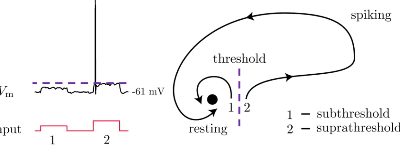
FIG. 4. The neuron as a dynamical system. Subthreshold inputs quickly return to rest, while suprathreshold inputs induce spiking behavior. Figure partially adapted from Izhikevich (2007).8
Previous studies have shown that tDCS modulates neuronal activity through subthreshold bidirectional modulation of the membrane potential.3 This effect is amplified for neurons with linear morphology; therefore, the main targets of tCS have traditionally been layer V (L5) pyramidal neuron cells, deep cortical neurons with distinct geometric polarization, shown in Figure 2.6In L5
pyramidal neurons, the dendrites are divided into two dif-ferent trees, apical and basal. The apical tree is further subdivided into a trunk region, which stems from the soma, and a tuft region, where the dendrites bifurcate and branch dramatically. Apical dendrites not explicitly part of the trunk or tuft are here referred to as auxiliary. The generally linear stretch of the neuron extending from the soma to the end of the apical tree is referred to as the somatodendritic axis.
tACS is the subject of ongoing research for its po-tential to modulate large-scale cortical network activity
by selectively enhancing or suppressing existing brain oscillations.4,5 However, not all frequencies appear to
be viable for use in tACS: for example, the tissue sur-rounding neurons has a low-pass filtering effect that makes high-frequency tACS impractical.9In addition,
5
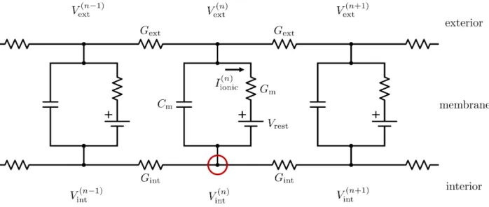
FIG. 5. A finite-difference approximation of the electrical properties of a neuron’s membrane. Kirchhoff’s current law is applied at the indicated node.
III. NEURONAL DYNAMICS
A. Electrophysiology
The neuron’s electrical behavior arises from the con-trolled flow of ions across its semi-permeable membrane and the diffusion of these ions along the inside of the cell, illustrated in Figure 3. The membrane naturally prevents the passage of these charged molecules, but embedded proteins act as selective ion channels and pumps that al-low the formation of electrochemical gradients across the membrane. For example, sodium (Na+), chloride (Cl−), and calcium (Ca2+) ions are all held at higher concentra-tions outside of the cell, while potassium (K+) ions are held at a higher concentration inside the cell. The re-sulting separation of charge creates an electric potential difference between the interior and exterior of the cell, referred to as the membrane potential (Vm).
Functionally, neurons act as analog-to-digital convert-ers: they sum incoming electrical signals, which shift the membrane potential until a threshold is reached and a fixed signal, called an action potential, is emitted. Positive shifts in the membrane potential, towards this threshold, are called depolarizations, while shifts away from the threshold to more negative values are called hy-perpolarizations.
These dynamics are generally describable by the pres-ence of two equilibria, one stable and unstable, shown in Figure 4. The stable equilibrium is called the rest-ing membrane potential (Vrest), with a typical value of
-60 mV, the negative sign indicating a more negatively-charged interior. The unstable equilibrium, already hinted at, is the action potential threshold. Mem-brane potentials below the threshold quickly return to the resting potential, while values above the threshold cause a nonlinear spike with an eventual return to rest.
These two responses are referred to as subthreshold and suprathreshold, respectively. In typical tCS therapies, the strength of the electric field is tailored such that the effects on neurons is entirely subthreshold: the external field acts to modulate the proximity of the stable and unstable equilibria of the neuron, thereby altering the dynamics of the neuron.
B. Multicompartmental modeling
Consider, as in Figure 5, a finite-difference approxi-mation of a thin, cylindrical section of the neuron’s cell membrane as a series of discrete electrical circuits. A transmembrane capacitance Cm represents the build-up
of ionic charge on each side of the membrane, a trans-membrane conductanceGmrepresents the ability of ions
to flow across the membrane through channels, and a voltage sourceVrest represents the resting membrane
po-tential. Internal and external conductancesGintandGext
represent the ability of ions to move parallel to the mem-brane on either side.
We can apply Kirchhoff’s law to the internal node of the nth compartment and show that the current flow at the interior node is
Cm
d dt(V
(n) int −V
(n) ext)
−Gint(V (n+1) int −2V
(n) int +V
(n−1) int ) +I
(n)
ionic= 0 (1)
DefiningVmas
6
FIG. 6. Illustration of the dipole induced across the neuron in response to an applied electric field. As one pole of the neuron is depolarized (top), the opposite pole is hyperpolarized (bottom).
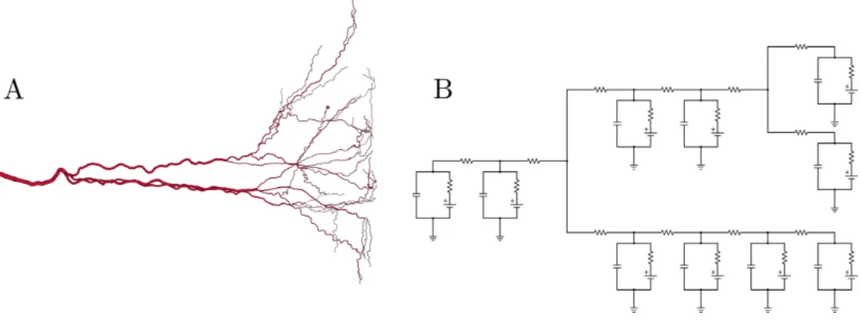
FIG. 7. Multicompartmental models represent neuron branching with bifurcating electrical circuits. A. Branching in a dendritic tree. B. Electrical circuit representation of tree.
and substituting into (1) yields
Cm
dVm(n)
dt −Gint(V
(n+1) m −2V
(n) m +V
(n−1) m
+Vext(n+1)−2Vext(n)+Vext(n−1)) +Iionic(n) = 0 (3)
Here, it is useful to take advantage of the fact that, for a thin cylindrical section of membrane,
Gint=
πd2 4ρint∆x
(4)
and
Cm=πd∆xcm (5)
where dis the diameter of the section, ρint is the
resis-tivity of the fluid inside the cell, ∆xis the length of the compartment parallel to the membrane, and cm is the
7
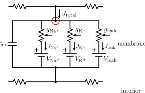
FIG. 8. The more complex Hodgkin-Huxley compartment model, with transmembrane conductance split into three components corresponding to sodium, potassium, and passive ion flow.
(3), we find a discretized variant of the cable equation:10
cm
dVm(n)
dt − d 4ρint
(V
(n+1)
m −2Vm(n)+Vm(n−1)
∆x2
+V
(n+1) ext −2V
(n) ext +V
(n−1) ext
∆x2 ) +I
(n)
ionic= 0 (6)
In the case where ∆x→0, the terms in (6) dependent on the external electric potential simplify to the second partial derivative of the external electric potential with respect to x, which is the negative of the first partial derivative of the external electric field with respect tox:
∂2V ext
∂x2 =−
∂Eext
∂x (7)
Remaining in the finite ∆x case, evidently the con-tribution of any spatially-uniform external electric field will be zero for all compartments except for at endpoints of homogenous sections - for example, at the branching points of dendrites in the apical tree.10This is consistent
with effects observed in vitro: neurons are polarized by static external electric fields, charge accumulating at each end of the neuron such that an electric dipole is formed parallel to the direction of the field, as in Figure 6, an effect that corresponds to the external field affecting the endpoints of the neuron itself, as in Equation 6.6
A multicompartmental model of a neuron chains to-gether the discrete circuits, or compartments, complete with branching that mimics the complex bifurcation pat-terns seen in real neurons, especially in dendritic trees, as shown in Figure 7.
The size of these compartments is determined by the maximum length along the membrane where the
mem-brane potential can be considered equipotential, approx-imated by the electrotonic lengthλ:
λ=
rg
m
gint
(8)
where gm is the total transmembrane conductance
den-sity and gint is the intracellular conductance density. λ
is equivalent to the length parallel to the membrane over which the steady-state solution to the electric potential decreases by a factor ofe. The compartmental approach is considered a good approximation when the length of all compartments is less thanλ.
C. Hodgkin-Huxley dynamics
The contribution of the ionic current to the mem-brane potential is of special importance; it is, in fact, nonlinearly-dependent on the membrane potential, giv-ing rise to the spikgiv-ing behavior of the neuron. Gmis more
accurately represented as several parallel conductances, one for each type of ion channel. The Hodgkin-Huxley representation, shown in Figure 8, is therefore more ac-curate, although it is also a simplification.11
In this circuit,cmis again the capacitance of the
mem-brane per unit area and gj is the conductance of the
membrane per unit area for the ion speciesj. Vj is the
membrane potential value at which the net flow of the ion speciesj due to electrostatic forces and diffusion is equal to zero, referred to as the equilibrium potential. The total current densityJtotalis equivalent to
Jtotal=cm
dVm
8
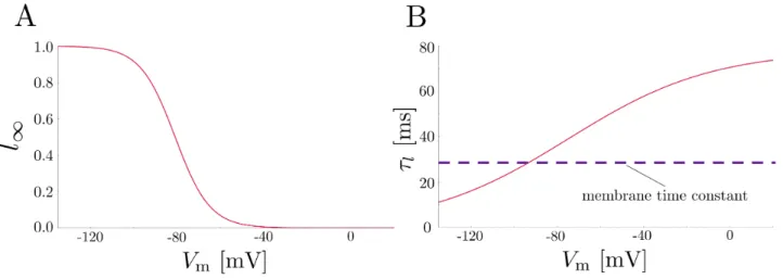
FIG. 9. Biophysical characteristics of the hyperpolarization-activated cation (Ih) channel. Steady-state (A) and time constant (B) values of modeled Ih channels at 34◦C, as originally described in Magee (1998).12 For most membrane potential values, the time constant of the channel is greater than the membrane time constant, indicating that it will activate slowly relatively to changes in the membrane potential itself.
where eachJjis the ion current density for the ion species
j:
JNa+ =gNa+(Vm−VNa+) (10)
JK+=gK+(Vm−VK+) (11)
Jleak=gleak(Vm−Vleak) (12)
referring to the inward flow of sodium ions, the outward flow of potassium ions, and the passive leakage of both sodium and potassium ions, respectively.
The nonlinearity in the system arises from gNa+ and gK+ - these terms are dependent on the membrane po-tential in the form of gating variables, determined exper-imentally by Hodgkin and Huxley:
gNa+ = ¯gNa+m3h (13)
gK+= ¯gK+n4 (14)
where ¯gj is a constant;nandmare probabilities of
indi-vidual potassium or sodium channels activating (or open-ing), respectively; and his the probability of individual sodium channels inactivating (or closing). These gating variables are defined by the equation
dx dt =
x∞−x
τx
forx∈n,m, or h (15)
wherex∞is the steady-state value ofxfor a given
mem-brane potential and τx is a time constant representing
the response time of the channel for a given membrane potential and temperature.
In summary, the basic equations governing the ionic current flow into a given section of the neuron’s mem-brane are
Jtotal=cm
dVm
dt + ¯gNa+m
3h(V
m−VNa+)
+ ¯gK+n4(Vm−VK+) +gleak(Vm−Vleak) (16)
dx dt =
x∞−x
τx
forx∈n,m, orh (17)
Additional channel types and ion species are added in a similar fashion, with independently-determined equi-librium potentials and conductances:
Jtotal=cm
dVm
dt +
X
i
gi(Vm−Vi) (18)
For example, hyperpolarization-activated cation (Ih)
channels modeled in our work are characterized by an equilibrium potential of -34 mV and a conductance den-sity
gh= ¯ghl(Vm−Vh) (19)
dl dt =
l∞−l
τl
(20)
wherel∞andτlare shown in Figure 9 for a temperature
of 34◦C.
Due to its depolarized (relative to the resting mem-brane potential) equilibrium potential and activation in the hyperpolarized regime near -70 mV, the Ih channel
9
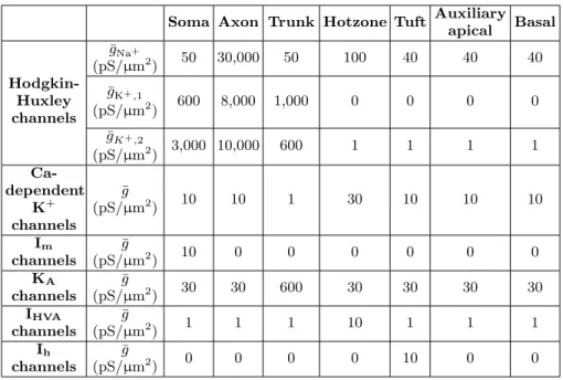
Soma Axon Trunk Hotzone Tuft Auxiliary apical Basal
¯
gNa+
(pS/µm2) 50 30,000 50 100 40 40 40
Hodgkin-Huxley channels
¯
gK+,1
(pS/µm2) 600 8,000 1,000 0 0 0 0 ¯
gK+,2
(pS/µm2) 3,000 10,000 600 1 1 1 1
Ca-dependent K+ channels ¯ g
(pS/µm2) 10 10 1 30 10 10 10
Im
channels
¯
g
(pS/µm2) 10 0 0 0 0 0 0
KA
channels
¯
g
(pS/µm2) 30 30 600 30 30 30 30
IHVA
channels
¯
g
(pS/µm2) 1 1 1 10 1 1 1
Ih
channels
¯
g
(pS/µm2) 0 0 0 0 10 0 0
TABLE I. Selection of parameters applied to primary L5 pyramidal neuron model.
studies have demonstrated the Ihchannel’s role in
deter-miningVrest and generating rhythmic oscillations.13 The
channel also has a high-pass filtering effect on the mem-brane potential in the subthreshold regime: its activa-tion time constant is large relative to the time constant of the membrane for most membrane potential values, meaning that fast oscillations in the membrane potential are generally unaffected while slower oscillations are op-posed by the channel’s activation. Previous papers have shown that Ih channels are involved in suprathreshold
resonance, tailoring spiking response to a specific input frequency, typically in the 2 - 3 Hz range.14
Hodgkin-Huxley style ion channels have conductances which depend on the cell’s membrane potential; there-fore, we will refer to these type of biophysical parameters as “active” and biophysical parameters that are constant and intrinsic to the membrane material itself, such ascm,
as “passive.”
IV. SIMULATIONS
A. The NEURON environment
In vitroexperimentation with neurons presents a num-ber of complications not present in computational model-ing, including time to prepare for each experiment and to maintain animal colonies or tissue cultures. These exper-iments are also limited by techniques to record the neu-ronal response itself: measuring subthreshold responses from individual neurons is nontrivial, especially from multiple locations. Using models, we were able to simul-taneously record subthreshold membrane potential shifts from every point in a given model. We were also able to manipulate the biophysics and morphology of the models
at ease, even creating conditions not replicable in vitro, such as the isolation of sections from the rest of the neu-ron without otherwise altering function.
We used NEURON for the development and test-ing of all models. NEURON is a powerful open-source environment for simulating biologically-realistic models of neurons that enables fine control over morphology (three-dimensional geometry and compartmentalization) and biophysical properties (eg. conductance parameters and types of channels), modeled with Hodgkin-Huxley dynamics.15At the most basic level, NEURON integrates the differential equations presented earlier across the en-tire spatial domain of a given model using either a back-wards Euler or a Crank-Nicolson solver. Both methods are technically uncondtionally stable, but for cases with high spatial resolution, Crank-Nicholson solvers require a very small time step size to avoid spurious oscillations. For our simulations, we found that the backwards Eu-ler method with a time step of 0.1 ms provided the best balance between stability, accuracy, and efficiency.
NEURON natively divides models into unbranched “sections,” which are in turn divided into “segments.” These segments correspond to the previously described compartments. Segment length may be modified to meet the desired spatial resolution of the model.
B. Layer V pyramidal neuron model
10
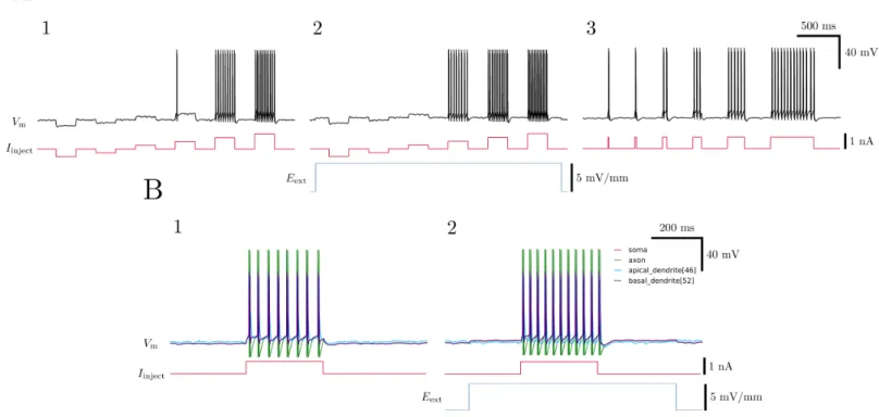
FIG. 10. Validation of model with current injection experiments. A. Somatic current injections show that the strength of current injected (1) and the duration of the injection (3) both accurately influence spiking behavior. 2. A static external electric field oriented such that it depolarizes the soma pushes the cell’s resting potential closer to its threshold, making a previous subthreshold current injection suprathreshold. B.1. Different sections of the neuron show different behaviors during spiking. 2. Application of a static external electric field causes more spikes in response to the same stimulus.
11
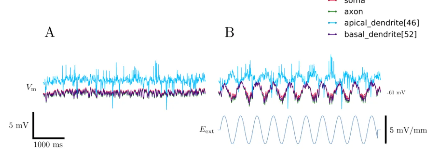
FIG. 12. Effects of an oscillatory electric field applied to a neuron model. A. Resting membrane potential with noisy synaptic input. B. Membrane potential under the influence of an electric field oscillating at 2 Hz.
dendritic tree. This tree was divided into trunk, hotzone, and tuft regions for the purpose of assigning realistic dis-tributions of biophysical parameters, examples of which are shown in Table I. All sections in our model, including the soma, axon, and each individual dendrite, were mod-eled with 11 segments, such that all segment lengths were smaller than the electrotonic length of the given section, with a maximum segment length of 27µm. Randomly-generated synaptic inputs were used to create noise in the model: 4,000 excitatory and 1,000 inhibitory synapses spread over the apical tree with randomly-generated on-sets, numbers of inputs, intervals between inputs, and strengths of inputs were used.
To validate the neuron models, we simulated current injection experiments, which are commonly usedin vitro
to hyperpolarize or depolarize the membrane potential and induce spiking. The results of these experiments are shown in Figure 10, and are in agreement with the results published in the original papers describing the models.7 A somatic current injection of 750 pA for 300 ms was sufficient to induce action potentials, which is well within expectations.
C. Validation of electric field effects
To model the effects of an externally-applied electric field, we made a few key approximations: namely, in the range of frequencies used (<1000 Hz) and in the spatial domain of the model (approximately 1 mm in length), the electric field is spatially uniform and unidirectional. NEURON provides access to the extracellular electric po-tential at each segment; given a time-evolving electric field waveform pointing along the x axis, we described the extracellular electric potential at a point (x,y,z) as
Vext(x,y,z,t) =−xEext(t) (21)
With a static external electric field present, we con-firmed a modulation of the neuron’s excitability,
consis-tent with previous experiments conductedin vitro, shown in Figure 10. This modulation of excitability was af-fected by a depolarization or hyperpolarization of the cell’s membrane potential at the point of current injec-tion, the magnitude of which was linearly related to the magnitude and orientation of the electric field relative to the somatodendritic axis of the cell, as described in Fig-ure 11. This is another example of the dipole formation across the neuron in response to an external electric field: the apical dendrite is on the opposite “pole” of the neu-ron as the other reported sections, therefore showing an opposite effect on its membrane potential in response to the same field strength and direction.
Having confirmed the effects of a static external electric field,3 we introduced an oscillatory field and observed the effect on the cell’s membrane potential at various sections, shown in Figure 12. This oscillatory external electric field induced oscillations of the same frequency across the neuron, with the phase and amplitude of these responses dependent varying from section-to-section.
D. Frequency response of model sections
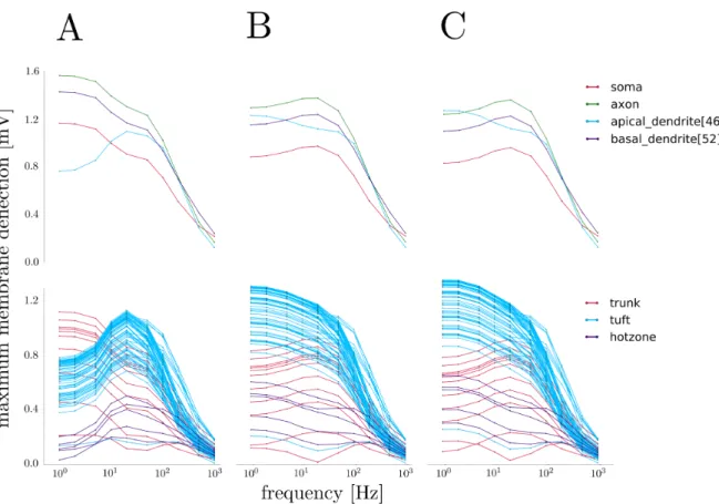
In order to measure the frequency-dependent response of the models to an oscillatory electric field, we measured the membrane potential at the center of various sections of the neuron under stimulation by an electric field os-cillating with a frequency ranging from 1 to 1000 Hz. The maximum deflection (either depolarizing or hyper-polarizing) from the resting membrane potential of each section was recorded, with results shown in Figure 13 for the intact neuron.
12
FIG. 13. Frequency-dependent response of several model variants to an oscillatory electric field. Field strength is 5 mV/mm for all experiments. A. Top: Frequency response of several sections of the normal model. Note the general low-pass behavior. Bottom: Frequency response of the entire apical tree, with regions color-coded. The tuft dendrite frequency response resembles a band-pass filter centered around 20 Hz. B. Frequency response of the model with all active biophysics removed. The band-pass effect has disappeared in the tuft dendrites, which now behave as low-band-pass filters. C. Frequency response of the model with Ih channels removed from the tuft dendrites. Note the similarity to the passive model’s response.
the tuft region displayed a slight dip in sensitivity at this same frequency. In general, the magnitude of the frequency response of each section corresponded to the section’s proximity to the poles of the neuron, matching expectations.
In order to distinguish between effects due to the bio-physical properties and morphology of the neuron model, we removed all active biophysical parameters from the model, leaving only a constant transmembrane capaci-tance, conduccapaci-tance, and voltage source to establish a re-alistic resting membrane potential at -70 mV. Shown in Figure 13, this passive model exhibited a low-pass be-havior in all sections, including the tuft dendrites, con-sistent with the expectations for the corresponding cir-cuit. Clearly, one of the ion channels removed is at least partially responsible for the frequency response of the neuron.
Previous studies have shown that Ih channels are
re-sponsible for suprathreshold resonance near 2 - 3 Hz in response to oscillating current injections.14 This
stimu-lation technique is quite different from an external elec-tric field; current injections necessarily involve ion flow into or out of the neuron, while electric field stimulation
does not directly cause transmembrane ion flow. Cur-rent injections are also local perturbations, in contrast to external electric fields which affect the entire neuron simultaneously. Nevertheless, in order to investigate any similar role in response to oscillating electric fields, we se-lectively removed all Ihchannels from the tuft dendrites.
The resulting frequency response of the modified model closely resembled that of the passive neuron, removing bandpass behavior from the tuft dendrites, as seen in Figure 13.
Having identified a significant contribution from the Ih
channels to the neuron’s overall frequency response, we modified the conductance density of the channels in the tuft region (previously 10 pS/µm2) to observe any effect
on the cell’s filtering, shown in Figure 14. Decreasing the conductance density to 1 pS/µm2 shifted the peak
frequency to around 10 Hz, while increasing the conduc-tance density to a value of 50 pS/µm2 had the effect
of shifting the peak frequency of the tuft dendrites to a value between 20 and 50 Hz.
As further evidence of the contribution of the Ih
fluctua-13
FIG. 14. Changing the density of Ih channels in the tuft shifts the peak frequency of the tuft response. Field strength is 5 mV/mm for all experiments. A. Top: Frequency response of several sections of the normal model. Bottom: Frequency response of the entire apical tree, with regions color-coded. B. Frequency response of the model with Ih conductance reduced to 1 pS/µm2. The peak frequency of the tuft response has shifted to around 10 Hz. C. Frequency response of the model with Ihconductance increased to 50 pS/µm2. Note the new location of the peak frequency around 20 - 50 Hz.
tions of non-apical sections disappeared when the chan-nels were removed completely, shown in Figure 15. That is, Ihchannels in the tuft region shaped not only the
fre-quency response of the tuft itself, but that of sections across the entire neuron.
Next, we examined the contribution of the branching of the apical tree to the entire neuron’s frequency response. This was done by progressively shortening the apical tree, removing first non-apical sections, then auxiliary apical dendrites, and finally trunk dendrites (Figure 2). At each step, we measured the maximum membrane potential de-flection for a range of frequencies. The results, shown in Figure 16, illustrate a negligible effect from the removal of non-apical sections; however, removal of the auxiliary apical dendrites shifted the peak frequency of the tuft re-sponse in the normal model to a value between 10 and 20 Hz. This effect is likely explained by a decreased magni-tude in membrane potential oscillations under this condi-tion, such that Ihchannels are less activated at the same
frequencies relative to the intact model. Further removal of the trunk dendrites dramatically decreased the magni-tude of the response for the remaining tuft dendrites and removed any measurable indication of a bandpass effect. In general, as the length of the model was decreased,
the overall magnitude of the cell’s response was also de-creased, while as sections became more proximal to the “ends” of the model, the magnitude of their individual responses increased.
V. CONCLUSION
We modeled several L5 pyramidal neurons with com-plex morphology and biophysics, focusing on a sophisti-cated depiction of the apical dendritic tree, and showed that they responded realistically to stimuli. With the incorporation of spatially-uniform and unidirectional ex-ternal electric fields, we confirmed a bidirectional mod-ulation of the neuron’s membrane potential dependent on the orientation of the field and location of the section relative to the field.
We then introduced oscillatory electric fields and char-acterized the frequency response of variants of these mod-els. In doing so, we identified two primary factors con-tributing to the entire cellular frequency-dependent re-sponse: the presence of Ihchannels in the tuft region and
14
FIG. 15. Ih channels in the tuft region shape the entire neuron’s response to oscillatory electric fields. A. Application of a 2 Hz electric field illustrates the asymmetric response of the soma, axon, and basal dendrite to the electric field in the normal model (1), with the difference in membrane potential between the three sections increasing in the hyperpolarized region and decreasing in the depolarized region. This asymmetry disappears when Ih channels are removed (2). B. The same effect is observed at 10 Hz.
FIG. 16. The connectivity of the apical dendritic tree shapes the frequency response of the entire neuron. This response is different with all sections intact (A), with non-apical sections removed (B), with auxiliary apical dendrites removed (C), and with tuft dendrites isolated (D).
of these attributes are necessary for the unique band-pass filtering of oscillating electric fields seen in tuft dendrites, centered around 20 Hz. We showed that changing the conductance density of the Ihchannels changes the
loca-tion of the peak response frequency of the tuft dendrites, and that these channels are responsible for the asymmet-ric oscillatory response of non-apical sections.
Our results have significant implications in the devel-opment of noninvasive electrical stimulation techniques.
advan-15
tage of the cell’s heightened sensitivity in this frequency range. Examination of the physiological significance of this peak in the larger context of the brain is warranted in order to further investigate the apical tree as a target of tACS. This work is crucial to designing effective
-and patient-specific - tACS treatments. For example, Ih
channel mutations have been implicated in certain types of epilepsy and neuropathic pain.16 We need to
under-stand how these mutations change neuronal response to tACS in order to successfully treat these patients.
1 M. F. Kuo, W. Paulus, and M. A. Nitsche, Neuroimage
85, 948 - 960 (2014). 2
M. T. Berlim, F. Van den Eynde, and Z. J. Daskalakis, J Psychiatr Res47, 1 - 7 (2013).
3 M. A. Nitsche and W. Paulus, J Physiol 527, 633 - 639 (2000).
4
J. Vosskuhl, R. J. Hunter, and C. S. Herrmann, Front Hum Neurosci9, 257 (2015).
5
M. M. Ali, K. K. Sellers, and F. Fr¨ohlich, J Neurosci33, 11262 - 11275 (2013).
6 T. Radman, R. L. Ramos, J. C. Brumberg, and M. Bikson, Brain Stimul2, 215 - 228 (2009).
7
M. E. Larkum, T. Nevian, M. Sandler, A. Polsky, and J. Schiller, Science325, 756 - 760 (2009).
8 E. M. Izhikevich,Dynamical Systems in Neuroscience: The
Geometry of Excitability and Bursting (The MIT Press,
Cambridge, 2007), p. 9.
9 C. B´edard, H. Kr¨oger, and A. Destexhe, Phys Rev E73, 051911 (2006).
10
F. Rattay, IEEE Trans Biomed Eng36, 676 - 682 (1989). 11 A. L. Hodgkin and A. F. Huxley, J Physiol117, 500 - 544
(1952). 12
J. C. Magee, J Neurosci18, 7613 - 7624 (1998).
13 A. L¨uthi and D. A. McCormick, Neuron21, 9 - 12 (1998). 14
S. L. Schmidt, C. R. Dorsett, A. K. Iyengar, and F. Fr¨ohlich, Cereb Cortex1, 1 - 15 (2016).
15 M. L. Hines and N. T. Carnevale, Neural Comput9, 1179 - 1209 (1997).
16
J. C. DiFrancesco and D. DiFrancesco, Front Cell Neurosci