The SysteMHC Atlas project
Wenguang Shao
1,†, Patrick G.A. Pedrioli
1,†, Witold Wolski
2, Cristian Scurtescu
3,
Emanuel Schmid
3, Juan A. Vizca´ıno
4, Mathieu Courcelles
5, Heiko Schuster
6,7,
Daniel Kowalewski
6,7, Fabio Marino
8,9,10, Cecilia S.L. Arlehamn
11, Kerrie Vaughan
11,
Bjoern Peters
11, Alessandro Sette
11, Tom H.M. Ottenhoff
12, Krista E. Meijgaarden
12,
Natalie Nieuwenhuizen
13, Stefan H.E. Kaufmann
13, Ralph Schlapbach
2, John C. Castle
14,
Alexey I. Nesvizhskii
15,16, Morten Nielsen
17,18, Eric W. Deutsch
19, David S. Campbell
19,
Robert L. Moritz
19, Roman A. Zubarev
20, Anders Jimmy Ytterberg
20,21, Anthony W. Purcell
22,
Miguel Marcilla
23, Alberto Paradela
23, Qi Wang
24, Catherine E. Costello
24, Nicola Ternette
25,
Peter A. van Veelen
26, C ´ecile A.C.M. van Els
27, Albert J.R. Heck
9,10, Gustavo
A. de Souza
28,29, Ludvig M. Sollid
28, Arie Admon
30, Stefan Stevanovic
6,7,
Hans-Georg Rammensee
6,7, Pierre Thibault
5, Claude Perreault
5,
Michal Bassani-Sternberg
8, Ruedi Aebersold
1,31,*and Etienne Caron
1,*1
Department of Biology, Institute of Molecular Systems Biology, ETH Zurich, Zurich 8093, Switzerland,
2Functional
Genomics Center Zurich, ETH Zurich and University of Zurich, Zurich 8057, Switzerland,
3Scientific IT Services
(SIS), ETH Zurich, Zurich 8093, Switzerland,
4European Molecular Biology Laboratory, European Bioinformatics
Institute (EMBL-EBI), Wellcome Trust Genome Campus, Hinxton, Cambridge, CB10 1SD, UK,
5Institute for Research
in Immunology and Cancer, Universit ´e de Montr ´eal, Montreal, H3T 1J4, Canada,
6Department of Immunology,
Interfaculty Institute for Cell Biology, University of T ¨ubingen, T ¨ubingen, 72076, Germany,
7German Cancer
Consortium (DKTK), DKFZ partner site T ¨ubingen, T ¨ubingen, 72076, Germany,
8Ludwig Institute for Cancer
Research, University Hospital of Lausanne, Lausanne 1011, Switzerland,
9Biomolecular Mass Spectrometry and
Proteomics, Bijvoet Center for Biomolecular Research and Utrecht Institute for Pharmaceutical Sciences, Utrecht
University, 3584 CH, The Netherlands,
10Netherlands Proteomics Centre, Utrecht, 3584 CH, The Netherlands,
11La
Jolla Institute for Allergy and Immunology, La Jolla, CA 92037, USA,
12Department of Infectious Diseases, Leiden
University Medical Center, Leiden, 2333 ZA, The Netherlands,
13Department of Immunology, Max Planck Institute for
Infection Biology, Berlin 10117, Germany,
14Vaccine Research and Translational Medicine, Agenus Switzerland Inc.,
4157 Basel, Switzerland,
15Department of Pathology, BRCF Metabolomics Core, University of Michigan, Ann Arbor,
MI 48109, USA,
16Department of Computational Medicine and Bioinformatics, University of Michigan, Ann Arbor, MI
48109, USA,
17Instituto de Investigaciones Biotecnol ´ogicas, Universidad Nacional de San Mart´ın, Buenos Aires,
1650, Argentina,
18Department of Bio and Health Informatics, Technical University of Denmark, 2800 Kgs. Lyngby,
Denmark,
19Institute for Systems Biology, Seattle, WA 98109, USA,
20Division of Physiological Chemistry I,
Department of Medical Biochemistry and Biophysics, Karolinska Institutet, Stockholm, SE-171 77, Sweden,
21
Rheumatology Unit, Department of Medicine, Solna, Karolinska Institutet, Stockholm, SE-171 77, Sweden,
22Infection and Immunity Program, Department of Biochemistry and Molecular Biology, Monash Biomedicine
Discovery Institute, Monash University, Clayton 3800, Australia,
23Proteomics Unit, Spanish National Biotechnology
Centre, Madrid 28049, Spain,
24Center for Biomedical Mass Spectrometry, Department of Biochemistry, Boston
University School of Medicine, Boston, MA 02118, USA,
25The Jenner Institute, Target Discovery Institute Mass
Spectrometry Laboratory, University of Oxford, Oxford, OX3 7FZ, UK,
26Center for Proteomics and Metabolomics,
Leiden University Medical Center, Leiden, 2333 ZA, The Netherlands,
27Centre for Infectious Disease Control,
National Institute for Public Health and the Environment, Bilthoven, 3720 BA, The Netherlands,
28Centre for Immune
*To whom correspondence should be addressed. Tel: +41 44 633 26 97; Fax: +41 44 633 10 51; Email: [email protected]
Correspondence may also be addressed to Ruedi Aebersold. Tel: +41 44 633 31 70; Fax: +41 44 633 10 51; Email: [email protected] †These authors contributed equally to the paper as first authors.
C
The Author(s) 2017. Published by Oxford University Press on behalf of Nucleic Acids Research.
This is an Open Access article distributed under the terms of the Creative Commons Attribution License (http://creativecommons.org/licenses/by-nc/4.0/), which permits non-commercial re-use, distribution, and reproduction in any medium, provided the original work is properly cited. For commercial re-use, please contact [email protected]
Regulation, Department of Immunology, University of Oslo and Oslo University Hospital-Rikshospitalet, Oslo 0372,
Norway,
29The Brain Institute, Universidade Federal do Rio Grande do Norte, 59056–450, Natal-RN, Brazil,
30Department of Biology, Technion, Israel Institute of Technology, Haifa 3200003, Israel and
31Faculty of Science,
University of Zurich, 8006 Zurich, Switzerland
Received June 13, 2017; Revised July 16, 2017; Editorial Decision July 18, 2017; Accepted July 21, 2017
ABSTRACT
Mass spectrometry (MS)-based immunopeptidomics
investigates the repertoire of peptides presented at
the cell surface by major histocompatibility complex
(MHC) molecules. The broad clinical relevance of
MHC-associated peptides, e.g. in precision medicine,
provides a strong rationale for the large-scale
gener-ation of immunopeptidomic datasets and recent
de-velopments in MS-based peptide analysis
technolo-gies now support the generation of the required data.
Importantly, the availability of diverse
immunopep-tidomic datasets has resulted in an increasing need
to standardize, store and exchange this type of data
to enable better collaborations among researchers,
to advance the field more efficiently and to establish
quality measures required for the meaningful
com-parison of datasets. Here we present the SysteMHC
Atlas (https://systemhcatlas.org), a public database
that aims at collecting, organizing, sharing,
visu-alizing and exploring immunopeptidomic data
gen-erated by MS. The Atlas includes raw mass
spec-trometer output files collected from several
labo-ratories around the globe, a catalog of
context-specific datasets of MHC class I and class II peptides,
standardized MHC allele-specific peptide spectral
li-braries consisting of consensus spectra calculated
from repeat measurements of the same peptide
se-quence, and links to other proteomics and
immunol-ogy databases. The SysteMHC Atlas project was
cre-ated and will be further expanded using a uniform
and open computational pipeline that controls the
quality of peptide identifications and peptide
annota-tions. Thus, the SysteMHC Atlas disseminates
qual-ity controlled immunopeptidomic information to the
public domain and serves as a community resource
toward the generation of a high-quality
comprehen-sive map of the human immunopeptidome and the
support of consistent measurement of
immunopep-tidomic sample cohorts.
INTRODUCTION
T cells have the ability to eliminate abnormal cells through
recognition of short peptides presented at the cell surface by
major histocompatibility complex (MHC) molecules
(hu-man leukocyte antigen [HLA] molecules in hu(hu-man). In
mammals, cells are decorated by thousands of such
pep-tides, which are collectively referred to as the MHC class
I and class II immunopeptidome (1–3). The MHC class I
immunopeptidome is composed predominantly of peptides
of 8–12 amino acids in length that are presented at the
sur-face of virtually any cell- and tissue-type in the body. The
MHC class II immunopeptidome is composed of peptides
of 10–25 amino acids in length that are mainly found on
a subset of professional antigen presenting cells, reviewed
in (4,5). The amino acid sequence of those peptides is not
random. In fact, individual peptides have the ability to
bind MHC molecules via specific anchor residues that
de-fine a MHC binding motif (6). Such motifs are generally
MHC allele-specific, thereby limiting the pool of peptides
that can be presented on the surface of a specific cell for
scrutiny by T cells. In humans, this limitation is
counter-acted by the very high diversity of HLA alleles. In fact,
each individual can express up to six different HLA class
I allotypes and typically eight different HLA class II
al-lotypes, and more than 16 700 allelic forms are expressed
at the human population level (http://www.ebi.ac.uk/ipd/
imgt/hla/stats.html; May 2017). Thus, the composition of
the human immunopeptidome is tremendously complex (7).
Describing and understanding the complexity of the
im-munopeptidome and its functional implications is a central
and fundamental challenge of immunology with important
clinical implications in precision medicine (8).
Mass spectrometry (MS) is a powerful unbiased method
to explore the composition of the immunopeptidome (9).
Following pioneering work by Hans-Georg Rammensee
(10) and Donald Hunt (11) in the early 90’s, the
analyti-cal performance of this technique has rapidly evolved and
currently enables the identification of thousands of
HLA-associated peptides from a single MS measurement (12–22).
Notably, the use of MS techniques to conduct
‘immunopep-tidomic’ studies has become increasingly popular over
re-cent years, thanks to technical advances and breakthroughs
in the field of immuno-oncology (23). As a consequence,
huge amounts of immunopeptidomic data have been and
continue to be generated at significant expense.
Immunopeptidomics is an expanding field driven
by
a
rapidly
growing
community
of
researchers
and
deep
technology
platforms.
In
2015,
a
Hu-man
Immuno-Peptidome
Project
(HIPP;
https:
//www.hupo.org/Human-Immuno-Peptidome-Project)
was created as a new initiative of the Biology
/
Disease-Human Proteome Project (B
/
D-HPP)––a program
un-der the umbrella of the Human Proteome Organization
(HUPO) (24). The long-term goal of this initiative is
to make the robust analysis of immunopeptidomes
ac-cessible to any immunologist, clinical investigator and
other researchers by the generation and dissemination of
new methods
/
technologies and informational resources
(25–27). Participants in this initiative recognized the need
for an open immunopeptidomics repository in which
output files of mass spectrometric measurements of
im-munopeptidome samples would be annotated, stored
and shared without restriction. Here, we introduce the
SysteMHC Atlas project, the first public repository
de-voted to MS-based immunopeptidomics. In brief, the
SysteMHC Atlas uploads raw immunopeptidomics MS
data originally deposited into the PRIDE database along
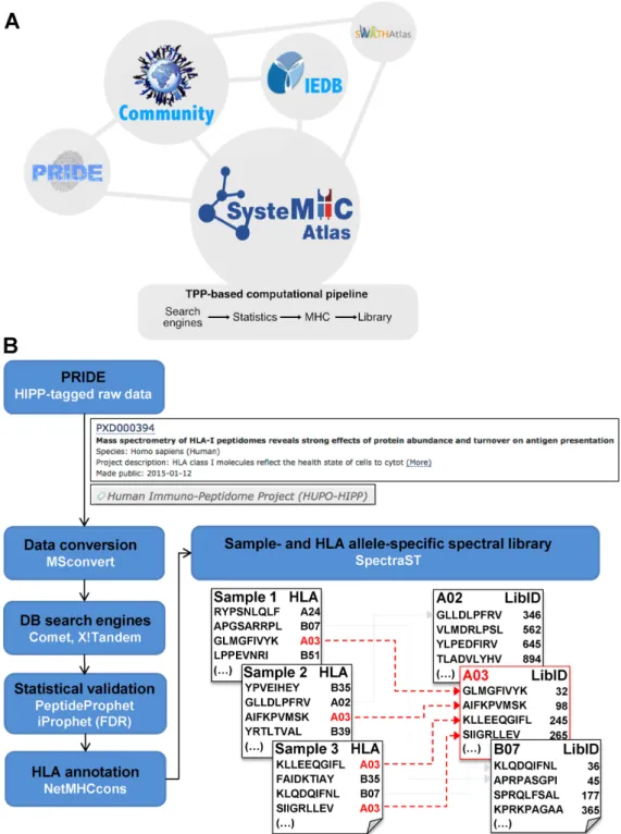
with the metadata associated with the experiment (Figure
1) (28). Each project is labeled with the HIPP tag as a
B
/
D-HPP subproject. Raw MS data are then processed
through a uniform computational pipeline for MHC
peptide identification, annotation (29) and statistical
vali-dation (30,31) (Figure
1B). Lists of MHC peptide ligands
as well as sample
/
context- and allele-specific peptide
spectral libraries (32) are generated and presented in the
Atlas in a way that they can be searched and browsed
by researchers via a web interface. Allele-specific peptide
spectral libraries can be further converted into formats
that are compatible for uploads into the SWATHAtlas
database in order to support immunopeptidomic analyses
by SWATH-MS
/
DIA (Data-Independent Acquisition)
methods. Importantly, the SysteMHC Atlas aims to be an
open and active repository in which raw MS data can be
periodically reprocessed with more advanced informatics
tools for peptide identification, statistical validation, HLA
peptide annotation and library generation, as these become
available to the community––a procedure that has been
successfully applied in the field of proteomics to ensure
high-quality peptide identification with well-understood
false discovery rates (FDR) and quality controls (33). The
community is expected to benefit from the SysteMHC
Atlas at various levels: (i) basic scientists and clinicians
can navigate within a large catalog of high-quality
context-specific HLA-associated peptides to gain new insights into
the composition of the immunopeptidome, (ii)
computa-tional scientists find a rich source of data to develop or test
new algorithms for immunopeptidomic analyses and (iii)
access to HLA peptide assay spectral libraries facilitates
next-generation MS analysis of immunopeptidomes (i.e.
SWATH-MS
/
DIA) (34).
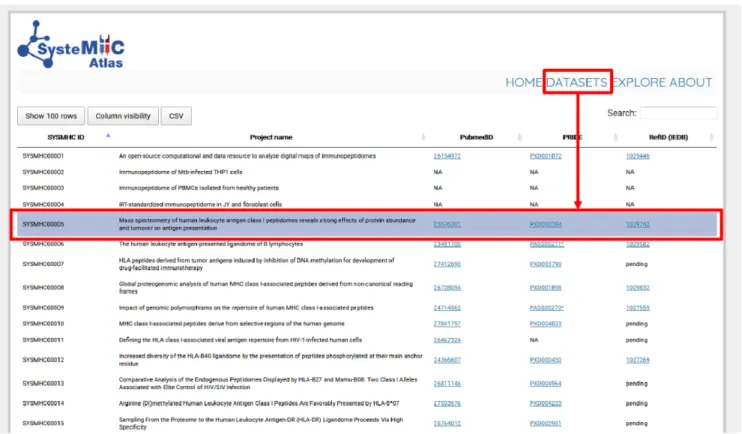
CONTENT AND FUNCTIONALITIES OF THE ATLAS
The first version of the SysteMHC Atlas (February 2017)
contains raw and processed MS data derived from 16
pub-lished human immunopeptidomics projects
/
datasets
(Fig-ure
2). It also contains information from seven unpublished
datasets that were released by the data producers. The
num-ber of MS output files per project ranges between 1 and 192
for a total of 1184 raw files. All datasets were generated in
data-dependent acquisition (DDA) mode using different
in-struments and fragmentation methods, including
collision-induced dissociation (CID), higher energy collisional
dis-sociation (HCD), electron transfer disdis-sociation (ETD) and
electron transfer and higher energy collision dissociation
(EThcD) (21). Several laboratories used the spiked-in
land-mark indexed Retention Time (iRT) peptides for retention
time normalization (35,36). Each dataset is labeled with a
unique and permanent SYSMHC number. Direct links to
PubMed, PRIDE and Immune Epitope Database (IEDB)
are also provided if applicable (Figure
2). We briefly
de-scribe below the content and functionalities of the
Sys-teMHC Atlas.
A catalog of context- and allele-specific MHC class I and
class II peptides
The SysteMHC Atlas contains mainly naturally presented
human MHC class I and class II peptides. Natural
MHC-associated peptides were extracted by immunoaffinity
pu-rification or mild acid elution from cell lines, primary
cells and tissues––i.e. peripheral blood mononuclear cells
(PBMCs), T cells, B cells, dendritic cells, macrophages,
fibroblasts, colon carcinoma, breast cancer and
glioblas-toma. All biological sources were HLA typed and peptides
from 67 HLA class I and 27 HLA class II alleles are
rep-resented in the current version of the database (February
2017). A full listing of all the samples and
correspond-ing metadata (i.e. organism, tissue and cell type, culture
conditions, disease state, HLA type, antibody used for
im-munoaffinity purification, LC-MS/MS parameters etc.) can
be found next to the raw MS files at the project website.
In May 2017,
∼
29.5 million MS/MS spectra were
searched using a uniform and well-tested computational
pipeline and yielded 250, 768 and 1458, 698 distinct peptides
with iProphet probability
P
≥
0.9 and
P
>
0.0, respectively.
After applying strict confidence filters for the identification
of class I and class II peptides, 119 073 high-confidence
HLA class I peptides (peptide FDR 1%, 8–12 amino acids)
were identified and annotated to specific HLA-A, -B or -C
alleles using an automated annotation strategy as described
(34) (see Supplementary Figure S1 for statistics). For class II
molecules, 73 465 high-confidence peptides were identified
(peptide FDR 1%, 10–25 amino acids, belonging to groups
of two or more peptides with an overlap of at least four
amino acids). Of note, the assignment of peptides to
spe-cific HLA class II alleles will be considered in the future as
soon as robust bioinformatics tools for class II peptide
an-notation become openly available (26). The high-confidence
class I and class II peptides were mapped onto 13, 132 and
7704 of the human UniProtKB
/
Swiss-Prot proteins,
respec-tively.
An important goal in MS-based immunopeptidomics is
to assess the size of the human immunopeptidome at the
population level. To answer this question, we plotted the
cu-mulative number of distinct HLA class I peptides as a
func-tion of the addifunc-tion of identified spectra at FDR 1% (Figure
3). Each data point on the curve represents an added
exper-iment, and the experiments are presented in chronological
order of data acquisition. When looking at the combined
data from all HLA class I alleles in the Atlas, our analysis
suggests that for the presently available technology the
satu-ration level might already be reached (Figure
3A). However,
this observation might be biased given the limited number
of HLA alleles as well as the limited number of cell and
tis-sue types that were sampled until now. In addition, when
individual HLA class I alleles were considered, the number
of distinct peptides continued to steeply increase for several
HLA class I allele such as -A02, -C02 and C16 (Figure
3B)
indicating that for these alleles, saturation had not yet been
achieved as the curves are expected to reach saturation only
Figure 1. Overview of the SysteMHC Atlas project. (A) The SysteMHC Atlas aims to be a long-term data-driven project that serves the community. It is linked to other repositories of proteomic data and consists of two main components: (i) a uniform computational pipeline for processing raw MS files and (ii) a web interface with storage, searching and browsing capabilities. First, shotgun/DDA-MS experimental data generated for specific projects are submitted by the data producers to PRIDE. Raw MS data are then uploaded into the SysteMHC Atlas and processed through a consistent and open computational pipeline (B) that controls the quality of peptide identification and peptide annotation to specific HLA alleles. Spectral libraries are generated and can be converted into high-quality HLA allele-specific peptide assay libraries, also available at SWATHAtlas. All the results generated by the computational pipeline are made available to the public domain via the SysteMHC Atlas web-based interface, which provides links to the Immune Epitope Database (IEDB) for accessing lists of peptides originally identified and published by the data producers. (B) Current computational pipeline used for generating the immunopeptidome- and spectral database for different HLA allotypes. MS output files generated from several types of instruments are first converted into mzXML file format and then searched using several open-source database search engines. The resulting peptide identifications are combined and statistically scored using PeptideProphet and iProphet within the Trans-Proteomic Pipeline (TPP) (30,31). The identified peptides are next annotated to their respective HLA allele in a fully automated fashion using the stand-alone software package of NetMHCcons 1.1 (29). Spectral libraries are generated using SpectraST (32). Allele-specific peptide spectral libraries are generated from multiple samples––an example for HLA-A03 is highlighted in red. Each HLA peptide is labeled with a unique and permanent library identifier (LibID). Details regarding the computational pipeline and how the data were processed are available at the SysteMHC Atlas website in the ‘ABOUT’ section.
Figure 2. Immunopeptidomics datasets used for building the first version of the SysteMHC Atlas. Data from 23 projects that collectively generated 1184 raw MS files constitute the initial contents of the SysteMHC Atlas. Each project is labeled with a unique SYSMHC identifier and linked to its corresponding PubMed, PRIDE and IEDB ID. For unpublished projects, IDs are not applicable (NA).
when most peptides observable with the applied
technol-ogy will have been cataloged. Altogether, our current
anal-ysis suggests that the Atlas data are not yet comprehensive.
In the future, collecting additional MS
/
MS data from new
experiments––including from new HLA alleles, new cell
ori-gins, new experimental conditions, new protocols and new
MS technologies––will be necessary to properly assess the
size and complexity of the human immunopeptidome.
In additional to naturally processed ligands, the
Sys-teMHC Atlas also contains data for synthetic peptides
predicted to bind specific HLA alleles. Datasets
gener-ated from synthetic peptides might be particularly
use-ful for benchmarking new software tools (37) and to
ex-tend the contents of libraries derived from native
pep-tides for targeted analysis of immunopeptidomes (3,38,39).
To date, SysteMHC Atlas contains four datasets
com-posed of synthetic peptides: SYSMHC00001 contains
data generated from a large collection of synthetic HLA
class II peptides encoded by
Mycobacterium
tuberculo-sis
(Mtb) (34,40); SYSMHC00020, SYSMHC00021 and
SYSMHC00022 contain data obtained from synthetic HLA
class I peptides encoded by Mtb (41,42), Epstein–Barr virus
(EBV) and Homo sapiens, respectively.
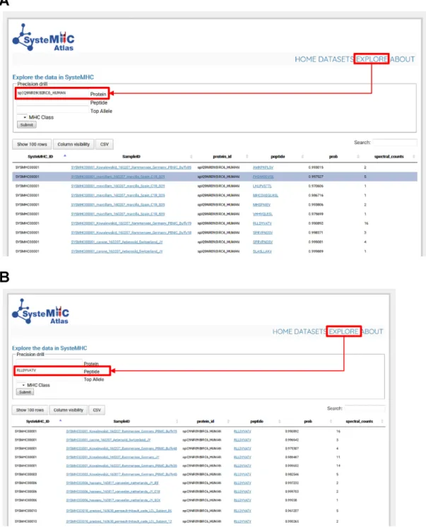
The SysteMHC Atlas user interface
Researchers can browse, search and download
informa-tion using query interfaces available at the website (https:
//systemhcatlas.org). In particular, the ‘EXPLORE’ link
leads to a page where immunopeptidomic data are
search-able on numerous levels, including peptide sequence, source
protein, as well as HLA class and type. For instance, the user
can query the data to specifically identify (i) all class I
pep-tides derived from a specific source protein (e.g. BIRC6 in
Figure
4), (ii) the repertoire of peptides presented by a
spe-cific HLA type and/or (iii) in which tissues or experimental
conditions have specific peptides been observed etc. Thus,
the SysteMHC user interface enables large
immunopep-tidomics datasets to be explored in a user-specifiable
fash-ion.
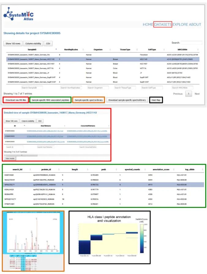
An important function of the SysteMHC Atlas is to serve
as a repository devoted to immunopeptidomics MS-related
data at several levels of processing. Specifically, we provide
raw and converted mzXML files, iProphet results and HLA
peptide spectral libraries, all available for download at the
website (Figure
5). In the current version of the Atlas, a
total of 539 sample
/
context- and 37 HLA allele-specific
peptide spectral libraries were made available and can be
visualized using the respective links from the web
inter-face. Three new allele-specific spectral libraries (i.e.
HLA-B15, -C03 and -C07) were also converted into TraML files
for SWATH-MS/DIA analysis of immunopeptidomes, as
previously described (34,36). These standardized libraries
contained the iRT peptides for retention time
normaliza-tion and data analysis. TraML files are directly available for
download at SWATHAtlas.
Figure 3. Cumulative number of MS/MS spectra versus cumulative number of distinct peptides for HLA class I alleles at FDR 1%. (A) All HLA class I peptides were combined. (B) HLA class I alleles that were frequently found in various datasets. Eventually, the curves are expected to reach saturation when most observable peptides will have been cataloged at 1% peptide FDR.
Figure 4.Explore page in the SysteMHC Atlas web-based interface. HLA allele-specific peptide spectral libraries can be downloaded here. The web interface can also be used to query the SysteMHC Atlas and find specific information. (A) As an example the source protein BIRC6 was searched and the Atlas returned back all HLA-associated peptides originating from this protein as well as the context (i.e. SysteMHC ID, Sample ID, iProphet score, HLA annotation score, spectral counts, assigned HLA type and class) in which this peptide was observed. Then, the user can click on a specific Sample ID hyperlink and be redirected to the corresponding raw MS files and metadata (e.g. tissue type, cell type, culture condition, purification method, antibody used, mass spectrometer used etc). (B) The peptide RLLDYVATV was searched and the Atlas returned back the datasets in which this peptide was observed. By clicking on the peptide sequence hyperlink, the user is redirected to a new page in which the LibID information is available for MS/MS spectra visualization. Information can be downloaded as .csv files for further analysis.
Figure 5. Data storage and visualization. To access information about specific datasets, the user selects a specific SYSMHC ID/Project name (e.g. SYSMHC00005) and clicks on ‘view dataset’ at the bottom left of the screen. The samples related to this project are then listed and linked to the number of replicates, organism, tissue and cell type of origin as well as the HLA typing information (upper panel). The user can then click on a specific Sample ID to visualize the metadata and to download the raw or converted mzXML MS files (red squares). A list of sample-specific HLA-associated peptides can be visualized at 1% peptide-level FDR (green squares). Sample-specific spectral libraries, including consensus fragment ion spectra, can be visualized and downloaded (orange and blue squares). Heat maps (black squares) are used to visualize the annotation of individual peptides to their respective HLA allele (dark blue peptides are predicted to be strong HLA binders according to NetMHCcons).
FUTURE DIRECTIONS
Data sharing, public resources and large-scale/community
projects are growing in popularity and necessity in life
sciences (43–48), and specifically in proteomics (49,50)
where public data sharing is growing exponentially in
re-cent years. Along this line, the SysteMHC Atlas
repre-sents the first community-driven resource devoted to
col-lect, store, organize and share large immunopeptidomics
datasets generated by MS methods––an important
contri-bution to the Human Immuno-Peptidome Project (25,27).
The SysteMHC Atlas will be further developed and
en-hanced to enable public dissemination of uniform and
high-quality immunopeptidome data generated by an open and
ever-improving computational pipeline. To this end, raw
MS data will be reprocessed periodically using novel
high-performance software tools as they are made available to the
community. Future software tools are expected to
outper-form current algorithms for (i) MHC peptide identification,
(ii) MHC peptide FDR estimation in large
immunopep-tidomic datasets and (iii) class I and class II peptide
an-notation to specific HLA alleles, as described (http://www.
biorxiv.org/content/early/2017/05/13/098780) (51). In the
near future, we aim at providing the necessary tools to
re-trieve information on post-translationally modified
MHC-associated peptides: phosphopeptides, Arg-methylated
pep-tides and proteasome-generated spliced peppep-tides in
par-ticular, as those might be of particular relevance for the
rational design of immunotherapeutic interventions (52–
56). We also plan to identify the potential for large-scale
integration and interoperability of all immunopeptidomic
data with PRIDE (28), IEDB (57) and SWATHAtlas (34).
Thus, we intend the SysteMHC Atlas to become a
grow-ing community-driven database and an interoperable,
high-performance infrastructure for systematic analysis of
ter-abytes of immunopeptidomic big data. If successful in
longer term, we anticipate that the SysteMHC Atlas project
will provide key insights to the immunology community and
will foster the development of vaccines and
immunother-apies against various immune-related diseases such as
au-toimmunity, allergies, infectious diseases and cancers.
SUPPLEMENTARY DATA
Supplementary Data are available at NAR Online.
ACKNOWLEDGEMENTS
We thank Layla Lang (www.laylalang.com) for illustrating
the immunopeptidomic landscape of cells on the website
home page and An Guo for designing the logo of the
Sys-teMHC Atlas. We thank Lorenz Blum and Pascal K¨agi for
computational assistance. We also thank all members of the
Aebersold and Wollscheid laboratories for discussions.
FUNDING
TBVAC2020 [2–73838-14 to R.A.]; ERC grant Proteomics
v3.0 [ERC-2008-AdG 20080422 to R.A.]; ERC Proteomics
4D [670821 to R.A.]; Swiss National Science
Founda-tion [3100A0–688 107679 to R.A.]; Netherlands
Organi-zation for Scientific Research (NWO) (Roadmap Initiative
Proteins@Work (project number 184.032.201)) (to F.M.,
A.J.R.H.); EC HOR2020 project TBVAC2020 [643381
to T.H.M.O.]; Wellcome Trust [WT101477MA to J.A.V.];
National Institute of Allergy and Infectious Diseases
[HHSN272201200010C to A.S.]; National Health and
Medical Council of Australia Senior Research Fellowship
[APP1044215 to A.W.P.]; ERC [AdG339842 to H.-G.R.];
Mutaediting (to H.-G.R.); NIH, National Institute of
Gen-eral Medical Sciences [R01 GM087221 to E.W.D., R.L.M.];
2P50 GM076547/Center for Systems Biology (to R.L.M.);
Research Council of Norway [179573
/
V40 to A.G.deS.,
L.M.S., in part]; NIH National Institute of General
Medi-cal Sciences [P41 GM104603 to C.E.C.]. Funding for open
access charge: TBVAC2020 [2-73838-14].
Conflict of interest statement.
None declared.
REFERENCES
1. Istrail,S., Florea,L., Halld ´orsson,B.V., Kohlbacher,O., Schwartz,R.S., Yap,V.B., Yewdell,J.W. and Hoffman,S.L. (2004) Comparative immunopeptidomics of humans and their pathogens.Proc. Natl. Acad. Sci. U.S.A.,101, 13268–13272.
2. Caron,E., Vincent,K., Fortier,M.-H., Laverdure,J.-P., Bramoull´e,A., Hardy,M.-P., Voisin,G., Roux,P.P., Lemieux,S., Thibault,P.et al. (2011) The MHC I immunopeptidome conveys to the cell surface an integrative view of cellular regulation.Mol. Syst. Biol.,7, 533–533. 3. Caron,E., Kowalewski,D.J., Koh,C.C., Sturm,T., Schuster,H. and
Aebersold,R. (2015) Analysis of major histocompatibility complex (MHC) immunopeptidomes using mass spectrometry.Mol. Cell. Proteomics,14, 3105–3117.
4. Neefjes,J., Jongsma,M.L.M., Paul,P. and Bakke,O. (2011) Towards a systems understanding of MHC class I and MHC class II antigen presentation.Nat. Rev. Immunol.,11, 823–836.
5. Rock,K.L., Reits,E. and Neefjes,J. (2016) Present yourself! by MHC class I and MHC class II molecules.Trends Immunol.,37, 724–737. 6. Falk,K., R ¨otzschke,O., Stevanovic,S., Jung,G. and Rammensee,H.-G.
(1991) Allele-specific motifs revealed by sequencing of self-peptides eluted from MHC molecules.Nature,351, 290–296.
7. Cole,D.K. (2015) The ultimate mix and match: making sense of HLA alleles and peptide repertoires.Immunol. Cell Biol.,93, 515–516. 8. Bassani-Sternberg,M. and Coukos,G. (2016) Mass
spectrometry-based antigen discovery for cancer immunotherapy. Curr. Opin. Immunol.,41, 9–17.
9. Mann,M. (2016) Origins of mass spectrometry-based proteomics. Nat. Rev. Mol. Cell Biol.,17, 678.
10. R ¨otzschke,O., Falk,K., Deres,K., Schild,H., Norda,M., Metzger,J., Jung,G. and Rammensee,H.G. (1990) Isolation and analysis of naturally processed viral peptides as recognized by cytotoxic T cells. Nature,348, 252–254.
11. Hunt,D.F., Henderson,R.A., Shabanowitz,J., Sakaguchi,K., Michel,H., Sevilir,N., Cox,A.L., Appella,E. and Engelhard,V.H. (1992) Characterization of peptides bound to the class I MHC molecule HLA-A2.1 by mass spectrometry.Science,255, 1261–1263. 12. Hassan,C., Kester,M.G.D., de Ru,A.H., Hombrink,P.,
Drijfhout,J.W., Nijveen,H., Leunissen,J.A.M., Heemskerk,M.H.M., Falkenburg,J.H.F. and van Veelen,P.A. (2013) The human leukocyte antigen-presented ligandome of B lymphocytes.Mol. Cell. Proteomics,12, 1829–1843.
13. Bergseng,E., Dørum,S., Arntzen,M.Ø., Nielsen,M., Nyg˚ard,S., Buus,S., de Souza,G.A and Sollid,L.M. (2014) Different binding motifs of the celiac disease-associated HLA molecules DQ2.5, DQ2.2, and DQ7.5 revealed by relative quantitative proteomics of endogenous peptide repertoires.Immunogenetics,67, 73–84. 14. Pearson,H., Daouda,T., Granados,D.P., Durette,C., Bonneil,E.,
Courcelles,M., Rodenbrock,A., Laverdure,J.-P., C ˆot´e,C., Mader,S. et al.(2016) MHC class I-associated peptides derive from selective regions of the human genome.J. Clin. Invest.,126, 4690–4701. 15. Abelin,J.G., Keskin,D.B., Sarkizova,S., Hartigan,C.R., Zhang,W.,
Sidney,J., Stevens,J., Lane,W., Zhang,G.L., Eisenhaure,T.M.et al. (2017) Mass spectrometry profiling of HLA-associated peptidomes in
mono-allelic cells enables more accurate epitope prediction. Immunity,46, 315–326.
16. Bassani-Sternberg,M., Br¨aunlein,E., Klar,R., Engleitner,T., Sinitcyn,P., Audehm,S., Straub,M., Weber,J., Slotta-Huspenina,J., Specht,K.et al.(2016) Direct identification of clinically relevant neoepitopes presented on native human melanoma tissue by mass spectrometry.Nat. Commun.,7, 13404.
17. Khodadoust,M.S., Olsson,N., Wagar,L.E., Haabeth,O.A.W., Chen,B., Swaminathan,K., Rawson,K., Liu,C.L., Steiner,D., Lund,P. et al.(2017) Antigen presentation profiling reveals recognition of lymphoma immunoglobulin neoantigens.Nature,543, 723–727. 18. Kowalewski,D.J., Schuster,H., Backert,L., Berlin,C., Kahn,S.,
Kanz,L., Salih,H.R., Rammensee,H.-G., Stevanovic,S. and Stickel,J.S. (2014) HLA ligandome analysis identifies the underlying specificities of spontaneous antileukemia immune responses in chronic lymphocytic leukemia (CLL).Proc. Natl. Acad. Sci. U.S.A.,
112, E116–E175.
19. Mommen,G.P.M., Marino,F., Meiring,H.D., Poelen,M.C.M., van Gaans-van den Brink,J.A.M., Mohammed,S., Heck,A.J.R. and van Els,C.A.C.M. (2016) Sampling from the proteome to the human leukocyte antigen-DR (HLA-DR) ligandome proceeds via high specificity.Mol. Cell. Proteomics,15, 1412–1423.
20. Schellens,I.M.M., Hoof,I., Meiring,H.D., Spijkers,S.N.M., Poelen,M.C.M., van Gaans-van den Brink,J.A.M., van der Poel,K., Costa,A.I., van Els,C.A.C.M., van Baarle,D.et al.(2015)
Comprehensive analysis of the naturally processed peptide repertoire: differences between HLA-A and B in the immunopeptidome.PLoS One,10, e0136417.
21. Mommen,G.P.M., Frese,C.K., Meiring,H.D., van Gaans-van den Brink,J., de Jong,A.P.J.M., van Els,C.A.C.M. and Heck,A.J.R. (2014) Expanding the detectable HLA peptide repertoire using
electron-transfer/higher-energy collision dissociation (EThcD).Proc. Natl. Acad. Sci. U.S.A.,111, 4507–4512.
22. Wang,Q., Drouin,E.E., Yao,C., Zhang,J., Huang,Y., Leon,D.R., Steere,A.C. and Costello,C.E. (2016) Immunogenic
HLA-DR-presented self-peptides identified directly from clinical samples of synovial tissue, synovial fluid, or peripheral blood in patients with rheumatoid arthritis or lyme arthritis.J. Proteome Res.,
16, 122–136.
23. Pardoll,D.M. (2012) The blockade of immune checkpoints in cancer immunotherapy.Nat. Rev. Cancer,12, 252–264.
24. van Eyk,J.E., Corrales,F.J., Aebersold,R., Cerciello,F., Deutsch,E.W., Roncada,P., Sanchez,J.-C., Yamamoto,T., Yang,P., Zhang,H.et al. (2016) Highlights of the Biology and Disease-driven Human Proteome Project, 2015–2016.J. Proteome Res.,15, 3979–3987. 25. Caron,E. and Aebersold,R. (2016) The Human Immuno-Peptidome
Project: a new initiative of B/D-HPP Program. In:15th Human Proteome Organization World Congress. Taipei.
26. Sette,A., Schenkelberg,T.R. and Koff,W.C. (2015) Deciphering the human antigenome.Expert Rev. Vaccines,15, 167–171.
27. Admon,A. and Bassani-Sternberg,M. (2011) The Human
Immunopeptidome Project, a suggestion for yet another postgenome next big thing.Mol. Cell. Proteomics,10, 1–4.
28. Vizca´ıno,J.A., Csordas,A., del-Toro,N., Dianes,J.A., Griss,J., Lavidas,I., Mayer,G., Perez-Riverol,Y., Reisinger,F., Ternent,T.et al. (2016) 2016 update of the PRIDE database and its related tools. Nucleic Acids Res.,44, D447–D456.
29. Karosiene,E., Lundegaard,C., Lund,O. and Nielsen,M. (2012) NetMHCcons: a consensus method for the major histocompatibility complex class I predictions.Immunogenetics,64, 177–186.
30. Shteynberg,D., Nesvizhskii,A.I., Moritz,R.L. and Deutsch,E.W. (2013) Combining results of multiple search engines in proteomics. Mol. Cell. Proteomics,12, 2383–2393.
31. Shteynberg,D., Deutsch,E.W., Lam,H., Eng,J.K., Sun,Z., Tasman,N., Mendoza,L., Moritz,R.L., Aebersold,R. and Nesvizhskii,A.I. (2011) iProphet: Multi-level integrative analysis of shotgun proteomic data improves peptide and protein identification rates and error estimates. Mol. Cell. Proteomics,10, 1–15.
32. Lam,H., Deutsch,E.W., Eddes,J.S., Eng,J.K., Stein,S.E. and Aebersold,R. (2008) Building consensus spectral libraries for peptide identification in proteomics.Nat. Methods,5, 873–875.
33. Desiere,F., Deutsch,E.W., King,N.L., Nesvizhskii,A.I., Mallick,P., Eng,J., Chen,S., Eddes,J., Loevenich,S.N. and Aebersold,R. (2006) The PeptideAtlas project.Nucleic Acids Res.,34, D655–D658.
34. Caron,E., Espona,L., Kowalewski,D.J., Schuster,H., Ternette,N., Alp´ızar,A., Schittenhelm,R.B., Ramarathinam,S.H., Lindestam Arlehamn,C.S., Chiek Koh,C.et al.(2015) An open-source computational and data resource to analyze digital maps of immunopeptidomes.Elife,4, e07661.
35. Escher,C., Reiter,L., MacLean,B., Ossola,R., Herzog,F., Chilton,J., MacCoss,M.J. and Rinner,O. (2012) Using iRT, a normalized retention time for more targeted measurement of peptides. Proteomics,12, 1111–1121.
36. Faridi,P., Aebersold,R. and Caron,E. (2016) A first dataset toward a standardized community-driven global mapping of the human immunopeptidome.Data Brief.,7, 201–205.
37. R ¨ost,H.L., Rosenberger,G., Navarro,P., Gillet,L., Miladinovi´c,S.M., Schubert,O.T., Wolski,W., Ben C,Collins, Malmstr ¨om,J.,
Malmstr ¨om,L.et al.(2014) OpenSWATH enables automated, targeted analysis of data- independent acquisition MS data.Nat. Biotechnol.,32, 219–223.
38. Croft,N.P., Purcell,A.W. and Tscharke,D.C. (2015) Quantifying epitope presentation using mass spectrometry.Mol. Immunol.,68, 77–80.
39. Gubin,M.M., Zhang,X., Schuster,H., Caron,E., Ward,J.P., Noguchi,T., Ivanova,Y., Hundal,J., Arthur,C.D., Krebber,W.-J.et al. (2014) Checkpoint blockade cancer immunotherapy targets tumour-specific mutant antigens.Nature,515, 577–581.
40. Lindestam Arlehamn,C.S., Gerasimova,A., Mele,F., Henderson,R., Swann,J., Greenbaum,J.A., Kim,Y., Sidney,J., James,E.A., Taplitz,R. et al.(2013) Memory T cells in latent Mycobacterium tuberculosis infection are directed against three antigenic islands and largely contained in a CXCR3+CCR6+ Th1 subset.PLoS Pathog.,9, e1003130.
41. Tang,S.T., van Meijgaarden,K.E., Caccamo,N., Guggino,G., Klein,M.R., van Weeren,P., Kazi,F., Stryhn,A., Zaigler,A., Sahin,U. et al.(2011) Genome-based in silico identification of new
mycobacterium tuberculosis antigens activating polyfunctional CD8+ T cells in human tuberculosis.J. Immunol.,186, 1068–1080. 42. Joosten,S.A., van Meijgaarden,K.E., van Weeren,P.C., Kazi,F.,
Geluk,A., Savage,N.D.L., Drijfhout,J.W., Flower,D.R., Hanekom,W.A., Klein,M.R.et al.(2010) Mycobacterium tuberculosis peptides presented by HLA-E molecules are targets for human CD8+ T-cells with cytotoxic as well as regulatory activity. PLoS Pathog.,6, e1000782.
43. Aebersold,R., Bader,G.D., Edwards,A.M., van Eyk,J.E., Kussmann,M., Qin,J. and Omenn,G.S. (2013) The
Biology/Disease-driven Human Proteome Project (B/D-HPP): enabling protein research for the life sciences community.J. Proteome Res.,12, 23–27.
44. Uhl´en,M., Oksvold,P., Fagerberg,L., Lundberg,E., Jonasson,K., Forsberg,M., Zwahlen,M., Kampf,C., Wester,K., Hober,S.et al. (2010) Towards a knowledge-based Human Protein Atlas.Nat. Biotechnol.,28, 1248–1250.
45. Kusebauch,U., Campbell,D.S., Deutsch,E.W., Chu,C.S., Spicer,D.A., Brusniak,M.-Y., Slagel,J., Sun,Z., Stevens,J., Grimes,B.et al.(2016) Human SRMAtlas: a resource of targeted assays to quantify the complete human proteome.Cell,166, 766–778.
46. Whiteaker,J.R., Halusa,G.N., Hoofnagle,A.N., Sharma,V., MacLean,B., Yan,P., Wrobel,J.A., Kennedy,J., Mani,D.R., Zimmerman,L.J.et al.(2014) CPTAC Assay Portal: a repository of targeted proteomic assays.Nat. Methods,11, 703–704.
47. GTEx Consortium (2013) The Genotype-Tissue Expression (GTEx) project.Nat. Genet.,45, 580–585.
48. McKiernan,E.C., Bourne,P.E., Brown,C.T., Buck,S., Kenall,A., Lin,J., McDougall,D., Nosek,B.A., Ram,K., Soderberg,C.K.et al. (2016) How open science helps researchers succeed.Elife,5, e16800. 49. Vaudel,M., Verheggen,K., Csordas,A., Raeder,H., Berven,F.S.,
Martens,L., Vizca´ıno,J.A. and Barsnes,H. (2016) Exploring the potential of public proteomics data.Proteomics,16, 214–225. 50. Martens,L. and Vizca´ıno,J.A. (2017) A golden age for working with
public proteomics data.Trends Biochem. Sci.,42, 333–341. 51. Bassani-Sternberg,M. and Gfeller,D. (2016) Unsupervised HLA
peptidome deconvolution improves ligand prediction accuracy and predicts cooperative effects in peptide–HLA interactions.J. Immunol.,197, 2492–2499.
52. Zarling,A.L., Polefrone,J.M., Evans,A.M., Mikesh,L.M., Shabanowitz,J., Lewis,S.T., Engelhard,V.H. and Hunt,D.F. (2006)
Identification of class I MHC-associated phosphopeptides as targets for cancer immunotherapy.Proc. Natl. Acad. Sci. U.S.A.,103, 14889–14894.
53. Cobbold,M., De La Pe ˜na,H, Norris,A., Polefrone,J.M., Qian,J., English,A.M., Cummings,K.L., Penny,S., Turner,J.E., Cottine,J. et al.(2013) MHC class I-associated phosphopeptides are the targets of memory-like immunity in leukemia.Sci. Transl. Med.,5, 203ra125. 54. Marino,F., Mommen,G.P.M., Jeko,A., Meiring,H.D., van Gaans-van
den Brink,J.A.M., Scheltema,R.A., van Els,C.A.C.M. and Heck,A.J.R. (2016) Arginine (Di)methylated Human Leukocyte Antigen Class I Peptides Are Favorably Presented by HLA-B*07.J. Proteome Res.,16, 34–44.
55. Liepe,J., Marino,F., Sidney,J., Jeko,A., Bunting,D.E., Sette,A., Kloetzel,P.M., Stumpf,M.P., Heck,A.J. and Mishto,M. (2016) A large fraction of HLA class I ligands are proteasome-generated spliced peptides.Science,354, 354–358.
56. Delong,T., Wiles,T.A., Baker,R.L., Bradley,B., Barbour,G., Reisdorph,R., Armstrong,M., Powell,R.L., Reisdorph,N., Kumar,N. et al.(2016) Pathogenic CD4 T cells in type 1 diabetes recognize epitopes formed by peptide fusion.Science,351, 711–714.
57. Vita,R., Overton,J.A., Greenbaum,J.A., Ponomarenko,J., Clark,J.D., Cantrell,J.R., Wheeler,D.K., Gabbard,J.L., Hix,D., Sette,A.et al. (2015) The immune epitope database (IEDB) 3.0.Nucleic Acids Res.,
43, D405–D412.